

Abstract
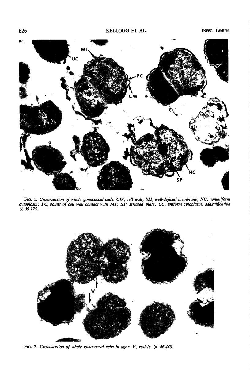
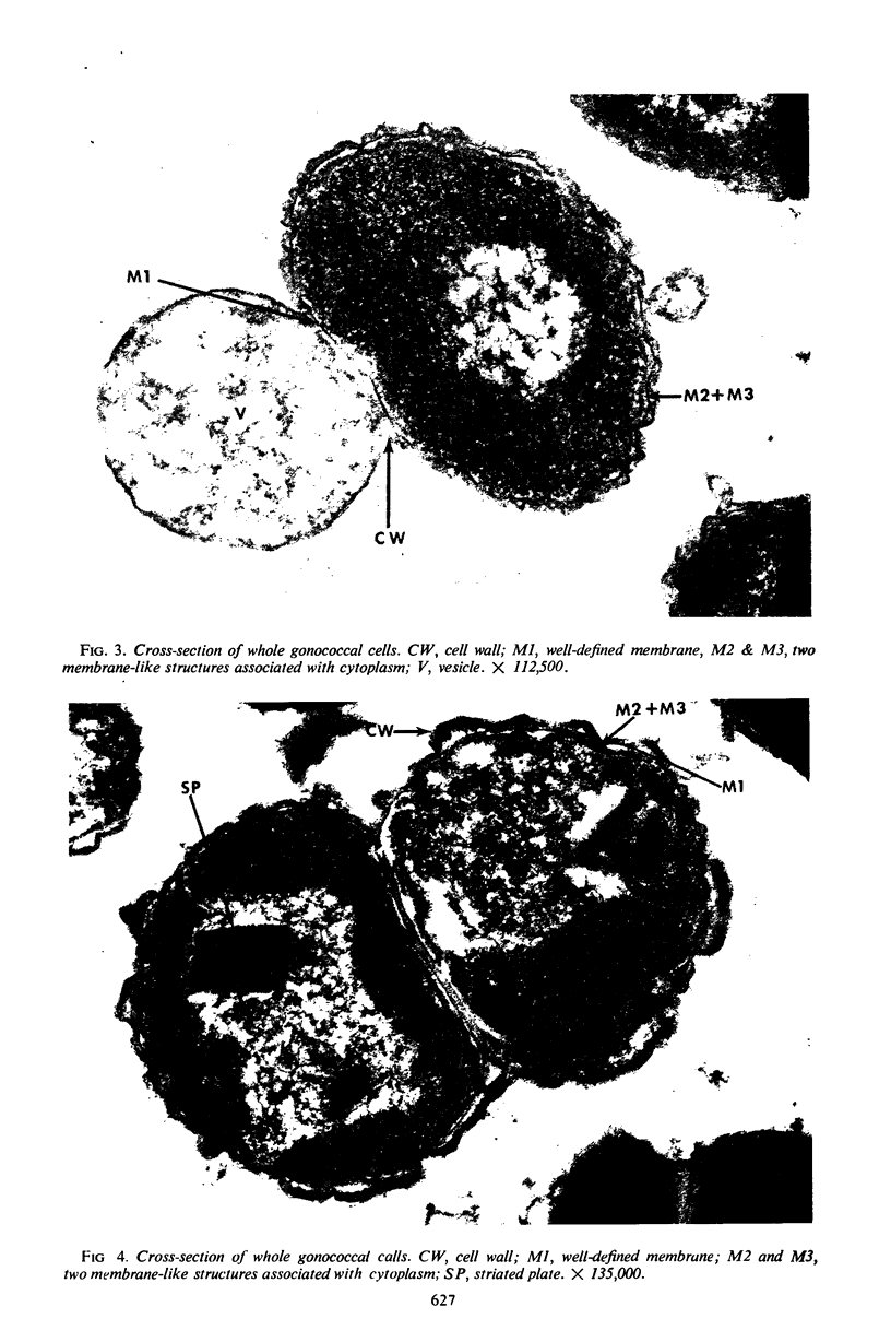
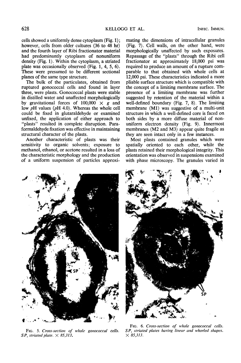
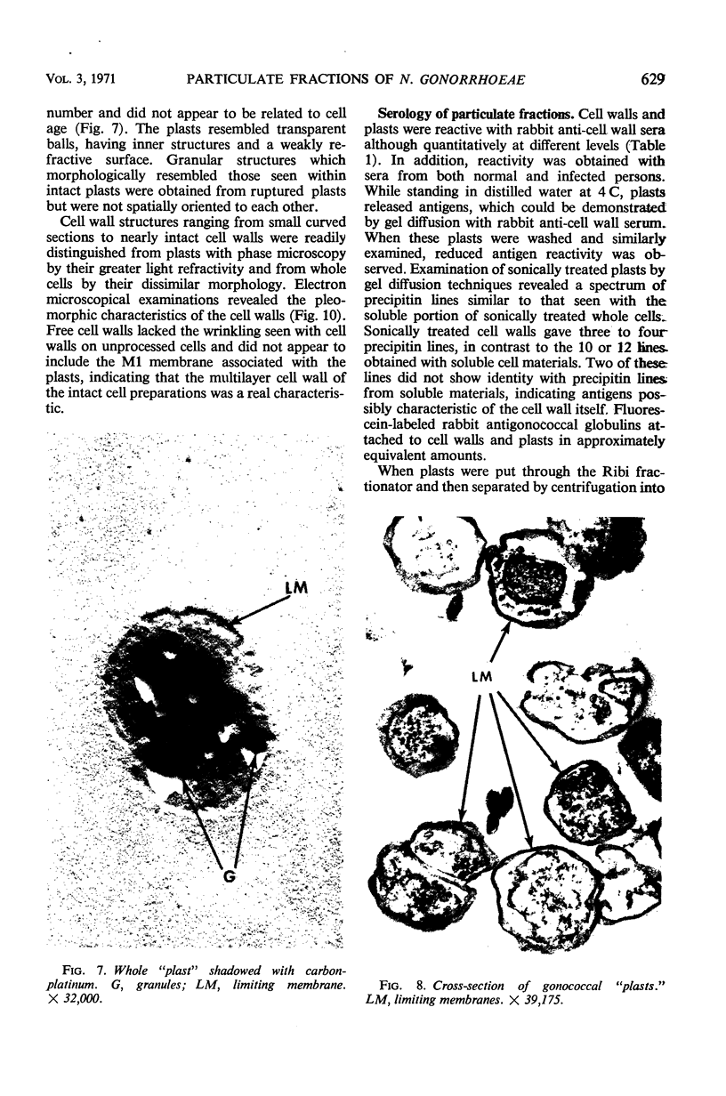
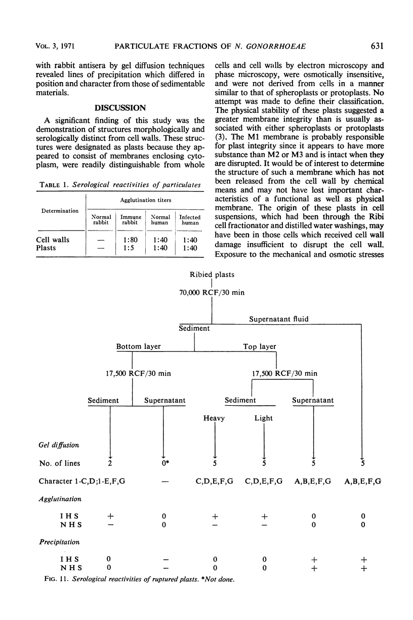
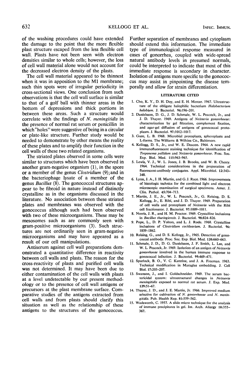
Passage of gonococcal cells through a Ribi cell fractionator produced a mixture of protoplasm and cell particulates, the latter being separable in sucrose solutions. Separation of these particulates resulted in the detection of objects identified as cell walls and “plasts.” The latter objects had an average diameter of 1.00 ± 0.05 μm and consisted of spatially oriented granules surrounded by a membrane-like structure only. Cell wall structures were not observed with these plasts. Plasts were morphologically stable in distilled water and at low pH values. Although 50% more pressure was required to rupture plasts than whole cells, plast morphology was destroyed by organic solvents. Whole cell and plast morphology was examined by electron and phase microscopy, and certain correlations were made between structure and morphological stability. Cell wall antiserum reactivities were quantitatively different for plasts and cell walls. Serological reactivities of plasts were separable by sonic oscillation and differential centrifugation of the sonically treated material.
Full text
PDF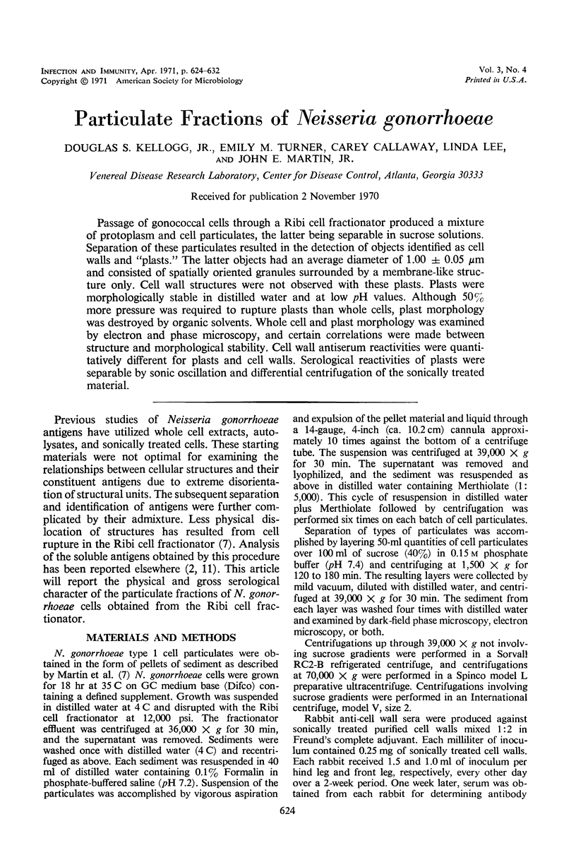


Images in this article


Selected References
These references are in PubMed. This may not be the complete list of references from this article.
- Cho K. Y., Doy C. H., Mercer E. H. Ultrastructure of the obligate halophilic bacterium Halobacterium halobium. J Bacteriol. 1967 Jul;94(1):196–201. doi: 10.1128/jb.94.1.196-201.1967. [DOI] [PMC free article] [PubMed] [Google Scholar]
- Danielsson D. G., Schmale J. D., Peacock W. L., Jr, Thayer J. D. Antigens of Neisseria gonorrhoeae: characterization by gel filtration, complement fixation, and agar-gel diffusion of antigens of gonococcal protoplasm. J Bacteriol. 1969 Mar;97(3):1012–1017. doi: 10.1128/jb.97.3.1012-1017.1969. [DOI] [PMC free article] [PubMed] [Google Scholar]
- KELLOGG D. S., Jr, DEACON W. E. A NEW RAPID IMMUNOFLUORESCENT STAINING TECHNIQUE FOR IDENTIFICATION OF TREPONEMA PALLIDUM AND NEISSERIA GONORRHOEAE. Proc Soc Exp Biol Med. 1964 Apr;115:963–965. doi: 10.3181/00379727-115-29090. [DOI] [PubMed] [Google Scholar]
- LEWIS V. J., JONES W. L., BROOKS J. B., CHERRY W. B. TECHNICAL CONSIDERATIONS IN THE PREPARATION OF FLUORESCENT-ANTIBODY CONJUGATES. Appl Microbiol. 1964 Jul;12:343–348. doi: 10.1128/am.12.4.343-348.1964. [DOI] [PMC free article] [PubMed] [Google Scholar]
- Lynn J. A., Martin J. H., Race G. J. Recent improvements of histologic technics for the combined light and electron microscopic examination of surgical specimens. Am J Clin Pathol. 1966 Jun;45(6):704–713. doi: 10.1093/ajcp/45.6.704. [DOI] [PubMed] [Google Scholar]
- Martin J. E., Jr, Peacock W. L., Jr, Reising G., Kellogg D. S., Jr, Ribi E., Thayer J. D. Preparation of cell walls and protoplasm of Neisseria with the Ribi cell fractionator. J Bacteriol. 1969 Mar;97(3):1009–1011. doi: 10.1128/jb.97.3.1009-1011.1969. [DOI] [PMC free article] [PubMed] [Google Scholar]
- Norris J. R., Proctor H. M. Crystalline inclusions in Bacillus thuringiensis. J Bacteriol. 1969 May;98(2):824–826. doi: 10.1128/jb.98.2.824-826.1969. [DOI] [PMC free article] [PubMed] [Google Scholar]
- Pope L., Yolton D. P., Rode L. J. Crystalline inclusions of Clostridium cochlearium. J Bacteriol. 1968 Nov;96(5):1859–1862. doi: 10.1128/jb.96.5.1859-1862.1968. [DOI] [PMC free article] [PubMed] [Google Scholar]
- Reising G., Kellogg D. S., Jr Detection of gonococcal antibody. Proc Soc Exp Biol Med. 1965 Dec;120(3):660–663. doi: 10.3181/00379727-120-30617. [DOI] [PubMed] [Google Scholar]
- Schmale J. D., Danielsson D. G., Smith J. F., Lee L., Peacock W. L., Jr Isolation of an antigen of Neisseria gonorrhoeae involved in the human immune response to gonococcal infection. J Bacteriol. 1969 Aug;99(2):469–471. doi: 10.1128/jb.99.2.469-471.1969. [DOI] [PMC free article] [PubMed] [Google Scholar]
- Swanson J., Goldschneider I. The serum bactericidal system: ultrastructural changes in Neisseria meningitidis exposed to normal rat serum. J Exp Med. 1969 Jan 1;129(1):51–79. doi: 10.1084/jem.129.1.51. [DOI] [PMC free article] [PubMed] [Google Scholar]
- Thayer J. D., Martin J. E., Jr Improved medium selective for cultivation of N. gonorrhoeae and N. meningitidis. Public Health Rep. 1966 Jun;81(6):559–562. [PMC free article] [PubMed] [Google Scholar]
- WADSWORTH C. A slide microtechnique for the analysis of immune precipitates in gel. Int Arch Allergy Appl Immunol. 1957;10(6):355–360. doi: 10.1159/000228394. [DOI] [PubMed] [Google Scholar]