Abstract
Background:
The aerobic Actinomycetes are a large group of soil-indwelling bacteria that are distributed in world-wide. These Gram-positive bacteria are most commonly associated with opportunistic infections in both immunocompromised and immunocompetent hosts.
Materials and Methods:
In this study, three phenotypic and deoxyribonucleic acid (DNA) extraction methods for isolation and identification of Nocardia genus were compared. Samples were taken in five different locations of Isfahan's suburb from hospitals area, parks, agricultural lands, gardens, arid lands with different soil temperature and pH.
Results:
In this study, showed that slip-buried-method was better than two other phenotypic methods; 14 out of 70 soil samples (20%) were positive for Nocardia spp. DNA of positive samples were extracted with three techniques and DNA extraction by microwave technique was better than others. This technique was confirmed with observation of DNA bands on 1% agarose gel.
Conclusions:
These bacteria are important in immune deficient patients such as cancer patients, transplant recipients, tuberculosis; acquired immunodeficiency syndrome etc., Their affluence is unsteady in different zones of the world. In this study, among the three phenotypic methods for the isolation of Nocardia slip-buried method was better than other methods. Among DNA extraction techniques, DNA extraction by microwave method would be selective method for DNA extraction of Nocardia spp. compared with others techniques.
Keywords: Deoxyribonucleic acid extraction techniques, Nocardia spp., phenotypic methods, soil
BACKGROUND
Nocardia spp. are aerobic, Gram-positive, modified acid fast, non-motile, non-spore - forming,[1,2] catalase and urease positive bacteria that belong to Actinomycetes group.[3] These bacteria are filamentous branched cells which fragmented into pleomorphic rod-shaped or coccoid elements and classified as Nocardiaceae family.[4] Soil is one of the best reservoir for keeping Nocardia. Nocardia species are determined by a number of chemical markers, entailing the presence of meso-diaminopimelic acid, arabinose and galactose, mycolic acids and deoxyribonucleic acid (DNA) G+C containing 64-72%.[5] However, these organisms are found in environments such as air, water, plants and rotten materials. Nocardia may be found in dry, dusty and often windy situations in that areas facilitate the aerosolization and scattering of fragmented nocardial cells and increase their acquisition through the respiratory path. Fewer infections are due to traumatic inoculation of organisms percutaneously.[6] Nocardia species are associated with opportunistic infections in human and animals that might become fatal. These bacteria are important in immune deficient patients such as cancer patients, transplant recipients, tuberculosis; acquired immunodeficiency syndrome etc., Their affluence is unsteady in different zones of the world.[1,6,7,8,9,10,11,12,13,14,15,16] Nocardiosis is sporadic in Iran, but in other countries, it has more rampancy. In USA, 500-1000 new case of nocardiosis had annually been reported.[1] Nocardia organisms found in different zones and situations like tropical and semi-tropical climates depending upon soil's temperature, pH and these effects on the abundance of these microorganisms in soil.[1] Until 2010, the National Center for Biotechnology Information itemizes 86 acknowledged species.[13] Until 2012, the list of prokaryotic names with standing in nomenclature itemizes 99 confirmed species. In this study, several phenotypic and DNA extraction techniques for isolation and identification of these bacteria from different environment have been compared. Three phenotypic methods in this study were perused: (1) Paraffin baiting technique, (2) paraffin coated slides and (3) slip-buried method. As well as three DNA extraction techniques were tested: (1) Genomic DNA purification kit (Fermentas), (2) cetyltrimethylammonium bromide (CTAB) method and (3) microwave oven.[17]
MATERIALS AND METHODS
A total 70 soil samples were collected from five different locations in Isfahan's suburb hospital areas, parks, agricultural lands, gardens and arid lands at different months of year. 50 g of soil samples were collected from 3 cm to 5 cm depth.[7] The samples were transported to laboratory in sterile tubes. Soil samples were suspended 1:5 in deionized water[1] and the pH were measured by pH meter or pH-indicator strips for 1-30 s (maximum 10 min). To determine the temperature, alcoholic thermometer was used. All samples were stored at low temperature (4°C) until tested.[1] In this study, three phenotypic methods were compared for isolation of Nocardia: (1) Paraffin baiting method,[8,9,18] (2) paraffin coated slides[12] and (3) slip-buried method.[1,7]
Paraffin baiting technique
Ten gram soil sample was added to 15 ml carbon free broth (containing NaNO3: 2 g, K2HPO4: 0.8 g, MgSO4.7H2O: 0.5 g, FeCL3: 10 mg, MnCl2.4H2O: 8 mg, ZnSO4: 2 mg, distilled water: 1000 ml; pH: 7.0) and mixed thoroughly. The mixture was pre-heated at 55°C for 6 min in water bath to remove some of the non-actinomycetic soil bacteria. The supernatant were transferred in equal volumes into two tubes. A paraffin waxed glass rod was soaked in each tube and its volume was made up to 10 ml by adding carbon-free broth. One tube was incubated at 35°C and others at 42°C for 3 weeks. Nocardia colonies were appeared white creamy to pinkish-orange. Then, the colonies were streaked on brain-heart infusion (BHI)-blood agar[8] or sabouraud dextrose agar (SDA) with cycloheximide (50 μg/ml) with or without chloramphenicol (50 μg/ml). The colonies were stained for Gram-positive and partial acid-fast characteristics. Afterward the colonies were streaked on BHI-blood agar or SDA with cycloheximide (50 μg/ml) with or without chloramphenicol (50 μg/ml). Identification was preceded by isolation of colonies.[8,9,18]
Paraffin coated slides
slides were coated with melted paraffin and 1 M NH4 Cl. Immediately, the slides put under the soil, for 1 day. Slides were incubated for 3-7 days in 37 and incubation was continued for 3 weeks. Then, the colonies were streaked on SDA medium with cycloheximide (50 μg/ml).[12]
Slip-buried method
Soil, 3-5 g, was added to 10 ml normal saline. Tubes were shaken for 3 min and the suspensions were incubated for 15 min in room temperature. Supernatant solution, 3-5 ml was transferred to another sterile tube by sterile pipette. The streptomycin/chloramphenicol solution (half of the total volume) was added to the supernatant. The mixture was incubated for ½ h. One drop (0.05 ml) of each sample was cultured on BHI agar with 5% human blood[8] medium and checked for hemolysis. Cycloheximide (0.5 g/l) and kanamycin (25 mg/l) was added to the tube or plate immediately. They were shacked and were incubated at 37°C for 2 weeks.[1]
DNA extraction techniques
In this study, three DNA extraction methods were tested: (1) Genomic DNA purification kit (Fermentas), (2) CTAB method[19] and (3) microwave oven.[17] All methods were tested with Nocardia asteroides DSM is German Collection of Microorganisms and Cell Cultures and DSM number is like ATCC number (DSM) 43757-type strain as standard. First method was performed based on kit instructions. In CTAB method,[19] suspensions were first heated at 80°C for 20 min, then incubated with lysozyme (10 mg lysozyme + 5.5 ml Tris-EDTA buffer) at 37°C for 1 h and finally treated with sodium dodecyl sulfate-proteinase K (10% SDS 700 μl + proteinase K 60 μl) at 65°C for 10 min. Afterward the CTAB-NaCl solution (80 μl) was added and the tube was incubated in at 65°C for 10 min. DNA purification and precipitation using chloroform - isoamylalcohol - isopropanol (chloroform - isoamylalcohol 700 μl + isopropanol 450 μl) extraction. In microwave method, based on studies Salgado et al., the suspension was made with a single bacterial colony in 20 μl of deionized water subjected to 800 W microwave oven for 10, 15 and 20 s at different potencies: 80, 160, 240, 320, 400, 560, 720 and 800 W. An electrophoresis in agarose 1% was applied to survey the presence and quality of the extracted DNA. In present study, the suspension was made with single bacterial colony in 70 μl of deionized water then suspensions were treated in a microwave oven for 10, 13, 20 s at 360 and 540 W after that some of the suspensions were put −20°C.
RESULTS
First and second phenotypic methods[12] were surveyed on soil samples, but no results were attained. Therefore, third method of phenotypic was tested. Utilizing the third technique, 14 out of 70 soil samples (20%) were positive for Nocardia spp. Hence, the best result in this study was slip-buried-method. Therefore, according to above results, phenotypic methods, paraffin baiting and paraffin coated slides were not satisfactory and slip-buried method was appropriate; hence, it used in latter steps from third method. After culture on BHI and SDA mediums with antibiotics by third method, the mediums kept for 2 weeks incubation at 37°C. After this time, colonies were observed to form chalky [Figure 1] and wrinkled [Figure 2]. The colonies stained with Kinyoun carbolfuchsin for microscopic observation.[10] With seeing of these colonies to color of red, orange, yellow and white to cream, within this period[1,7] and observation of partially acid-fast organisms to color reddish to purple filaments [Figure 3], we can suspicious to Nocardia and other Actinomycetes colonies.
Figure 1.
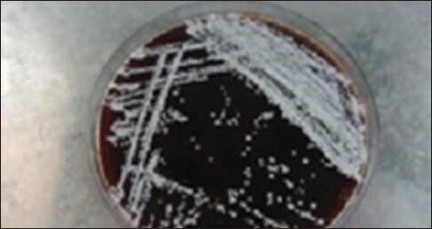
Chalky colonies of Nocardia
Figure 2.
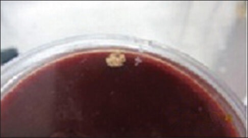
Wrinkled colonies of Nocardia
Figure 3.
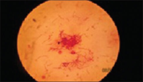
Partially acid-fast Nocardia organisms show reddish to purple filaments
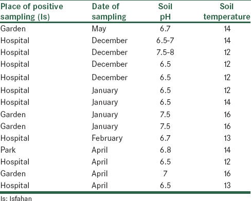
In association with DNA extraction techniques, the first technique of DNA extraction had no band and the second technique had pale band on 1% agarose gel; both techniques were time-consuming. Therefore, we decided to use third method for DNA extraction of all isolates [Figure 4] (microwave oven).[17] In this study, four and one Nocardia isolates were isolated from soil samples of different areas on January/April/December and May/February, respectively. Unfortunately, no survey was performed in the summer. More of these bacteria were isolated from places, which shadow exist, semi-desert climate to colder areas and dry regions.[1,20] The temperature of soil samples were between 12°C and 16°C. The pH of soil samples were between 6.5 and 8 [Table 1].
Figure 4.
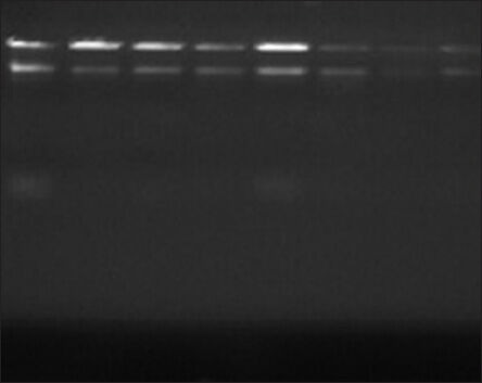
Deoxyribonucleic acid extraction using the microwave oven technique (540 W at 13 s)
Table 1.
Isolation of Nocardia spp. according to the types of place of positive sampling, date of sampling, soil pH, soil temperature
DISCUSSION
In the present study as mentioned, three techniques surveyed for isolation of Nocardia, in spite of the some disadvantages below mentioned, results showed that the third technique (slip-buried-method) was better than other techniques.[1,7] In some of the studies, MCclung method had acceptable result.[8,9] In third technique, BHI agar medium including two antibiotics (kanamycine and cycloheximide) were used and it showed that the growth of Nocardia and Actinomadura were provoked by kanamycin in most instances in positive samples.[1] In this study, Nocardia frequency in soil was 20%. Prevalence of Nocardia in various regions of the world is different and it has been reported to be 5-50%.[21,22,23] Furthermore, results have been found in Qazvin, Iran by Aghamirian and Ghiasian was 40.6% and other results by Kachuei et al.; in Isfahan was 19.1%.[1,7] Similar results with results in Qazvin have been reported in Argantina[24] and Tanzania.[25] These results are more than what found in Patiala, India that is 8%.[22] Isolation of Nocardia spp. from different zones of Isfahan, Iran and the effect of environmental factors such as soil pH and different climates were assayed Nocardia diversity in soil.[1] Molecular techniques such as polymerase chain reaction and gene sequencing techniques are certain methods than phenotypic methods for isolation of Nocardia from soil. In further studies, we intend to do molecular tests according to 16S rDNA sequencing, for precisely identification of Nocardia species. Our intention, in this study was a comparison of three phenotypic and DNA extraction methods for isolation and Nocardia spp.
There are several advantages and disadvantages about phenotypic methods that below is referred to: Paraffin baiting method has some of advantages, one of them is about paraffin it is as a sole carbon source for Nocardia,[26] other advantage is broth media that it can be decrease the numbers of contamination species and recovery of aerobic Actinomycetes.[26] Furthermore, this method has several disadvantages such as lack of incubator with two different temperatures in ordinary laboratory and cleaning of paraffin coated rods and tubes. Seeing of Nocardia weakly White colonies on the paraffin coated glass roads are difficult and they may lose. One of the benefits paraffin coated slides method is using 1 M NH4Cl to eliminate the soil microflora and this method other benefit is a reduction of time to isolate pathogenic strains of Nocardia from natural sources and Nocardia brasiliensis, Nocardia asteroids and Nocardia caviae strains have endurance to this salt (chemical inhibitors).[12] Paraffin coated slide method has disadvantages and one of the disadvantages can be found hardly slides from soil's layers and other disadvantage are weather changes and salt. Salt may lost its effect after melting of paraffin. (However, in paraffin bait method salt added to broth media with 0.22 μ filter and no melting). Contamination with fungi and other microorganisms in soil is others eventually; we reach advantages and disadvantage of slip-buried method. Third method advantages are more selective than other methods because of antibiotics, It will remove more fungi because of cycloheximide and Nocardia grows well in BHI medium specially BHI agar with 5% human blood. Amount of antibiotics is important especially chloramphenicol, extra usage of this antibiotic, may inhibit the growth of some Nocardia species and it is a disadvantage of this method.
According to the DNA extraction techniques, which has been mentioned, because of cell wall mycolic acids of Nocardia microwave oven technique[17] was better than others; for better result, microwave was adjusted to 540 W for suspension then it was put −20°C. As reports Butler et al. Nocardia species have mycolic acids of intermediate carbon chain length (C46-C60).[27] According to the studies Amaro et al. on CTAB technique, the best result was not achieved for Mycobacterium by this technique[19] because of mycolic acid carbons number in cell wall (C80). In present study, CTAB technique had no good result on Nocardia. Present study has suggestions for this technique such as boiling or microwave for breaking of Nocardia cell wall. We must mention that most of the Nocardia species are resistance to lysosyme and we must replace lysosyme with boiling or microwave in CTAB technique.[11] Eventually, our suggestion for using of Fermentas kit is breaking the Nocardia cell wall by boiling or freeze and thaw method.[28] One of the phenotypic errors for Nocardia isolation is false-positive because of resemblance Nocardia with other bacteria and fungi. In further studies, we must do molecular tests according to 16S rDNA sequencing, for precisely identification of Nocardia species.[13,29,30,31]
ACKNOWLEDGMENT
We wish to thank Isfahan University of Medical Sciences for supporting this assay vides Grant No. 390510.
Footnotes
Source of Support: Nil
Conflict of Interest: None declared.
REFERENCES
- 1.Kachuei R, Emami M, Mirnejad R, Khoobdel M. Diversity and frequency of Nocardia spp. in the soil of Isfahan Province, Iran. Asian Pac J Trop Biomed. 2012;2:474–8. doi: 10.1016/S2221-1691(12)60079-3. [DOI] [PMC free article] [PubMed] [Google Scholar]
- 2.Kämpfer P, Huber B, Buczolits S, Thummes K, Grün-Wollny I, Busse HJ. Nocardia acidivorans sp. nov., isolated from soil of the island of Stromboli. Int J Syst Evol Microbiol. 2007;57:1183–7. doi: 10.1099/ijs.0.64813-0. [DOI] [PubMed] [Google Scholar]
- 3.Sabuncuoğlu H, Cibali Açikgo ZZ, Caydere M, Ustün H, Semih Keskil I. Nocardia farcinica brain abscess: A case report and review of the literature. Neurocirugia (Astur) 2004;15:600–3. doi: 10.1016/s1130-1473(04)70453-4. [DOI] [PubMed] [Google Scholar]
- 4.Xing K, Qin S, Fei SM, Lin Q, Bian GK, Miao Q, et al. Nocardia endophytica sp. nov., an endophytic Actinomycete isolated from the oil-seed plant Jatropha curcas L. Int J Syst Evol Microbiol. 2011;61:1854–8. doi: 10.1099/ijs.0.027391-0. [DOI] [PubMed] [Google Scholar]
- 5.Xu P, Li WJ, Tang SK, Jiang Y, Gao HY, Xu LH, et al. Nocardia lijiangensis sp. nov., a novel Actinomycete strain isolated from soil in China. Syst Appl Microbiol. 2006;29:308–14. doi: 10.1016/j.syapm.2005.11.009. [DOI] [PubMed] [Google Scholar]
- 6.Saubolle MA, Sussland D. Nocardiosis: Review of clinical and laboratory experience. J Clin Microbiol. 2003;41:4497–501. doi: 10.1128/JCM.41.10.4497-4501.2003. [DOI] [PMC free article] [PubMed] [Google Scholar]
- 7.Aghamirian MR, Ghiasian SA. Isolation and characterization of medically important aerobic Actinomycetes in soil of Iran (2006-2007) Open Microbiol J. 2009;3:53–7. doi: 10.2174/1874285800903010053. [DOI] [PMC free article] [PubMed] [Google Scholar]
- 8.Khan ZU, Neil L, Chandy R, Chugh TD, Al-Sayer H, Provost F, et al. Nocardia asteroides in the soil of Kuwait. Mycopathologia. 1997;137:159–63. doi: 10.1023/a:1006857801113. [DOI] [PubMed] [Google Scholar]
- 9.Singh M, Sandhu RS, Randhawa HS. Comparison of paraffin baiting and conventional culture techniques for isolation of Nocardia asteroides from sputum. J Clin Microbiol. 1987;25:176–7. doi: 10.1128/jcm.25.1.176-177.1987. [DOI] [PMC free article] [PubMed] [Google Scholar]
- 10.Baron EJ, Peterson LR, Finegold SM. St. Louis: Mosby; 1994. Bailey and Scott's Diagnostic Microbiology. [Google Scholar]
- 11.Winn WC, Allen SD, Janda WM, Koneman EW, Schreckenberger PC, Procop GW, et al. Philadelphia: Lippincott Williams and Wilkins; Koneman's Color Atlas and Textbook of Diagnostic Microbiology. [Google Scholar]
- 12.van Gelderen de Komaid A, Durán EL. The efficiency of 1 M NH4Cl and 2 M NaCl for the isolation of pathogenic Nocardia from soil. Mycopathologia. 1989;108:117–23. doi: 10.1007/BF00436062. [DOI] [PubMed] [Google Scholar]
- 13.McTaggart LR, Richardson SE, Witkowska M, Zhang Sx. Phylogeny and identification of Nocardia species on the basis of multilocus sequence analysis. J Clin Microbiol. 2010;48:4525–33. doi: 10.1128/JCM.00883-10. [DOI] [PMC free article] [PubMed] [Google Scholar]
- 14.Kageyama A, Yazawa K, Kudo T, Taniguchi H, Nishimura K, Mikami Y. First isolates of Nocardia abscessus from humans and soil in Japan. Nihon Ishinkin Gakkai Zasshi. 2004;45:17–21. doi: 10.3314/jjmm.45.17. [DOI] [PubMed] [Google Scholar]
- 15.Rodríguez-Nava V, Couble A, Devulder G, Flandrois JP, Boiron P, Laurent F. Use of PCR-restriction enzyme pattern analysis and sequencing database for hsp65 gene-based identification of Nocardia species. J Clin Microbiol. 2006;44:536–46. doi: 10.1128/JCM.44.2.536-546.2006. [DOI] [PMC free article] [PubMed] [Google Scholar]
- 16.Sahathevan M, Harvey FA, Forbes G, O’Grady J, Gimson A, Bragman S, et al. Epidemiology, bacteriology and control of an outbreak of Nocardia asteroides infection on a liver unit. J Hosp Infect. 1991;18(Suppl A):473–80. doi: 10.1016/0195-6701(91)90059-h. [DOI] [PubMed] [Google Scholar]
- 17.Salgado A, Ramirez N, Sandoval E, Sandoval H. Fast method for DNA extraction in Nocardia and Saccharomonospora. J Mycol Med/J Med Mycol. 2008;18:100–2. [Google Scholar]
- 18.Ayyar S, Tendolkar U, Deodhar L. A comparison of three media for isolation of Nocardia species from clinical specimens. J Postgrad Med. 1992;38:70–2. [PubMed] [Google Scholar]
- 19.Amaro A, Duarte E, Amado A, Ferronha H, Botelho A. Comparison of three DNA extraction methods for Mycobacterium bovis, Mycobacterium tuberculosis and Mycobacterium avium subsp. avium. Lett Appl Microbiol. 2008;47:8–11. doi: 10.1111/j.1472-765X.2008.02372.x. [DOI] [PubMed] [Google Scholar]
- 20.Stapleton F, Keay LJ, Sanfilippo PG, Katiyar S, Edwards KP, Naduvilath T. Relationship between climate, disease severity, and causative organism for contact lens-associated microbial keratitis in Australia. Am J Ophthalmol. 2007;144:690–8. doi: 10.1016/j.ajo.2007.06.037. [DOI] [PubMed] [Google Scholar]
- 21.Vetlugina LA, Adiiatova ZhF, Khozhamuratova SSh, Rymzhanova ZA, Trenozhnikova LP, Kopytina MN. Isolation of Actinomycetales from the soil of Kazakhstan on selective media with antibiotics. Antibiot Khimioter. 1990;35:3–5. [PubMed] [Google Scholar]
- 22.Goel S, Kanta S. Prevalence of Nocardia species in the soil of Patiala area. Indian J Pathol Microbiol. 1993;36:53–60. [PubMed] [Google Scholar]
- 23.van Gelderen de Komaid A, Runco de Laborda R, Salim R. Natural occurrence of Nocardia in soil of Tucumán: Physiological characteristics. Mycopathologia. 1987;99:15–9. doi: 10.1007/BF00436675. [DOI] [PubMed] [Google Scholar]
- 24.Negroni R, López Daneri G, Arechavala A, Bianchi MH, Robles AM. Clinical and microbiological study of mycetomas at the Muñiz Hospital of Buenos Aires between 1989 and 2004. Rev Argent Microbiol. 2006;38:13–8. [PubMed] [Google Scholar]
- 25.Denguezli M, Kourda M, Ghariani N, Belajouza C, Mokni B, Chebil F, et al. Mycetomas in central Tunisia. Ann Dermatol Venereol. 2003;130:515–8. [PubMed] [Google Scholar]
- 26.Conville PS, Witebsky FG. Hodder Amold; Washington, DC: Distributed in the United States of America by ASM Press; 2005. Nocardia and other aerobic Actinomycetes. Topley and Wilson's Microbiology and Microbial Infections. [Google Scholar]
- 27.Butler WR, Kilburn JO, Kubica GP. High-performance liquid chromatography analysis of mycolic acids as an aid in laboratory identification of Rhodococcus and Nocardia species. J Clin Microbiol. 1987;25:2126–31. doi: 10.1128/jcm.25.11.2126-2131.1987. [DOI] [PMC free article] [PubMed] [Google Scholar]
- 28.Kim BY. Thesis. United Kingdom: School of Biology, Faculty of Science, Agriculture and Engineering, Newcastle University, Newcastle upon Tyne; 2010. Biosystematics of the genus Dactylosporangium and some other filamentous actinomycetes. [Google Scholar]
- 29.Naidoo D, Kumari S, Bux F. Characterization of Nocardia farcinica, a filamentous bacterium isolated from foaming activated sludge samples. Water Environ Res. 2011;83:527–31. doi: 10.2175/106143010x12851009156565. [DOI] [PubMed] [Google Scholar]
- 30.Lamm AS, Khare A, Conville P, Lau PC, Bergeron H, Rosazza JP. Nocardia iowensis sp. nov., an organism rich in biocatalytically important enzymes and nitric oxide synthase. Int J Syst Evol Microbiol. 2009;59:2408–14. doi: 10.1099/ijs.0.007427-0. [DOI] [PMC free article] [PubMed] [Google Scholar]
- 31.Reddy AK, Garg P, Kaur I. Spectrum and clinicomicrobiological profile of Nocardia keratitis caused by rare species of Nocardia identified by 16S rRNA gene sequencing. Eye (Lond) 2010;24:1259–62. doi: 10.1038/eye.2009.299. [DOI] [PubMed] [Google Scholar]


