

Background: Geobacillus stearothermophilus secretes an extracellular xylanase (Xyn10A, XT-6) for the utilization of xylan.
Results: The expression of xynA is regulated by XylR, CodY, and XynX, catabolite repression (CcpA), and quorum sensing.
Conclusion: The expression of the extracellular xylanase gene (xynA) is highly regulated by many transcriptional schemes.
Significance: The xynA gene is regulated by unique and not yet fully characterized mechanisms.
Keywords: Gene Regulation, Glycoside Hydrolase, Gram-positive Bacteria, Plant Cell Wall, Quorum Sensing, Thermophile, Transcription Repressor, Transformation, Nif3-like Family, Xylanase
Abstract
Geobacillus stearothermophilus T-6 produces a single extracellular xylanase (Xyn10A) capable of producing short, decorated xylo-oligosaccharides from the naturally branched polysaccharide, xylan. Gel retardation assays indicated that the master negative regulator, XylR, binds specifically to xylR operators in the promoters of xylose and xylan-utilization genes. This binding is efficiently prevented in vitro by xylose, the most likely molecular inducer. Expression of the extracellular xylanase is repressed in medium containing either glucose or casamino acids, suggesting that carbon catabolite repression plays a role in regulating xynA. The global transcriptional regulator CodY was shown to bind specifically to the xynA promoter region in vitro, suggesting that CodY is a repressor of xynA. The xynA gene is located next to an uncharacterized gene, xynX, that has similarity to the NIF3 (Ngg1p interacting factor 3)-like protein family. XynX binds specifically to a 72-bp fragment in the promoter region of xynA, and the expression of xynA in a xynX null mutant appeared to be higher, indicating that XynX regulates xynA. The specific activity of the extracellular xylanase increases over 50-fold during early exponential growth, suggesting cell density regulation (quorum sensing). Addition of conditioned medium to fresh and low cell density cultures resulted in high expression of xynA, indicating that a diffusible extracellular xynA density factor is present in the medium. The xynA density factor is heat-stable, sensitive to proteases, and was partially purified using reverse phase liquid chromatography. Taken together, these results suggest that xynA is regulated by quorum-sensing at low cell densities.
Introduction
Plant biomass degradation is a pivotal step in the carbon cycle on earth and is mediated mostly by microorganisms, which are found either free in the environment or part of the digestive track of higher animals (1–5). Plant-based biomass is considered a readily available renewable energy source that can be turned into liquid biofuel for transportation without contributing much net CO2 to the atmosphere (6, 7). Thus, there is a great incentive to explore the enzymology of biomass degradation (8, 9). The main sugar-based components of the plant cell wall are cellulose and hemicellulose. Hemicellulose is a heterogeneous group of branched and linear polysaccharides that can include up to 35% of the total plant cell wall dry mass, and one of its major constitutes is xylan (10). Xylan is a polysaccharide made of a backbone of β-1,4-xylose units that are decorated by side chains such as arabinose, methylglucuronic acid, and acetate groups (10). The complete degradation of xylan requires the action of specific glycoside hydrolases, including xylanases, which hydrolyze the main chain backbone, and accessory enzymes that act on the side chains, such as α-l-arabinofuranosidase, α-glucuronidase, β-xylosidase, and xylan acetyl esterase (11).
Utilization of high molecular weight polymers imposes several intrinsic challenges for the microorganism. First, the microorganism needs a mechanism to sense the presence of the extracellular polymer and to induce the expression of appropriate genes, which include hydrolytic enzymes and transport systems. Second, because the chemical structure of the polysaccharides is complex, efficient degradation requires the involvement of several hydrolytic enzymes with different specificities. Finally, because the polysaccharides are of high molecular weight, a minimum mass of enzymes is required to initiate growth based on the polymers, resulting in cell density-dependent growth (12).
Nature evolved a variety of enzymes and different physiological schemes for the microbial degradation of the plant cell wall polysaccharides (1, 13). Anaerobic bacteria, such as Clostridia, have evolved unique multienzyme complexes, named cellulosomes, that integrate many cellulolytic and hemicellulolytic enzymes (14–16). Cellvibrio japonicas, a Gram-negative soil bacterium, secretes around 30 enzymes that mediate the initial degradation of the integral plant cell wall polysaccharides. The resulting oligosaccharides are then hydrolyzed by surface-bound proteins (17). In both cases, the hydrolysis of polysaccharides occurs at the cell-substrate interface; as a result, the sugar products remain close to the cells and are maintained at relatively high concentrations. A different approach is to completely hydrolyze the polysaccharides outside of the cell. For example, aerobic filamentous fungi, such as Trichoderma and Aspergillus, secrete a large variety of free cellulases, hemicellulases, and ligninases that work synergistically to completely degrade the polymers into mono- and disaccharides (18). Consequently, these extracellular soluble products can also be utilized by other surrounding microorganisms. In Geobacillus spp. the initial breakdown of the polymer is executed by a limited number of extracellular endo-type enzymes that degrade the polysaccharide backbone into relatively large oligosaccharides (19–22). These oligosaccharides enter the cell via dedicated sugar transporters, and their final breakdown is carried out by intracellular enzymes (20, 22). In this strategy, much of the extracellular saccharide products are not available to most competing microorganisms, because they lack the appropriate sugar transporters. Recent studies demonstrated that microbial utilization of the plant cell wall biomass involves extracellular sensing systems for polysaccharides. In most cases, residual sugar monomers resulting from the hydrolytic activity of the surrounding microorganisms can induce the corresponding utilization systems. Other microorganisms have developed dedicated systems to sense the high molecular weight polysaccharides in the surrounding environments and to activate the corresponding genes. For example, in the rumen anaerobe Prevotella bryantii B14, the xylanase gene is up-regulated in response to xylan in the environment (23). This regulation is mediated by a multidomain protein, XynR, that contains a transmembrane domain, histidine kinase motif, response regulator sequence, and a DNA binding domain in its C terminus. In the human gut bacterium, Bacteroides thetaiotaomicron, part of the glycan degradation system is regulated by extra-cytoplasmic function σ factors and anti-σ factors and by a hybrid two-component system that incorporate all domains found in classical two-component environmental sensors into one polypeptide (24, 28). Alternative σ factors are also involved in the regulation of the cellulosomal genes in Clostridium thermocellum via an external carbohydrate-sensing mechanism (25–27).
Geobacillus stearothermophilus T-6 is a thermophilic soil bacterium that was isolated based on its ability to secrete thermostable alkaline-tolerant hemicellulases that could be used to bleach paper pulp (29, 30). The structure-function relationships of many of its hemicellulolytic enzymes were investigated intensively (31–39) together with its arabinan- and galactan-degrading systems (19, 20). The bacterium possesses two- and three-component oligosaccharide-sensing systems that activate the expression of dedicated ABC sugar transporters (19–22, 40), allowing the cell to rapidly respond to very low carbohydrates concentrations in the soil and to efficiently take up the oligosaccharides (20, 21). In strain T-6, most of the xylanolytic system is clustered on a 39.7-kb chromosomal segment containing 27 genes (Fig. 1) (22). When grown in the presence of xylan, strain T-6 secretes a single endo-1,4-β-extracellular xylanase (Xyn10A) that hydrolyzes the natural polymer's main backbone, producing short xylo-oligosaccharides decorated with various side chains, such as l-arabinose, 4-O-methylglucuronic acid, or acetate. These xylosaccharides enter the cell via specialized ABC sugar transporters and are hydrolyzed to xylose monomers by intracellular enzymes, including α-glucuronidase (Agu67A) (35, 39), two α-l-arabinofuranosidases (Abf51A and Abf51B) (36), three β-xylosidases (Xyn39B, Xyn52B2, and Xyn43B3) (33, 34, 37, 38, 41), two xylan acetylesterases (CE4) (31, 42), and an intracellular xylanase (Xyn10A2) (Fig. 1) (43).
FIGURE 1.

Schematic view of the xylanolytic system from G. stearothermophilus T-6. The utilization of xylan by G. stearothermophilus is initiated by residual levels of xylose or xylo-oligosaccharides present in the environment. These sugars interact with the extracellular domain of the class I histidine kinase sensor protein XynD, which triggers phosphorylation of the response regulator XynC (21). The phosphorylated XynC activates the expression of a dedicated ABC xylo-oligosaccharides transporter, XynEFG, which facilitated the entrance of xylosaccharides into the cell. Inside the cell, xylose serves as the molecular inducer and interacts with the master repressor XylR, which negatively regulates five transcriptional units. The expression of the extracellular xylanase provides the cell with ample amounts of decorated xylo-oligosaccharides that enter the cell via two dedicated ABC sugar transporters, XynEFG for xylooligosaccharides and AguEFG for aldotetrauronic acid. The decorated xylooligomers are hydrolyzed to their corresponding monomers by intracellular side chain-cleaving enzymes, including α-glucuronidase (Agu67A) (35, 39), two α-l-arabinofuranosidases (Abf51A and Abf51B) (36), two xylan acetylesterases (CE4) (31, 42), an intracellular xylanase (Xyn10A) (43), and three β-xylosidases (Xyn39B, Xyn52B2, and Xyn43B3) (33, 34, 37, 38, 41). The extracellular xylanase gene (xynA) is subjected to carbon catabolite repression mediated by the global negative repressor CcpA and most likely by CodY. Furthermore, xynA is also regulated by XynX and cell density by uncharacterized mechanisms.
The hallmark of the xylanolytic system in G. stearothermophilus T-6 is the extracellular xylanase, Xyn10A (also named XT-6) (29, 30, 44, 45). In this study, we demonstrate that the regulation of the extracellular xylanase (xynA) gene is mediated by several mechanisms that in part are connected to the xylan utilization strategy of this bacterium, thus allowing it to successfully compete in its natural niche.
MATERIALS AND METHODS
Growth Conditions
Growth media for G. stearothermophilus was modified Basic Salt Medium (mBSM)2 supplemented with 1% glucose or xylose. Liquid mBSM contained the following per liter: KH2PO4, 0.4 g; (NH4)2SO4, 2 g; MOPS-sodium salt, adjusted to pH 7.0, 10 g. After autoclaving, mBSM was supplemented with (per liter) the following: nitrilotriacetic acid, 0.2 g, NaOH, 0.125 g, 1 ml of amino acid stock solution (0.05 g/liter of 19 amino acids except tyrosine), and 1 ml of three different trace elements solutions. Solution I contained per liter the following: MgSO4·7H2O, 0.145 g. Solution II contained per liter the following: CaCl2·2H2O, 0.132 g. Solution III contained in g per liter the following: FeSO4·7H2O, 0.998 g; MnSO4·4H2O, 0.592 g; ZnCl2, 0.42 g; and CuSO4·5H2O, 0.624 g. The pH of the latter solution was adjusted to 2.0 with sulfuric acid. An additional source for amino acids was 0.3% of acid hydrolysate of casein (casamino acids) (Difco).
DNA and RNA Isolation and Manipulation
G. stearothermophilus T-6 genomic DNA was isolated according to the procedure published by Marmur (46) and as outlined by Johnson (47). Plasmid DNA was purified using the DNA Clean-Up System (Promega). DNA was manipulated by standard procedures (48). Total RNA was isolated with the RNeasy kit (Qiagen GmbH, Hilden, Germany) according to the manufacturer's protocol.
Transcriptional Analyses
Transcriptional analyses were performed on total RNA extracted from exponentially growing cells. Northern blot analysis was conducted following the procedure described by Moran et al. (49). For determination of the xynA transcription start site, primer extension reactions were carried out as described previously (50) with avian myeloblastosis virus reverse transcriptase (Promega), 40 μg of total RNA, and the relevant primers listed in Table 1. Rapid amplification of cDNA ends (RACE) technique was used to amplify portion of the putative transcript at xynX 5′-UTR (Clontech). Briefly, reverse transcription was generated using random hexamers using a reverse transcriptase (RT) that exhibits terminal transferase activity. The cDNA was amplified with universal primers as well as specific primer anti-parallel and complementary to a sequence downstream of the xynX gene (Table 1).
TABLE 1.
G. stearothermophilus strains and plasmids
| Strains/plasmids | Genotype and/or other relevant characteristics | Source or Ref. |
|---|---|---|
| G. stearothermophilus | ||
| T-6 | Wild type NCIBM 40222 | 30 |
| M-7 | Xylanase constitutive mutant | 30 |
| 18-2 | xynX null mutant, the entire xynX coding sequence was deleted | This work |
| Plasmids | ||
| pNW33N | Geobacillus-E. coli shuttle vector, Cmr (4217 bp) | Prof. Neil E. Welker |
| pNW35N | pNW33N containing deletion of 641 bp (positions 3576 to 4217) | This work |
| pNW36N | pNW35N containing DNA fragment with an entire deletion of xynX and 450 bp of flanking DNA upstream and downstream the deletion site | This work |
Continuous and Batch Cultures
Batch fermentations were conducted in a 10-liter fermenter (BIOSTAT ED, Braun) with 8-liter working volume or in 1-liter baffled flasks containing 100 ml of medium and reciprocally shaking at 250 rpm, 60 °C in a dry shaker (Lab-Line). The inoculum for all fermentations was originated from logarithmic cultures. Chemostat cultures were run in a 2-liter vessel fermenter (BIOSTAT MD, Braun) with a working volume of 1.5 liter, operating at agitation of 700–1200 rpm, air flow of 1.5 liters/min maintaining oxygen saturation over 80%. A pH of 7.0 was maintained by automatic addition of 5 m NaOH, and the growth temperature was 60 °C. The dilution rate was 0.2 h−1, and the turbidity was controlled by the concentration of the limited nutrient. The concentrations of O2 and CO2 in the outlet gas were measured by a gas analyzer URAS10E (Hartman and Braun). Samples were taken at steady state after determining that turbidity remained similar for over three generations.
Expression of XylR and Extraction of His6-tagged CodY and XynX
The xylR, codY, and xynX genes were cloned using two PCR primers (Table 1) that allow in-frame cloning of the gene into the T7 expression vector pET11d (Novagen) (linearized with BamHI and NcoI), resulting in pET11d-xylR, pET11d-codY, and pET11d-xynX. Sequences encoding a His6 tag peptide were added to the N-terminal primer of xynX and the C-terminal primer of codY. Expression of xylR was carried out by growing cultures of Escherichia coli JM109(DE3)(pLysS) (Promega) carrying pET11d-xylR in 200 ml of Terrific Broth (48) supplemented with chloramphenicol (25 μg/ml) and carbenicillin (50 μg/ml) in 2-liter shake flasks shaken at 230 rpm at 37 °C. To improve the solubility of XylR, growth was carried out at 18 °C in the presence of 0.4 mm isopropyl 1-thio-β-d-galactopyranoside for 48 h. CodY and XynX were produced in E. coli BL21(DE3) in 500 ml of Terrific Broth supplemented with ampicillin (100 μg/ml) in 2-liter shake flasks shaken at 230 rpm at 37 °C. Induction by 0.4 mm isopropyl 1-thio-β-d-galactopyranoside was carried out at a cell turbidity of 0.6 units of absorbance at 600 nm for 16 h. Cells expressing the xylR gene were harvested, suspended in 20 ml of solution containing 50 mm Tris-Cl (pH 7.5), 100 mm KCl, 10% glycerol, 1 mm EDTA, 0.5 mm phenylmethylsulfonyl fluoride, and 1 mm dithiothreitol, and disrupted by a single passage through a French press (Spectronic Instruments, Inc., Rochester, NY). Following centrifugation of the cell extract (14,000 × g for 15 min), the soluble fraction was used for gel retardation assays. The cultures expressing the codY and xynX genes were harvested, resuspended in 30 ml of buffer (20 mm imidazole, 20 mm phosphate buffer, 500 mm NaCl (pH 7.0)), disrupted by two passages through a French press, and centrifuged (14,000 × g for 15 min) to obtain soluble extracts. The His-tagged proteins, CodY and XynX, were isolated using a 1-ml HisTrap column (GE Healthcare) mounted on an AKTA-avant FPLC system (GE Healthcare), according to the manufacturer's instructions. The purified CodY and XynX proteins were dialyzed overnight against 2 liters of buffer containing 50 mm Tris-HCl (pH 7.0) and 100 mm KCl, followed by the addition of EDTA and glycerol to final concentrations of 1 mm and 10%, respectively. The proteins were stored at −80 °C in 100-μl aliquots.
Mobility Shift DNA Binding Assays
For binding analysis with XylR, four double-stranded DNA probes (corresponding to the xynA, xynX, xylA, and xylR promoter regions) were each composed of two synthetic complementary oligonucleotides (Table 1). The double-stranded probes contained two noncomplementary T nucleotides at the 5′ end for end labeling with Klenow fragment (Fermentas) in the presence of [α-32P]dATP. For DNA binding assays with His6-CodY, an 82-bp DNA probe (from position −96 to position −14 relative to the transcriptional start site of the xynA gene), was generated via PCR using a primer labeled at the 5′ end with biotin (Table 1). The PCR product was purified from 1% agarose gel. For His6-XynX, five DNA fragments of 212, 110, 123, 70, and 72 bp corresponding, respectively, to positions −191 to +21, −191 to −81, −102 to +21, −49 to +21, and −30 to −102 with respect to the transcriptional start site of xynA were generated via PCR with biotin 5′ end-labeled primers listed in Table 2. These PCR products were purified with the Wizard® SV Gel PCR Clean-Up system (Promega).
TABLE 2.
Oligonucleotides used in this study
The following abbreviations are used: N-ter, N-terminal; C-ter, C-terminal.

a |Boldface bases indicate engineered restriction sites.
b |Btn indicates biotin 5′ end-labeled primers.
The XylR-binding reaction mixture (30-μl total volume) contained 20 μl of binding solution (50 mm Tris-Cl (pH 7.5), 100 mm KCl, 10% glycerol, 1 mm EDTA, 2 μg of salmon sperm DNA, 0.66 mm dithiothreitol, 33 μg of bovine serum albumin, 0.08 ng of labeled probe), and the indicated concentration of crude protein extract. The binding mixture was incubated for 30 min at 45 °C, separated on a 6.6% nondenaturing polyacrylamide gel prepared in Tris borate/EDTA buffer (48), and run for 1 h. Gels were dried under vacuum and exposed to a phosphorimager screen before analysis with a Fuji BAS2000 phosphorimager. Binding of XylR to the xylR operator in the presence of various sugars was carried out under the same conditions described above but with 100 ng/μl of crude protein extract and 0.12 ng of labeled probe.
The binding reactions for CodY and XynX were carried out under the conditions described above but with 20 fmol of labeled probe. The binding reactions for CodY also included 2 mm GTP and 10 mm each of the three branched-chain amino acids (isoleucine, leucine, and valine). The binding mixture was incubated for 30 min at 45 °C and then separated on a 6.6% nondenaturing polyacrylamide gel prepared in Tris borate/EDTA buffer (48) and run for 1 h (set to 100 V for 8 × 10 × 0.1-cm gel). Binding reactions were transferred to a nylon membrane at 380 mA for 50 min and then was cross-linked with UV light at 0.32 J/cm2. For XynX, the detection of biotin-labeled DNA by chemiluminescence was performed using the Phototope Star Detection kit (N7020, New England Biolabs), according to the manufacturer's protocol, and for binding assays with CodY, the DNA migration was detected using Biotin Chromogenic Detection kit (Thermo Scientific).
Protoplast Transformation of G. stearothermophilus T-6
Protoplast plasmid DNA transformation to G. stearothermophilus T-6 was based on the protocol described by Wu and Welker (51) with the following modifications. Cells were converted to protoplasts by incubating in fresh lysozyme solution at a final concentration of 2.5 μg/ml. One microgram of plasmid DNA was gently mixed with 0.2 ml of protoplast suspension, and transformation was induced by adding 0.9 ml of 40% (w/v) PEG 6000 (Merck), filter-sterilized, and freshly prepared. Plating the transformants on regeneration plates was performed in two steps. First, transformants were plated on regeneration plates containing 0.4% agar and 10% lactose, and the colonies were transferred to regeneration plates with 0.4% agar and 4% lactose (instead of 10% lactose). Plates were incubated at 60 °C throughout the experiment. Regeneration plates contained the following (per liter): tryptone (Difco), 10 g; yeast extract (Difco), 5 g; NaCl, 5 g; and bacto agar (Difco), 4 g. After autoclaving, the medium was supplemented with the following (per liter): lactose, 100 g, nitrilotriacetic acid, 0.2 g, NaOH 0.125 g, and 1 ml of three different of the following trace elements solutions. Solution I contained per liter, MgSO4·7H2O, 0.145 g, Solution II contained per liter, CaCl2·2H2O, 0.132 g. Solution III contained per liter, FeSO4·7H2O, 0.998 g; MnSO4·4H2O, 0.592 g; ZnCl2, 0.42 g; and CuSO4·5H2O, 0.624 g. In addition, MgCl2·6H2O and CaCl2·2H2O were added to a final concentration of 0.01 and 0.02 m, respectively. Chloramphenicol was added to a final concentration of 7 μg/ml.
Construction of xynX Null Mutant of G. stearothermophilus T-6
To construct a xynX-null mutant, we designed a deletion using an overlap PCR technique resulting in removing the entire xynX gene (792 bp) and leaving 450 bp of flanking DNA upstream and downstream of the deletion site (52). The PCR product was digested with SacI and KpnI and ligated to pNW35N, which had been similarly digested to obtain pNW36N (Table 1). This plasmid was transformed to G. stearothermophilus T-6 by protoplast transformation and selection for Cmr clones. Cmr colonies were tested by colony PCR for a single crossover event in which pNW36N integrated into the chromosome and the presence of the vector. To allow a second double crossover event and thereby obtain a chloramphenicol-sensitive strain, a colony, positive for single integration event, was grown for five generations at 68 °C without selection and then plated to obtain single colonies and screened for sensitivity to chloramphenicol. About 8–10% of the Cms colonies were xynX null mutants, as confirmed by sizing and sequencing the PCR fragments flanking the xynX region.
Xylanase and Isocitrate Dehydrogenase Activity Assays
Xylanase activity was determined by measuring the hydrolysis of p-nitrophenyl cellobioside (Sigma) or p-nitrophenyl xylobioside (pNPX2) (Megazyme). The extracellular xylanase (Xyn10A) has a high specificity constant toward pNPX2 (kcat/Km = 8.6 × 10³ min−1 mm−1) (53). The reaction mixture contained 160 μl of an appropriately diluted enzyme, 160 μl of 0.1 m phosphate buffer (pH 7.0), and 80 μl of p-nitrophenyl cellobioside (4 mg/ml) in the same buffer. Tubes were incubated at 50 °C until a slight yellow color appeared, and then the reaction was terminated by the addition of 160 μl of 1 m Na2CO3. Absorbance at A420 was determined spectrophotometrically (Ultrospec 2100pro, Amersham Biosciences). One unit of activity was defined as (A420/min/ml of culture per A600) × 1000 (Miller units). Isocitrate dehydrogenase activity was assayed as described previously (54). Unit of activity was defined as nanomoles of NADPH per min per cell culture turbidity at A600.
RNase Protection Assays
Strain T-6 was grown in mBSM in a 10-liter fermenter (Braun ED). During the logarithmic growth, samples at different cell densities were collected, and total RNA was extracted with the RNeasy kit (Qiagen) following the supplier protocol. RNA from very low density cultures was extracted from at least 200 ml of culture volume. RNase protection assays were performed using the RPAII kit (Ambion Inc., Austin, TX). Radioactive antisense RNA probes were generated according to the Ambion MAXIscript T7/T3 in vitro transcription kit protocol. [α-32P]UTP (800 Ci/mmol) was obtained from Amersham Biosciences. The DNA fragment used for the transcription was cloned in pSL301 (Stratagene, La Jolla, CA), and the construct included the region from −87 bp to +227 relative to the initial ATG site of the xynA gene. The full-length antisense probes were isolated from polyacrylamide gels according to the supplier's instructions. RNase protection assays were performed with a 467-nt 32P-labeled antisense RNA probe, of which 227 nt were specific to the 5′ xylanase transcript and 240 nt were specific to chromosomal and vector sequences. The probe (0.5 ng) was hybridized with 4 μg of total RNA. The protected RNA was dissolved in 8 μl of sample buffer of which 6 μl was subjected to electrophoresis in a 5% polyacrylamide gel containing 7 m urea and visualized using a phosphorimager system.
Determination of the Amount of xynA mRNA Molecules per Cell
Total RNA for each of the cultures was determined using the reagent orcinol monohydrate (Sigma) (55). To determine the amount of total RNA per cell, total cell number was estimated microscopically using a Petroff-Hausser counting chamber. The number of transcripts per cell of the xynA mRNA was derived based on the counts/min per μg of total RNA, the specific activity of the probe, the molecular weight of the probe, and the average amount of total RNA per cell (1.78 × 10⁻⁷ μg). To quantify the results and to ensure that the amount of the probe in the hybridization reaction was not a limiting factor, different amounts of isolated RNA were hybridized with the 32p-labled antisense probe. Following digestion with RNase, the RNase protection assay product was precipitated, and radioactivity was assessed in a scintillation counter. A linear correlation was obtained between the amount of total RNA used in the assay and radioactivity of the protected product.
Preparation of Conditioned Medium
Conditioned medium was prepared by growing strain T-6 in mBSM containing 1% xylose to a density of ∼0.8 A600. The cells were removed by centrifugation; the supernatant was passed through a 0.45-μm pore size filter (Millipore). The soluble content was concentrated 2-fold using a freeze dryer and then boiled for 10 min (to inactivate residual extracellular xylanase and proteases).
Cell Density Activation Assay in a 96-Well Plate
Xylanase activity was assayed following the hydrolysis of pNPX2. A xylose-induced logarithmic culture at 0.1 A600 turbidity was diluted 200-fold in mBSM supplemented with 0.5% xylose to a final turbidity of ∼0.001 A600. Cells were then transferred to a 96-well plate containing 0.5 mm pNPX2 and conditioned medium (10–30 μl). The plate (200 μl total volume per well) was incubated with shaking at 60 °C, and monitored simultaneously for both cell growth (A600) and p-nitrophenol release (A420) in a microplate spectrophotometer (EON, BioTek).
Partial Purification of Xylanase Density Factor (XDF)
Partial purification of the putative XDF was as follows. A T-6 culture was grown on mBSM in a 2.5-liter fermenter (KLF, Bioengineering) to a turbidity of 0.7–0.8 A600. The culture was centrifuged (7,000 rpm, 5 min); the supernatant was filtered via a 0.4-μm pore size filters (Millipore), and the filtrate was loaded on an XAD-2 column (5 × 20 cm) at a flow rate of 2.5 ml min−1. After washing the column with 700 ml of 10% ethanol, the remaining content was eluted with 80% ethanol. The eluate was evaporated to 200 ml in a rotary evaporator and then further concentrated in a freeze dryer to ∼5 ml, resulting in a 500-fold concentration. Aliquots of 0.25 ml of active samples were subjected to reverse phase liquid chromatography using a C18 column (Kromasil, 5-μm particle size, pore size 100 Å, 250 mm × 4.6 mm) mounted on an AKTA-Basic FPLC system (Amersham Biosciences). Fractionation was carried out by washing the column with 6% acetonitrile (ACN) in 0.1% aqueous trifluoroacetic acid (TFA), followed by a linear gradient of ACN (6% to 65% in 120 min) in 0.1% aqueous (TFA) at a flow rate of 0.15 ml min−1. Eluent absorbance was monitored at 215 nm. Samples (1.5 ml) were lyophilized, re-dissolved in 50 μl of 0.1 m MOPS (pH 7.0), and tested for xylanase activation at low density cultures.
Real Time RT-PCR Analysis
Total RNAs from different cell density cultures were extracted with an RNeasy kit (Qiagen) following the supplier's protocol. Extraction of RNA from very low density cultures was done by collecting at least 100 ml of culture volume. The cDNA was obtained with the Verso cDNA kit (Thermo Fisher Scientific) using 1 μg of total RNA and random hexamers as primers. To test for DNA contamination, control reactions were carried out in the absence of reverse transcriptase. Real time RT-PCR primers for the xynA, xynA2, xynX, axe2, xynB1, xynB2, and xynB3 and citC (isocitrate dehydrogenase) genes were designed with the aid of the Primers Express 2.0 software (Applied Biosystems) (Table 1). Relative quantification was performed with the Applied Biosystems 7300 real time PCR system. Each 20-μl reaction mixture included template cDNA, 300 nm of each primer, and Power SYBR Green PCR Master Mix (Applied Biosystems). The amplification conditions for all reactions were one cycle at 95 °C for 15 min followed by 40 cycles of 95 °C for 10 s, 60 °C for 20 s, and 72 °C for 15 s. Melting curves were analyzed to ensure specificity of primer annealing and lack of primer secondary structure. Data analysis was carried out with the 7300 system software (Applied Biosystems), using citC for normalization.
Nucleotide Sequence Accession Number
The 77,747-bp sequence containing the arabinan, xylan, xylose utilization region, and the XylR repressor from G. stearothermophilus T-6 have been deposited in the GenBankTM under accession numbers DQ868502 and DQ868501.1, respectively.
RESULTS
The efficient utilization of xylan by G. stearothermophilus T-6 is initiated and controlled by the extracellular xylanase Xyn10A (XT-6). Previous studies from our laboratory suggested that the expression of the xylanase gene is regulated at several levels, including induction by xylose and catabolite repression by glucose (56). In this study, we demonstrate that additional regulatory mechanisms control the expression of the xynA gene, and the roles of some of these mechanisms can be explained based on the biomass utilization strategy used by the bacterium.
Transcriptional Analyses of xynA and xynX-axe2-xynB3 Operon
To map the transcriptional start site of xynA, total RNA was extracted from cultures grown in the presence of xylose or glucose and was used for primer extension analysis (Fig. 2A). The apparent transcriptional start site point of the xynA gene was identified and assigned to a G nucleotide, 90 bases upstream from the ATG initiation codon. The apparent −35 sequence, TTTACA, resembles the σA consensus and is separated by 17 bp from the potential −10 region (TAAAAT), which differs by one nucleotide from the σA consensus sequence, TATAAT (49). An inverted repeat sequence (5′-TTAGTATatttaatttacAAACAAA-3′) resembling the xylR operator site from Bacillus and other Gram-positive bacteria (57) overlaps the −10 region (Fig. 2B). In addition, three potential operator sites for CcpA-mediated, catabolite-responsive regulation (catabolite-responsive elements) (58) were identified between position −130 with respect to the transcriptional start point and the ATG initiation codon (Fig. 2B). The xynA gene lies upstream of and is divergently transcribed with respect to a three-gene cluster, xynX-axe2-xynB3, that potentially constitutes a polycistronic operon. To determine whether the cluster constitutes a single transcriptional unit, Northern blot analysis was performed using a DNA probe for xynB3. The probe hybridized to RNAs of many different sizes, with a maximum length of about 3 kb, which is close to the calculated size of the xynX-axe2-xynB3 transcript (3.2 kb), suggesting that the three genes are co-transcribed from the xynX promoter (results not shown). Using 5′-RACE analysis, the apparent transcriptional start point of the xynX-axe2-xynB3 transcript was identified and assigned to a G nucleotide (Fig. 2B). The apparent −35 sequence, TTGAAA, differs by one nucleotide from the σA consensus sequence and is separated by 17 bp from the potential −10 region (TATAAT) that matches the σA consensus perfectly. It should be noted that there were no detectable transcripts from this region when total RNA was isolated from cultures grown without xylose, indicating that xylose is the molecular inducer of both the xynX-axe2-xynB3 operon and xynA gene (56).
FIGURE 2.

Transcriptional analyses of the xynA and xynX-axe2-xynB3 transcripts. A, mapping the 5′ termini of the xynA transcript by primer extension analysis. Extension products resulting from RNA obtained from cultures grown on 0.5% glucose (lane 1) or with 0.5% xylose (lane 2) as the carbon source are shown. Dideoxynucleotide sequencing reactions were carried out with the same primer used for the reverse transcriptase reactions. The position of the transcriptional start point is indicated with an asterisk on the inferred nontemplate strand sequence. B, sequence data for the xynA and xynX regulatory regions. The 5′ end of the xynX gene was determined by 5′-RACE analysis. Total RNA was isolated from mid-exponential phase cultures of G. stearothermophilus T-6 grown on minimal media supplemented with 0.5% xylose as a sole carbon source. Vertical arrow indicates the transcriptional start point (+1). The potential inverted repeats binding site for the xylose repressor XylR is indicated by horizontal arrows. The −35 and −10 regions, the proposed ribosome binding site (RBS), the initiating methionine codon, and the potential catabolite-responsive elements (CRE) are in boldface. The proposed catabolite-responsive element sequence is TG(T/A)AANC|GNTN(A/T) CA, where underlined letters represent the most critical bases; N is any base, and the vertical line catabolite-responsive element denotes an axis of symmetry (68). A putative CodY-binding site is underlined and bold letters are identical to the canonical 15-bp motif (AATTTTCWGAAAATT) (61). P indicates the proposed promoter regions; Ω indicates Rho-independent terminator-like transcription terminator.
XylR Binds to the Promoters of Xylose and Xylan-utilization Genes in Vitro
The xylR gene from G. stearothermophilus T-6 encodes a 395-amino acid protein and is not part of the xylanolytic gene cluster. The gene was previously cloned and expressed, and we have demonstrated the ability of XylR to bind the promoter region of the two-component, xylo-oligosaccharide-sensing system encoded by xynDC (21). To verify the ability of XylR to bind additional xylan utilization-related promoters, we applied gel retardation analyses (Fig. 3). The mobility of DNA fragments containing the xynA, xynX, xylA, and xylR promoter regions was retarded when the fragments were incubated with a cell extract of E. coli expressing XylR (Fig. 3). No retardation was observed with an unrelated 32-bp fragment containing a 14-bp inverted repeat (the GlcUA operator) (Fig. 3A, lane 6), demonstrating the specificity of XylR. In addition, no DNA-XylR complex was formed when pET11d-containing extract was used (Fig. 3A, lane 7). XylR was also able to bind its own promoter, suggesting that xylR is negatively auto-regulated (Fig. 3D). To test whether xylose is the molecular inducer of xynA, the binding of XylR to the promoter was assayed in the presence of xylose, xylotetraose, and glucose. Binding was completely prevented in the presence of 10 mm xylose; xylotetraose reduced the binding only partially, whereas glucose showed no effect (Fig. 3E). These results are consistent with previous xynA-Northern blot analysis (56) and the primer extension results (Fig. 2A), indicating that xylose is the molecular inducer of the extracellular xylanase gene. Sequence analysis of the promoter regions of xynD, xynA, xynX, xylA, xylR as well as of the uncharacterized genes xylM and xylE revealed a similar inverted repeat sequence resembling an operator sequence motif (Fig. 3F).
FIGURE 3.

XylR binds to the promoter regions of xylose and xylan-utilization genes in gel retardation assays. All lanes contained about 0.15 ng of radioactively labeled DNA fragment. A, XylR binds to the xynA promoter region. Lanes 1–5 contained different amounts (in micrograms) of crude extracts of E. coli cells producing XylR. Lane 6 contained an unrelated 32-bp fragment containing a 14-bp inverted repeat of the GlcUA operator. Lane 7 contained micrograms of crude extract from cells carrying only the vector (pET11d). The shifted bands are indicated by arrows: B, higher mobility band; a, lower mobility band. XylR binds to the promoter regions of xynX (B), xylA (C), and xylR (D). Lanes 1–4 contained different amounts (in micrograms) of crude extracts of E. coli cells producing XylR. E, binding of XylR to the xynA promoter in the presence of xylose, glucose, and xylotetraose (Xyl4). All lanes contained 0.12 ng of labeled xynA promoter DNA and crude extract of E. coli cells producing XylR (100 ng/μl) together with different concentrations (in millimolar) of xylose (lanes 1 and 2), glucose (lanes 3–5), and Xyl4 (lanes 6 and 7). F, alignment of XylR-binding sites in the xylose and xylan-utilization genes promoter regions.
Expression of the Extracellular Xylanase Is Repressed by Glucose and Casamino Acids
The effect of glucose on extracellular xylanase activity was demonstrated on strain T-6 and its constitutive mutant M-7 (30) using continuous cultures (Chemostat System). Continuous culture allows control of the growth rate (via the dilution rate) and maintenance of the limiting nutrient and cell density at steady state conditions. This technique is advantageous (compared with batch cultures) when examining carbon catabolite repression using different carbon sources, because the growth rate is not affected by the specific carbon source, and the composition of the growth medium remains constant at steady state. Thus, this technique allows the demonstration of catabolite repression independently of the growth rate. G. stearothermophilus T-6 was grown under nitrogen limitation with an excess of xylose at a growth rate of 0.3 h−1 (doubling time of 140 min). Upon reaching steady state (no change in turbidity), small amounts of either glucose or arabinose were added, and the enzymatic activity of the extracellular xylanase Xyn10A was measured (Fig. 4A). The addition of glucose or arabinose resulted in ∼70 and 30% decrease in Xyn10A-specific activity, respectively. After several generations, the concentration of the sugars was reduced due to dilution and consumption by the cells, and the specific activity was restored to its initial values. In batch cultures of the wild type strain grown in medium containing xylose and glucose, xylanase activity was repressed by about 50% (Fig. 4B). Interestingly, in the xylanase constitutive mutant M-7 (30), which codes for a defective XylR (59), glucose repression was only about 35%, suggesting that catabolic repression of xylan utilization genes is mediated in part by XylR. The expression of the extracellular xylanase was also repressed by about 3-fold (60%) in defined medium containing casamino acids and highly repressed (87%) in rich medium such as Luria-Bertani (LB) broth (Fig. 4B). In B. subtilis, the expression of many genes is regulated by CodY, a global transcriptional regulator, which is highly active as a repressor in rapidly growing cells in rich medium (60). Indeed, the xynA gene possesses three putative binding sites resembling the CodY-binding consensus motif (AATTTTCWGAAAATT) (61). Two sequences are located between positions −70 and −10 with respect to the transcriptional start point (Fig. 2B), and an additional sequence with four mismatches (AACTGTATGAAAAGT) is located inside the xynA gene.
FIGURE 4.

Extracellular xylanase activity is repressed during growth with glucose or in nutrient-rich medium. A, effect of glucose or arabinose on xylanase activity in a Chemostat operating under nitrogen limitation. The dilution rate was 0.3 h−1, and the medium contained 0.2 g/liter ammonium sulfate (the limiting nutrient) and 0.4% xylose as sole carbon source. Glucose or arabinose (0.2%) was added directly to the fermenter (indicated by arrows), and the effect on xylanase-specific activity was determined. B, repression of xylanase activity at different nutritional conditions. Cells were grown in minimal medium supplemented with xylose (mBSMX), xylose and glucose (mBSMXG), xylose and casamino acids (mBSMXCA), and on rich medium with xylose (mLBX). When reaching mid-exponential phase (A600 ranging from 0.7–0.8 to 2 for mLBX media), the specific activity of the extracellular xylanase was measured. Briefly, 1 ml of culture was clarified by centrifugation, and 80 μl of the supernatant fluid was mixed with 80 μl of 50 mm phosphate buffer and 80 μl of chromogenic substrate, p-nitrophenol-cellobioside. The increase in absorbance at 420 nm was measured at 60 °C in a plate reader. The specific activity was calculated dividing A420 min−1 per cell turbidity (A600).
CodY Binds to the xynA Promoter Region
The xynA promoter region contains sequences that resemble the CodY-binding site (Fig. 2B), suggesting that CodY can bind the xynA promoter and regulates its transcription. The codY gene from G. stearothermophilus T-6 codes for a 259-amino acid protein that shares high similarity with CodY proteins from other Gram-positive bacteria. The codY gene was fused at its C-terminal sequence to a six-histidine tag, and the gene product was purified from E. coli by nickel-nitrilotriacetic acid chromatography. To test the ability of CodY to bind the putative “codY box” within the xynA promoter, we performed gel mobility shift assays. An 82-bp DNA fragment corresponding to positions −96 to −14 with respect to the xynA transcriptional start site was used as a probe for testing CodY-DNA interaction (Fig. 2B). Binding of CodY to the 82-bp xynA promoter fragment, in the presence of 2 mm GTP and 10 mm of mix of the three branched-chain amino acids, was observed at 100 nm of purified His6-CodY (Fig. 5A). The binding is specific because no retardation was observed at a concentration of 600 nm CodY with a 79-bp DNA fragment (positions −172 to −93), which lacks potential binding sites for CodY (Fig. 5A, lane 6). These results suggest that CodY is a repressor of xynA and may regulate its transcription in response to nutrient excess. Interestingly, at least 21 hemicellulolytic genes contain putative binding sites for Cody (Fig. 5B).
FIGURE 5.

Gel retardation assay demonstrating the binding of CodY to the xynA promoter region. A, CodY and a biotin-labeled 82-bp DNA fragment corresponding to positions −96 to −14 with respect to the xynA transcriptional start site (shown at the bottom) were incubated with increasing concentrations of His6-CodY and 2 mm GTP and 10 mm of each of the three branched-chain amino acids (isoleucine, leucine, and valine) (lanes 1–5). Lane 6 contained a 79-bp DNA fragment corresponding to positions −172 to −93 with respect to the xynA transcriptional start site, which lacks potential binding sites for CodY. The shifted bands are indicated by arrows: B, CodY-DNA complex; a, unbound DNA. B, motif logo for the putative CodY-binding site identified in hemicellulolytic genes from G. stearothermophilus T-6. Searching for sequences containing the CodY-binding motif (AATTTTCWGAAAATT) was done using the program Gene Runner version 3.05 allowing five mismatches. The logo was generated by the MEME function of the Galaxy site (96).
XynX Binds to the xynA Promoter Region
The xynX gene is located adjacent to xynA in an opposite direction of transcription. Based on sequence homology, the xynX gene product belongs to the Nif3-like protein family (Ngg1p interacting factor 3), which is highly conserved from bacteria to human (62). In yeast, the Nif3 protein is involved in glucose repression of galactose-utilization genes, and in mouse Nif3L1 mediates neural differentiation in the early developmental stages (63). In both cases, the Nif3 proteins cooperate with additional transcriptional factors. In prokaryotes, the biological function of the Nif3 family is enigmatic. Because the xynX gene is induced by xylose and is part of a xylan utilization operon, it is likely that XynX is a transcriptional regulator of xynA. The ability of XynX to bind DNA was tested first by gel mobility shift assays. The tests were conducted using four DNA probes representing different upstream regions of the apparent xynA transcriptional start site (Fig. 6A). XynX binds specifically, albeit weakly, to a 72-bp DNA fragment corresponding to positions −102 to −30, suggesting it can function as a transcriptional factor of the xynA gene (Fig. 6B). The 72-bp DNA fragment contained an inverted repeat sequence positioned at −30 to −68 with respect to the transcriptional start point (Fig. 6C). Examining the promoter regions of other xylan-utilization genes in G. stearothermophilus T-6 did not reveal sequences resembling this inverted repeat, suggesting that XynX specifically regulates xynA.
FIGURE 6.

XynX binds to the xynA promoter region in gel retardation assay. A, His6-XynX and a biotin-labeled 212-bp DNA fragment containing the xynA promoter were incubated as described under “Materials and Methods.” Lane 1 contained no protein. Lanes 2–4 contained increasing concentrations of purified XynX. XynX and a biotin-labeled 72-bp DNA fragment positioned (−102 to −30) (B) or with 70-bp DNA fragment (−49 to +21) with respect to the xynA transcriptional start site (C) were incubated with increasing concentrations of His6-XynX. D, schematic map summarizing the gel mobility experiments. The regulatory region that binds to XynX is shown by gray box, and black horizontal bars indicate the DNA fragments (a, b, c, and d) tested by gel mobility analysis. The asterisks indicate fragments retarded by XynX. The −35 is underlined, and the inverted repeat sequence is indicated by horizontal arrowheads.
Xylanase Activity in xynX Null Mutant
To assess the role of XynX in regulating the xynA gene, we deleted the xynX gene (Fig. 7) and examined the expression of extracellular xylanase in xynX null mutants under different nutritional conditions. At a minimal xylose-containing medium, the level of xynA expression was higher in the mutant compared with the wild type (Fig. 8A), suggesting that XynX either directly or indirectly represses xynA expression. To determine whether the nature of the carbon source influences xynA expression in a xynX null background, we measured the activity of the extracellular xylanase in a xylose-minimal medium supplemented with either glucose or casamino acids. Both wild type and the xynX null mutant cells grown in xylose-containing medium supplemented with glucose exhibited a 2–3-fold decrease in extracellular xylanase activity compared with xylose-grown cells (Fig. 8B). Similar results were obtained when cultures were grown in xylose-containing medium supplemented with casamino acids (data not shown). Therefore, it appears that XynX is not mediating directly the repression of xynA caused by glucose or casamino acids.
FIGURE 7.

Confirmation of xynX deletion by genomic PCR. Deletion of the entire xynX coding sequence (264 amino acid residues) was confirmed by PCR using genomic DNA and primers 1 and 2 (Table 2). In the wild type strain the PCR fragment is indicates by dashed line and is expected to be 1502 bp (lane 1) and in xynX-null mutant it is 710 bp.
FIGURE 8.

Extracellular xylanase activity in xynX null mutant. G. stearothermophilus T-6 and its xynX null mutant were grown in a 0.5-liter fermenter (Multifors, INFORS HT, Basel, Switzerland) in mBSM supplemented with xylose (mBSMX) (A) or with xylose and glucose (mBSMXG) (B). Samples were taken from exponentially growing cells at the indicated time points, and xylanase extracellular activity was measured. Briefly, 1 ml of culture was clarified by centrifugation, and 80 μl of the supernatant fluid was mixed with 80 μl of 50 mm phosphate buffer, and 80 μl of substrate, p-nitrophenol-cellobioside. The increase in absorbance at 420 nm was measured at 50 °C using a plate reader. Specific activity was calculated dividing A420 min−1 per cell turbidity (A600). Error bars present S.D. of the means of three independent experiments.
Expression of the Extracellular Xylanase Gene xynA Is Controlled by Cell Density
In early studies on the expression of xynA in strain T-6, we suggested that the expression of xynA occurs toward and during the stationary phase (56), similar to extracellular enzymes in Bacillus subtilis (64). In these early experiments, growth was carried out in shake flasks, and xylanase activity was determined by a reducing sugar assay using the dinitrosalicylic acid reagent (29). However, when these experiments were modified and growth was carried out in a well aerated 10-liter fermenter that allows maintaining logarithmic growth for more generations, together with improving the sensitivity of the enzymatic assay using the chromogenic substrate pNPX2 (53), the specific activity of Xyn10A appeared to increase with cell density even during the very early logarithmic phase (Fig. 9A). The same results were obtained with the xylanase constitutive mutant strain M-7, indicating that this effect is not related to induction. These results suggest that xynA could be regulated by cell density (quorum sensing). If cell density mediates the expression of xynA, it is possible to revert a high xynA-expressing culture (at high cell density) to a low xynA-expressing culture by diluting the high cell density culture with fresh medium. A typical result from such experiments is presented in Fig. 8B. Indeed, following a 500-fold dilution of a 0.1 A600 culture (grown on minimal medium with xylose), the xylanase-specific activity first decreased and then started to increase again with the increase in cell density (Fig. 9B). Thus, cell density-dependent activation of xynA is a reversible process. To rule out that the increase in xynA expression during batch growth is caused by the changes in medium composition or the growth rate, we measured the expression of xynA in continuous cultures operating at a constant growth rate but at different cell densities (Fig. 9C). As a control, the specific activity of an unrelated intracellular enzyme, isocitrate dehydrogenase, was determined. Again, Xyn10A-specific activity increased with cell density, whereas the specific activity of isocitrate dehydrogenase remained constant (Fig. 9C). Similar results were obtained when the Chemostat cultures were operated under magnesium limitation, showing that the cell density activation is not influenced by the limiting nutrient. Taken together, it appears that the extracellular xylanase gene (xynA) is regulated by cell density (quorum sensing) during early logarithmic phase.
FIGURE 9.

Cell density affects extracellular xylanase-specific activity. A, effect of cell turbidity on xylanase T-6 production in batch growth. Growth was carried out in a well aerated 10-liter fermenter in mBSM with 1% xylose. B, xylanase activity in cycled batch culture of G. stearothermophilus T-6. Strain T-6 culture was grown in mBSM with 1% xylose, and the culture was repeatedly diluted 500-fold into fresh medium. C, effect of cell turbidity on xylanase-specific activity in a carbon-limited Chemostat. Growth was carried out on defined medium at a constant dilution rate (D = 0.2 h−1). The specific activity of xylanase (●) and the intracellular enzyme isocitrate dehydrogenase (○) were determined at various culture densities at steady state. Xylanase-specific activity was determined by hydrolytic activity on the synthetic substrate pNPX2 at 50 °C, measured by absorbance at A420 nm (OD420). One unit of activity was defined as (A420/min/ml of culture per A600) × 100 (Miller units).
Cell Density Regulation of xynA and xynX-axe2-xynB3 Is on the Transcriptional Level
To study the mRNA expression profiles of specific genes as a function of cell density, total RNA was extracted from T-6 cultures at different cell densities, and the expression levels of the genes coding for the extracellular xylanase xynA, the intracellular xylanase xynA2, the intracellular xylosidases xynB1 and xynB2, the xynX-axe2-xynB3 operon, and the isocitrate dehydrogenase gene citC (used for normalization) were evaluated by real time RT-PCR (Fig. 10). The mRNA expression level of xynA increased with cell density, whereas the expression levels of the intracellular xylanase (xynA2) and the two xylosidases (xynB1 and xynB2) remained unchanged (Fig. 10A). Interestingly, the xynX-axe2-xynB3 transcript level was also activated by cell density (Fig. 10B). Cell density-dependent expression of the xylanase gene was also verified by RNase protection assay using 32P-labeled antisense RNA probes. The protected RNA products were separated on a polyacrylamide gel, visualized by a phosphorimager, and quantified based on direct reading of the radioactive samples (Fig. 11A). The RNase protection data allowed calculating the absolute amount of xynA-mRNA molecules per cell. On average, noninduced cultures, induced cultures at low densities, and high density cultures contained 0.7, 3, and 75 xynA-mRNA transcripts per cell, respectively (Fig. 11B). These results indicate that cell density regulation of xynA is at the transcriptional level.
FIGURE 10.

Expression of extracellular xylanase gene xynA and xynX, axe2, and xynB3 genes is increased by cell density. Strain T-6 cultures were grown in mBSM supplemented with 1% xylose and amino acids in a 10-liter fermenter. Samples (up to 100 ml) were collected at various cell densities along the logarithmic phase, and total RNA was extracted. Relative mRNA levels of the indicated genes at different cell densities were measured by quantitative real time RT PCR using the isocitrate dehydrogenase gene for normalization. A, xynA, xynA2, xynB1 and xynB2; B, xynX, axe2, xynB3.
FIGURE 11.

Quantitative xynA-mRNA levels at different cell densities from strain T-6 cultures grown on medium containing xylose or glucose. A, strain T-6 cultures were grown in a 10-liter fermenter, and samples (up to 200 ml) were drawn at various cell densities along the logarithmic growth phase for total RNA isolation. RNase protection assays were performed with a 467-nt 32P-labeled antisense RNA probe. The probe was hybridized with 4 μg of total RNA. The protected RNAs were loaded on a 5% polyacrylamide gel, which was further visualized using a phosphorimager system. Mr is the labeled DNA fragments of pUC19 digested with Sau3A. Lane 1, probe hybridized with 10 μg of nonspecific yeast RNA incubated without RNase; lane 2, same as lane 1 but with RNase digestion; lanes 3–8, RNA from cultures grown on xylose, lanes 9–13, RNA from cultures grown on glucose. Cell turbidities at A600 (OD600): 3–0.02; 4–0.03; 5–0.05; 6–0.1; 7–0.23; 8–0.47; 9–0.008; 10–0.02; 11–0.04; 12–0.23; 13–0.65. B, xynA mRNA molecules per cell as function of cell density. The number of xylanase transcripts per cell was calculated based on the following: total RNA per cell is 1.78 × 10⁻⁷ μg, 1 A600 culture contains 2.6 × 10⁸ cells/ml, and probe molecular weight is 75,240. Xylanase mRNA levels at different cell densities from three independent experiments. (○ indicates glucose; ●, ■, ▴ xylose).
Preliminary Characterization and Partial Purification of XDF
Cell density-dependent regulation is usually mediated by one or more extracellular factors that accumulate in the local environment (65). Because the extracellular factor is present at a high concentration in conditioned medium, cultures grown in this medium should exhibit high specific activity even at low density. To follow the presence of the XDF for the purification process, we developed a simple and sensitive cell density activation assay in a 96-well plate. In this assay, cells from low density culture were placed in the wells in the presence of a xylanase chromogenic substrate, pNPX2, and different elution fractions (200-μl total volume). The 96-well plate was incubated with shaking at 60 °C in a microplate reader, and growth (turbidity at A600) and extracellular xylanase activity (absorbance at A420) were monitored simultaneously. From the resulting curves, xylanase-specific activity at different stages of growth could be readily deduced. To inactivate the extracellular xylanases, the conditioned medium was heated to 100 °C for 10 min. As shown in Fig. 11A, adding boiled-conditioned medium to low cell density cultures resulted in high activity of Xyn10A even at low cell turbidities, indicating that a diffusible heat-stable extracellular xynA density factor (XDF) is present in the medium. XDF appears to be hydrophobic because passing the conditioned medium via a hydrophobic matrix such as Sep-Pak C18 cartridge (Waters Corp.) or Amberlite XAD-2 (Sigma) resulted in the loss of cell density activity (Fig. 12A). Treating the conditioned medium with a protease (Staphylococcus aureus V8 attached to agarose beads, Sigma) abolished the induction of extracellular xylanase expression at low cell turbidities (Fig. 12B). Based on these results, it is likely that XDF is a small hydrophobic peptide or peptides. Initial attempts toward XDF identification were performed with a relatively small volume of conditioned medium, which was first concentrated 500-fold by absorption on XAD-2 matrix. Further purification was executed using reverse phase liquid chromatography on a C18 column, and fractions containing XDF activity were eluted with 20–25% acetonitrile (Fig. 12C). Analyzing the active fraction using MALDI-TOF mass spectrometry revealed about 300 peptides of which only 10% were small molecular weight peptides. The majority of the peptides were part of a single protein that showed high homology to bacterial flagellin protein. We are now applying additional chromatography steps to further purify XDF to homogeneity.
FIGURE 12.

Partial characterization and purification of XDF. A, activation of xylanase using conditioned medium before and after absorption to XAD-2 matrix. 100 ml of conditioned medium was concentrated 2-fold using freeze dryer and then boiled for 10 min. Cells from early logarithmic growth phase were diluted 200-fold (∼0.001 A600 (OD600)) in fresh mBSM, conditioned medium (10–30 μl), and pNPX2 and placed in a 96-well plate. The plate is then incubated with shaking at 60 °C and monitored simultaneously for both cell growth (A600) and p-nitrophenol release (A420). Specific activity = A420/(A600 × min). The remaining medium was loaded on XAD-2 column and the flow-through was used to activate xylanase production at low cell-density. B, effect of proteinase K treatment of conditioned medium on xylanase cell density activation. Conditioned medium was incubated with proteinase K-beaded agarose (Sigma) for 1 h at 37 °C, subsequent by bead removal by centrifugation. The treated conditioned medium was used to assay xylanase activation at low cell density. C, elution pattern of the active XDF fraction by reverse phase HPLC on C18 column. 2.5 liters of conditioned medium were concentrated 500-fold by absorption on XAD-2 column. Aliquots of 0.25 ml of concentrated active sample were fractionated by reverse-phase chromatography on C18 column with a linear gradient of ACN in 0.1% aqueous TFA at 0.5% min−1. Fraction containing XDF activity was eluted with 20–25% ACN, and it is marked with dashed circle. mAU, milliabsorbance units.
DISCUSSION
The xylan utilization strategy of G. stearothermophilus is based on several elements. The bacterium first senses the presence of xylan in the environment by detecting minute amounts of xylose, which presumably exist as a by-product of xylan hydrolysis by neighboring microorganisms. Xylose is most likely detected by the two-component system, XynDC, comprised of a class I histidine kinase xylose-sensor protein, XynD, and the response regulator XynC (Fig. 1). In the presence of extracellular xylose, XynD phosphorylates XynC, and as a result, the phosphorylated form activates the expression of a dedicated xylo-oligosaccharide ABC transporter XynEFG (21). The facilitated entrance of xylosaccharides into the cell and subsequently their hydrolysis to xylose allows the induction of the system by xylose via inactivation of the XylR repressor.
XylR Is the Master Repressor of the Xylanolytic System
XylR is the master regulator of both xylose and xylan utilization genes and regulates at least six transcriptional units, including the two-component system (xynDC), an uncharacterized xylose epimerase (xylM), an acetyl xylan esterase (axe1), the extracellular xylanase (xynA), the xynX-axe2-xynB3 operon, a xylose kinase, and a xylose isomerase operon (xylAB) as well as its own gene (xylR). The XylR-binding site consensus sequence was found to be TT(A/T)GTTT-(3–11)n(A/T)(A/T)ACAAA (Fig. 3F). The fact that XylR regulates its own transcription (autoregulation) allows the cells to respond more gradually to increasing concentrations of xylose. This type of autoregulation was also observed with the related repressors AraR (20) and UxuR (22), which negatively regulate the arabinan and glucuronic acid utilization systems in G. stearothermophilus, respectively. Interestingly, the xylR gene from G. stearothermophilus M-7, a xylanase constitutive mutant, has a single G to A transition resulting in the substitution of the highly conserved Gly-146 by Asp. Indeed, XylR (G146D) failed to bind a 90-bp DNA fragment containing the xyl operator (66), indicating that Gly-146 is critical for operator binding.
In strain T-6, the molecular inducer of the xylan-utilization genes is xylose, based on its ability to induce the target genes and more specifically its ability to prevent binding of XylR to the corresponding operators as demonstrated by gel retardation assays (Fig. 3E). Interestingly, G. stearothermophilus grows very well on xylose with growth rates approaching those observed on glucose. This is in contrast to B. subtilis that is capable of growing on xylan but grows very poorly on xylose due to the fact that it lacks a dedicated xylose transporter (57).
Xylan Utilization Genes Are Repressed by Glucose
An additional layer of regulation that coordinates the expression of the xylan utilization genes is the presence of a preferred carbon source, such as glucose. The xynA gene is repressed in media that contain glucose, as was demonstrated in continuous cultures operating at steady state (Fig. 4A). In many Gram-positive bacteria, carbon catabolite repression is mediated by the global regulator CcpA and a seryl-phosphorylated form of Hpr, which binds to the cre (catabolite-responsive elements) sites located in or near the promoter regions of the target operons (67, 68). The xynA gene contains several potential cre sites, suggesting that xynA is subject to CcpA-dependent catabolite repression (56). G. stearothermophilus strain T-6 encodes a putative ccpA gene whose product shows 68% identity to B. subtilis CcpA (69). The xylanase constitutive mutant M-7, which produces a defective XylR, has a lower level of glucose repression (Fig. 4B). Indeed, induction of the xyl operon in B. subtilis is prevented by glucose 6-phosphate, which was shown to function as an anti-inducer of XylR (59). Furthermore, analysis of the phylogeny and specificities of the ROK family suggests that XylR together with glucose-responsive regulators evolved from a common ancestral glucose-responsive regulator (70). Taken together, it is likely that, in strain T-6, carbon catabolite repression is partially mediated by XylR.
Extracellular Xylanase Gene Is Regulated by CodY
In addition to glucose, the extracellular xylanase activity is reduced in rich medium or minimal medium supplemented with casamino acids (Fig. 4B). CodY, a global transcriptional regulator, is active as a regulator in cells growing in an excess of casamino acids or cells that have a high intracellular level of GTP, as found in rapidly growing cells (71). However, at the entrance to stationary phase, when cells are nutritionally limited, many genes repressed by CodY become expressed (60, 72, 73). Our results suggest that expression of xynA and of additional hemicellulolytic systems is subject to CodY control. Thus, in nutrient excess, the hemicellulolytic genes from G. stearothermophilus T-6 are repressed. In B. subtilis the arabinogalactan oligomer permease (ganP) gene contains a putative binding site for CodY (74), suggesting that additional bacterial hemicellulolytic systems are regulated by CodY.
Extracellular Xylanase Gene Is Regulated by XynX, a Novel Transcriptional Factor
The extracellular xylanase gene xynA is adjacent to the xynX-axe2-xynB3 operon that is negatively regulated by XylR and induced by xylose (Fig. 3). Whereas both axe2 and xynB3 code for hydrolases with known functions (31, 33, 42), the role of xynX is unknown. Based on sequence homology, the xynX gene product belongs to the Nif3-like protein family (Ngg1p-interacting factor 3, Pfam family PF01784), which is highly conserved from bacteria to humans (75).
Despite the fact that XynX has no recognizable DNA-binding motif found in bacterial regulators, it was able to shift a 72-bp DNA fragment in gel retardation assays (Fig. 5). This region, corresponding to positions −30 to −102, is AT-rich and contains an inverted repeat sequence that is highly conserved in several Geobacillus spp. (Fig. 13). A similar sequence can be found upstream of xynX, thus raising the possibility that XynX regulates its own expression (Fig. 13). To date, the ability of a Nif3-like protein to bind DNA was demonstrated only with the TTHA1606 protein from Thermus thermophilus HB8 (76).
FIGURE 13.
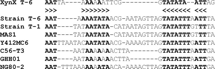
Alignment of regulatory regions upstream to xylanases genes from Geobacillus spp. The sequences upstream the putative −10 regions of several xylanases genes from Geobacillus strains: G. stearothermophilus T-6, G. stearothermophilus T-1, Geobacillus sp. MAS1, Geobacillus sp. Y412MC6, Geobacillus sp. C56-T3, Geobacillus sp. GHH01 and G. thermodenitrificans NG80–2. The first sequence aligned is the sequence upstream of the xynX gene from G. stearothermophilus T-6. Conserved nucleotides are shown in bold, and the inverted repeat sequence, the putative XynX-binding site, is indicated by arrowheads.
In the xynX null strain, xylanase-specific activity was higher than in the wild type, indicating that XynX represses the expression of xynA. However, the xynX null strain exhibited normal expression of the xynA gene during the logarithmic growth phase and the same level of repression by glucose or casamino acids as in the wild type strain. Taken together, these results indicate that XynX is involved in xynA regulation but does not contribute directly to carbon catabolite repression. In summary, the mechanism by which XynX controls xynA gene expression is elusive as well as the additional factors that may mediate its effect. Characterization of the XynX mode of action might reveal a novel pathway for the Nif3-like proteins in prokaryotes.
The Extracellular Xylanase Gene Is Regulated by Cell Density (Quorum Sensing)
The least expected result presented here is quorum-sensing regulation of the extracellular xylanase (xynA) gene. In batch cultures, the specific activity of the extracellular xylanase increases over 50-fold during exponential growth, suggesting cell density regulation. This phenomenon is not related to induction or cell adaptation as was demonstrated in continuous cultures operating at different cell densities and with a xylanase-constitutive mutant. Quantitative real time RT-PCR and quantitative measurements of xynA-mRNA using RNase protection assays indicated that the regulation is at the transcriptional level.
In Gram-positive bacteria, quorum-sensing regulation is mediated by small modified peptides and controls many physiological processes, including biofilm formation (77), virulence factor regulation (65, 78), competence development (79, 80), sporulation (81, 82), and induction of conjugation (83, 84). Our initial attempts to purify the extracellular XDF suggest that it is heat-stable (100 °C for 10 min) and pH-stable (2.0), yet it is sensitive to protease treatment. The factor can be absorbed on a hydrophobic matrix and eluted with 20–25% acetonitrile using a C18 reverse phase HPLC column. All of these results are consistent with Gram-positive quorum-sensing peptide factors (85–88), suggesting that XDF is a small hydrophobic peptide.
Analysis of the draft genome of G. stearothermophilus strain T-6 reveals sequence homology to well characterized quorum-sensing pathways for competence and sporulation processes, including ComQ and ComC, and the modified peptides ComX and CSF. Strain T-6 also possesses the NrpR-NrpX quorum-sensing system, and therefore we tested whether the predicted pentapeptide (AKDEH) can activate xynA expression in low cell density cultures. The synthetic peptide failed to activate xynA expression (data not shown), indicating that strain T-6 utilizes a different cell density regulatory system to regulate xynA. The secreted cell-extracellular signaling peptides can bind to a specific sensor component of a two-component signal transduction system (89) or can be import to the cell via specific oligopeptide transport system (90). Bialaphos is a toxic tri-peptide that is known to enter bacteria via oligopeptide permeases (91). We have isolated bialaphos-resistant T-6 mutants, suspected to code for a defective oligopeptide transport system. These bialaphos-resistant mutants exhibited typical activation of xylanase expression during logarithmic growth, suggesting that in strain T-6 cell density activation of xynA is not solely based on an oligopeptide transport system.
What can be the biological benefit of regulating the expression of a vegetative extracellular enzyme by quorum sensing? Quorum-sensing regulation provides a mechanism by which secretion and production of exoproducts (“public goods”) are regulated to benefit the cells in the local population (92–95). In many Gram-positive bacteria the regulation of “public goods” production is associated with late exponential phase and stationery phase and occurs at high cell densities. During this period, and before the cell is committed to sporulation, it expresses proteins that allow it to adapt to poor nutritional conditions and maintain growth, for example, by degrading macromolecules via expression of extracellular enzymes. However, the quorum-sensing regulation of the extracellular xylanase appears to function at very low cell densities (<0.01 A600) at the early exponential phase of growth and not at the entrance to the stationary phase. To the best of our knowledge, regulation at these low cell densities has never been reported. A possible biological rationale for cell density activation of the extracellular xylanase stems from the fact that the utilization of high molecular weight polymers, such as xylan, requires a threshold concentration of extracellular enzymes. At very low cell densities, the effective concentration of diffusible extracellular enzymes is too low to support growth because the xylo-oligosaccharides products are too large to import into the cells, resulting in “cell density-dependent growth.” Only at moderate or “high cell densities” does the culture produce sufficient amounts of extracellular hydrolyzing enzymes that provide suitable degradation products. Thus, it is advantageous for the cell to produce the extracellular enzymes only when cell density can support growth. Cell density growth dependence was demonstrated elegantly with Myxococcus xanthus, a Gram-negative topsoil bacterium that forms swarms and fruiting bodies (12). When grown on casein, the cells exhibited cooperative behavior by pooling their extracellular digestive enzymes. The extracellular proteinase activity was directly proportional to cell number, and the growth rate was correlated with increased concentrations of hydrolyzed casein.
In summary, we have shown that the expression of the extracellular xylanase gene, xynA, from G. stearothermophilus T-6 is intricate and involves multiple regulatory mechanisms (Fig. 14). This regulation is well suited to the physiological scheme the bacterium use for degrading polysaccharides in the environment. The xynA gene is negatively controlled by the xylose repressor, XylR, and is induced by xylose. Expression of the gene is also repressed by glucose via carbon catabolite repression, most likely mediated by CcpA and casamino acids via the transcriptional regulator CodY. In addition, xynA is regulated by two yet uncharacterized mechanisms, a NIF3-like protein, XynX, and by cell density. The quorum-sensing regulation is effective at very low cell densities and most likely represents a novel system.
FIGURE 14.

Schematic representation of multiple regulatory mechanisms controlling the expression of xynA. Xylose and short xylo-oligomers are sensed by the two-component system XynDC, which in turn activates the ABC xylo-oligosaccharide transporter, XynEFG, facilitating the entrance of xylose and xylosaccharides into the cell. Inside the cell, xylose serves as the molecular inducer and interacts with the master repressor XylR, which negatively regulates the transcription of the xynA gene. XylR can also interact with glucose 6-phosphate, which exerts inducer prevention. Expression of the xynA gene is also repressed by glucose via carbon catabolite repression, most likely mediated by CcpA. In rapidly growing cells in high nutrition availability, xynA is repressed by CodY, which is subjected to regulation by GTP and branched-chain amino acids. Additionally, xynA is regulated by two yet uncharacterized mechanisms, a NIF3-like protein, XynX, and by cell density. The xynA gene is induced via quorum sensing regulation at relatively low cell density. The XDFs are most likely short modified peptides that can either bind to a specific sensor component of a two-component signal-transduction system or can be imported into the cell by a dedicated peptide transporter. The DNA sequence corresponds to position −131 bp with respect to the transcriptional start point (+1). The proposed catabolite-responsive element (cre) is presented in red letters. The potential inverted repeat binding site for the xylose repressor, XylR, and the putative XynX-binding site are indicated by arrows. Two of the proposed CodY-binding site sequences are shown in green letters. The table below summarizes the proposed regulatory proteins and their effectors involved in the regulation of the xynA gene.
This work was supported in part by United States-Israel Binational Science Foundation Jerusalem, Israel, Grant 96-178 (to A. L. S. and Y. S.), Israel Science Foundation Grants 500/10 and 152/11, the I-CORE Program of the Planning and Budgeting Committee, the Ministry of Environmental Protection and the Grand Technion Energy Program, and part of The Leona M. and Harry B. Helmsley Charitable Trust reports on Alternative Energy series of the Technion, Israel Institute of Technology, and the Weizmann Institute of Science.
The nucleotide sequence(s) reported in this paper has been submitted to the GenBankTM/EBI Data Bank with accession number(s) DQ868502 and DQ868501.1
- mBSM
- modified Basic Salt Medium
- XDF
- xylanase density factor
- pNPX2
- p-nitrophenyl xylobioside
- nt
- nucleotide
- RACE
- rapid amplification of cDNA end
- ACN
- acetonitrile.
REFERENCES
- 1. Shallom D., Shoham Y. (2003) Microbial hemicellulases. Curr. Opin. Microbiol. 6, 219–228 [DOI] [PubMed] [Google Scholar]
- 2. Yu H., Zeng G., Huang H., Xi X., Wang R., Huang D., Huang G., Li J. (2007) Microbial community succession and lignocellulose degradation during agricultural waste composting. Biodegradation 18, 793–802 [DOI] [PubMed] [Google Scholar]
- 3. Taylor L. E., 2nd, Henrissat B., Coutinho P. M., Ekborg N. A., Hutcheson S. W., Weiner R. M. (2006) Complete cellulase system in the marine bacterium Saccharophagus degradans strain 2-40T. J. Bacteriol. 188, 3849–3861 [DOI] [PMC free article] [PubMed] [Google Scholar]
- 4. Ouyang J., Yan M., Kong D., Xu L. (2006) A complete protein pattern of cellulase and hemicellulase genes in the filamentous fungus Trichoderma reesei. Biotechnol. J. 1, 1266–1274 [DOI] [PubMed] [Google Scholar]
- 5. Gilbert H. J., Stålbrand H., Brumer H. (2008) How the walls come crumbling down: recent structural biochemistry of plant polysaccharide degradation. Curr. Opin. Plant Biol. 11, 338–348 [DOI] [PubMed] [Google Scholar]
- 6. Kopetz H. (2013) Renewable resources: Build a biomass energy market. Nature 494, 29–31 [DOI] [PubMed] [Google Scholar]
- 7. Ragauskas A. J., Williams C. K., Davison B. H., Britovsek G., Cairney J., Eckert C. A., Frederick W. J., Jr., Hallett J. P., Leak D. J., Liotta C. L., Mielenz J. R., Murphy R., Templer R., Tschaplinski T. (2006) The path forward for biofuels and biomaterials. Science 311, 484–489 [DOI] [PubMed] [Google Scholar]
- 8. Brunecky R., Alahuhta M., Xu Q., Donohoe B. S., Crowley M. F., Kataeva I. A., Yang S. J., Resch M. G., Adams M. W., Lunin V. V., Himmel M. E., Bomble Y. J. (2013) Revealing nature's cellulase diversity: the digestion mechanism of Caldicellulosiruptor bescii CelA. Science 342, 1513–1516 [DOI] [PubMed] [Google Scholar]
- 9. Himmel M. E., Ding S. Y., Johnson D. K., Adney W. S., Nimlos M. R., Brady J. W., Foust T. D. (2007) Biomass recalcitrance: engineering plants and enzymes for biofuels production. Science 315, 804–807 [DOI] [PubMed] [Google Scholar]
- 10. Scheller H. V., Ulvskov P. (2010) Hemicelluloses. Annu. Rev. Plant Biol. 61, 263–289 [DOI] [PubMed] [Google Scholar]
- 11. Gilbert H. J. (2010) The biochemistry and structural biology of plant cell wall deconstruction. Plant Physiol. 153, 444–455 [DOI] [PMC free article] [PubMed] [Google Scholar]
- 12. Rosenberg E., Keller K. H., Dworkin M. (1977) Cell density-dependent growth of Myxococcus xanthus on casein. J. Bacteriol. 129, 770–777 [DOI] [PMC free article] [PubMed] [Google Scholar]
- 13. Wilson D. B. (2008) Three microbial strategies for plant cell wall degradation. Ann. N.Y. Acad. Sci. 1125, 289–297 [DOI] [PubMed] [Google Scholar]
- 14. Bayer E. A., Belaich J. P., Shoham Y., Lamed R. (2004) The cellulosomes: multienzyme machines for degradation of plant cell wall polysaccharides. Annu. Rev. Microbiol. 58, 521–554 [DOI] [PubMed] [Google Scholar]
- 15. Bayer E. A., Shoham Y., Lamed R. (2000) in Glycomicrobiology (Doyle R. J., ed) pp. 387–439, Kluwer Academic/Plenum Publishers, New York [Google Scholar]
- 16. Shoham Y., Lamed R., Bayer E. A. (1999) The cellulosome concept as an efficient microbial strategy for the degradation of insoluble polysaccharides. Trends Microbiol. 7, 275–281 [DOI] [PubMed] [Google Scholar]
- 17. Larsbrink J., Izumi A., Ibatullin F. M., Nakhai A., Gilbert H. J., Davies G. J., Brumer H. (2011) Structural and enzymatic characterization of a glycoside hydrolase family 31 α-xylosidase from Cellvibrio japonicus involved in xyloglucan saccharification. Biochem. J. 436, 567–580 [DOI] [PubMed] [Google Scholar]
- 18. Glass N. L., Schmoll M., Cate J. H., Coradetti S. (2013) Plant cell wall deconstruction by ascomycete fungi. Annu. Rev. Microbiol. 67, 477–498 [DOI] [PubMed] [Google Scholar]
- 19. Tabachnikov O., Shoham Y. (2013) Functional characterization of the galactan utilization system of Geobacillus stearothermophilus. FEBS J. 280, 950–964 [DOI] [PubMed] [Google Scholar]
- 20. Shulami S., Raz-Pasteur A., Tabachnikov O., Gilead-Gropper S., Shner I., Shoham Y. (2011) The l-arabinan utilization system of Geobacillus stearothermophilus. J. Bacteriol. 193, 2838–2850 [DOI] [PMC free article] [PubMed] [Google Scholar]
- 21. Shulami S., Zaide G., Zolotnitsky G., Langut Y., Feld G., Sonenshein A. L., Shoham Y. (2007) A two-component system regulates the expression of an ABC transporter for xylo-oligosaccharides in Geobacillus stearothermophilus. Appl. Environ. Microbiol. 73, 874–884 [DOI] [PMC free article] [PubMed] [Google Scholar]
- 22. Shulami S., Gat O., Sonenshein A. L., Shoham Y. (1999) The glucuronic acid utilization gene cluster from Bacillus stearothermophilus T-6. J. Bacteriol. 181, 3695–3704 [DOI] [PMC free article] [PubMed] [Google Scholar]
- 23. Miyazaki K., Miyamoto H., Mercer D. K., Hirase T., Martin J. C., Kojima Y., Flint H. J. (2003) Involvement of the multidomain regulatory protein XynR in positive control of xylanase gene expression in the ruminal anaerobe Prevotella bryantii B(1)4. J. Bacteriol. 185, 2219–2226 [DOI] [PMC free article] [PubMed] [Google Scholar]
- 24. Sonnenburg E. D., Sonnenburg J. L., Manchester J. K., Hansen E. E., Chiang H. C., Gordon J. I. (2006) A hybrid two-component system protein of a prominent human gut symbiont couples glycan sensing in vivo to carbohydrate metabolism. Proc. Natl. Acad. Sci. U.S.A. 103, 8834–8839 [DOI] [PMC free article] [PubMed] [Google Scholar]
- 25. Nataf Y., Bahari L., Kahel-Raifer H., Borovok I., Lamed R., Bayer E. A., Sonenshein A. L., Shoham Y. (2010) Clostridium thermocellum cellulosomal genes are regulated by extracytoplasmic polysaccharides via alternative σ factors. Proc. Natl. Acad. Sci. U.S.A. 107, 18646–18651 [DOI] [PMC free article] [PubMed] [Google Scholar]
- 26. Kahel-Raifer H., Jindou S., Bahari L., Nataf Y., Shoham Y., Bayer E. A., Borovok I., Lamed R. (2010) The unique set of putative membrane-associated anti-σ factors in Clostridium thermocellum suggests a novel extracellular carbohydrate-sensing mechanism involved in gene regulation. FEMS Microbiol. Lett. 308, 84–93 [DOI] [PubMed] [Google Scholar]
- 27. Yaniv O., Fichman G., Borovok I., Shoham Y., Bayer E. A., Lamed R., Shimon L. J., Frolow F. (2014) Fine-structural variance of family 3 carbohydrate-binding modules as extracellular biomass-sensing components of Clostridium thermocellum anti-σI factors. Acta Crystallogr. D Biol. Crystallogr. 70, 522–534 [DOI] [PubMed] [Google Scholar]
- 28. Martens E. C., Roth R., Heuser J. E., Gordon J. I. (2009) Coordinate regulation of glycan degradation and polysaccharide capsule biosynthesis by a prominent human gut symbiont. J. Biol. Chem. 284, 18445–18457 [DOI] [PMC free article] [PubMed] [Google Scholar]
- 29. Khasin A., Alchanati I., Shoham Y. (1993) Purification and characterization of a thermostable xylanase from Bacillus stearothermophilus T-6. Appl. Environ. Microbiol. 59, 1725–1730 [DOI] [PMC free article] [PubMed] [Google Scholar]
- 30. Shoham Y., Schwartz Z., Khasin A., Gat O., Zosim Z., Rosenberg E. (1992) Delignification of wood pulp by a thermostable xylanase from Bacillus stearothermophilus strain T-6. Biodegradation 3, 207–218 [Google Scholar]
- 31. Lansky S., Alalouf O., Solomon H. V., Alhassid A., Govada L., Chayen N. E., Belrhali H., Shoham Y., Shoham G. (2014) A unique octameric structure of Axe2, an intracellular acetyl-xylooligosaccharide esterase from Geobacillus stearothermophilus. Acta Crystallogr. D Biol. Crystallogr. 70, 261–278 [DOI] [PubMed] [Google Scholar]
- 32. Alhassid A., Ben-David A., Tabachnikov O., Libster D., Naveh E., Zolotnitsky G., Shoham Y., Shoham G. (2009) Crystal structure of an inverting GH 43 1,5-α-l-arabinanase from Geobacillus stearothermophilus complexed with its substrate. Biochem. J. 422, 73–82 [DOI] [PubMed] [Google Scholar]
- 33. Brüx C., Ben-David A., Shallom-Shezifi D., Leon M., Niefind K., Shoham G., Shoham Y., Schomburg D. (2006) The structure of an inverting GH43 β-xylosidase from Geobacillus stearothermophilus with its substrate reveals the role of the three catalytic residues. J. Mol. Biol. 359, 97–109 [DOI] [PubMed] [Google Scholar]
- 34. Czjzek M., Ben David A., Bravman T., Shoham G., Henrissat B., Shoham Y. (2005) Enzyme-substrate complex structures of a GH39 β-xylosidase from Geobacillus stearothermophilus. J. Mol. Biol. 353, 838–846 [DOI] [PubMed] [Google Scholar]
- 35. Golan G., Shallom D., Teplitsky A., Zaide G., Shulami S., Baasov T., Stojanoff V., Thompson A., Shoham Y., Shoham G. (2004) Crystal structures of Geobacillus stearothermophilus α-glucuronidase complexed with its substrate and products: mechanistic implications. J. Biol. Chem. 279, 3014–3024 [DOI] [PubMed] [Google Scholar]
- 36. Hövel K., Shallom D., Niefind K., Belakhov V., Shoham G., Baasov T., Shoham Y., Schomburg D. (2003) Crystal structure and snapshots along the reaction pathway of a family 51 α-l-arabinofuranosidase. EMBO J. 22, 4922–4932 [DOI] [PMC free article] [PubMed] [Google Scholar]
- 37. Bravman T., Zolotnitsky G., Belakhov V., Shoham G., Henrissat B., Baasov T., Shoham Y. (2003) Detailed kinetic analysis of a family 52 glycoside hydrolase: a β-xylosidase from Geobacillus stearothermophilus. Biochemistry 42, 10528–10536 [DOI] [PubMed] [Google Scholar]
- 38. Bravman T., Belakhov V., Solomon D., Shoham G., Henrissat B., Baasov T., Shoham Y. (2003) Identification of the catalytic residues in family 52 glycoside hydrolase, a β-xylosidase from Geobacillus stearothermophilus T-6. J. Biol. Chem. 278, 26742–26749 [DOI] [PubMed] [Google Scholar]
- 39. Zaide G., Shallom D., Shulami S., Zolotnitsky G., Golan G., Baasov T., Shoham G., Shoham Y. (2001) Biochemical characterization and identification of catalytic residues in α-glucuronidase from Bacillus stearothermophilus T-6. Eur. J. Biochem. 268, 3006–3016 [DOI] [PubMed] [Google Scholar]
- 40. Salama R., Alalouf O., Tabachnikov O., Zolotnitsky G., Shoham G., Shoham Y. (2012) The abp gene in Geobacillus stearothermophilus T-6 encodes a GH27 β-l-arabinopyranosidase. FEBS Lett. 586, 2436–2442 [DOI] [PubMed] [Google Scholar]
- 41. Ben-David A., Shoham G., Shoham Y. (2008) A universal screening assay for glycosynthases: directed evolution of glycosynthase XynB2(E335G) suggests a general path to enhance activity. Chem. Biol. 15, 546–551 [DOI] [PubMed] [Google Scholar]
- 42. Alalouf O., Balazs Y., Volkinshtein M., Grimpel Y., Shoham G., Shoham Y. (2011) A new family of carbohydrate esterases is represented by a GDSL hydrolase/acetylxylan esterase from Geobacillus stearothermophilus. J. Biol. Chem. 286, 41993–42001 [DOI] [PMC free article] [PubMed] [Google Scholar]
- 43. Teplitsky A., Shulami S., Moryles S., Shoham Y., Shoham G. (2000) Crystallization and preliminary X-ray analysis of an intracellular xylanase from Bacillus stearothermophilus T-6. Acta Crystallogr. D Biol. Crystallogr. 56, 181–184 [DOI] [PubMed] [Google Scholar]
- 44. Teplitsky A., Feinberg H., Gilboa R., Lapidot A., Mechaly A., Stojanoff V., Capel M., Shoham Y., Shoham G. (1997) Crystallization and preliminary x-ray analysis of the thermostable alkaline-tolerant xylanase from Bacillus stearothermophilus T-6. Acta Crystallogr. D Biol. Crystallogr. 53, 608–611 [DOI] [PubMed] [Google Scholar]
- 45. Teplitsky A., Mechaly A., Stojanoff V., Sainz G., Golan G., Feinberg H., Gilboa R., Reiland V., Zolotnitsky G., Shallom D., Thompson A., Shoham Y., Shoham G. (2004) Structure determination of the extracellular xylanase from Geobacillus stearothermophilus by selenomethionyl MAD phasing. Acta Crystallogr. D Biol. Crystallogr. 60, 836–848 [DOI] [PubMed] [Google Scholar]
- 46. Marmur J. (1961) A procedure for isolation of deoxyribonucleic acid from micro-organisms. J. Mol. Biol. 3, 208–218 [Google Scholar]
- 47. Johnson J. L. (1981) in Manual of Methods for General Bacteriology (Gerhardt P., Murray R. G. E., Costilow E. W., Nester W. A., Wood N. R., Krieg R., Philips G. B., eds) pp. 450–472, American Society for Microbiology, Washington, D. C [Google Scholar]
- 48. Sambrook J., Russell D. W. (2001) Molecular Cloning: A Laboratory Manual, 3rd ed., Cold Spring Harbor Laboratories Press, Cold Spring Harbor, NY [Google Scholar]
- 49. Moran C. P., Jr., Lang N., LeGrice S. F., Lee G., Stephens M., Sonenshein A. L., Pero J., Losick R. (1982) Nucleotide sequences that signal the initiation of transcription and translation in Bacillus subtilis. Mol. Gen. Genet. 186, 339–346 [DOI] [PubMed] [Google Scholar]
- 50. Moran C. P. (1990) in Molecular Biological Methods for Bacillus (Harwood C. R., Cutting S. M., eds) pp. 267–293, John Wiley & Sons, Chichester, UK [Google Scholar]
- 51. Wu L. J., Welker N. E. (1989) Protoplast transformation of Bacillus stearothermophilus NUB36 by plasmid DNA. J. Gen. Microbiol. 135, 1315–1324 [DOI] [PubMed] [Google Scholar]
- 52. Heckman K. L., Pease L. R. (2007) Gene splicing and mutagenesis by PCR-driven overlap extension. Nat. Protoc. 2, 924–932 [DOI] [PubMed] [Google Scholar]
- 53. Mechaly A., Belakhov V., Shoham Y., Baasov T. (1997) An efficient chemical-enzymatic synthesis of 4-nitrophenyl β-xylobioside: a chromogenic substrate for xylanases. Carbohydr. Res. 304, 111–115 [Google Scholar]
- 54. Smith H., Bron S., Van Ee J., Venema G. (1987) Construction and use of signal sequence selection vectors in Escherichia coli and Bacillus subtilis. J. Bacteriol. 169, 3321–3328 [DOI] [PMC free article] [PubMed] [Google Scholar]
- 55. Ashwell G. (1957) in Methods in Enzymology (Colowick S. P., Kaplan N. O., eds) pp. 73–105, Academic Press, Inc., New York [Google Scholar]
- 56. Gat O., Lapidot A., Alchanati I., Regueros C., Shoham Y. (1994) Cloning and DNA sequence of the gene coding for Bacillus stearothermophilus T-6 xylanase. Appl. Environ. Microbiol. 60, 1889–1896 [DOI] [PMC free article] [PubMed] [Google Scholar]
- 57. Rodionov D. A., Mironov A. A., Gelfand M. S. (2001) Transcriptional regulation of pentose utilisation systems in the Bacillus/Clostridium group of bacteria. FEMS Microbiol. Lett. 205, 305–314 [DOI] [PubMed] [Google Scholar]
- 58. Hueck C. J., Hillen W. (1995) Catabolite repression in Bacillus subtilis: a global regulatory mechanism for the Gram-positive bacteria? Mol. Microbiol. 15, 395–401 [DOI] [PubMed] [Google Scholar]
- 59. Dahl M. K., Schmiedel D., Hillen W. (1995) Glucose and glucose-6-phosphate interaction with Xyl repressor proteins from Bacillus spp. may contribute to regulation of xylose utilization. J. Bacteriol. 177, 5467–5472 [DOI] [PMC free article] [PubMed] [Google Scholar]
- 60. Sonenshein A. L. (2005) CodY, a global regulator of stationary phase and virulence in Gram-positive bacteria. Curr. Opin. Microbiol. 8, 203–207 [DOI] [PubMed] [Google Scholar]
- 61. Belitsky B. R., Sonenshein A. L. (2008) Genetic and biochemical analysis of CodY-binding sites in Bacillus subtilis. J. Bacteriol. 190, 1224–1236 [DOI] [PMC free article] [PubMed] [Google Scholar]
- 62. Tascou S., Kang T. W., Trappe R., Engel W., Burfeind P. (2003) Identification and characterization of NIF3L1 BP1, a novel cytoplasmic interaction partner of the NIF3L1 protein. Biochem. Biophys. Res. Commun. 309, 440–448 [DOI] [PubMed] [Google Scholar]
- 63. Akiyama H., Fujisawa N., Tashiro Y., Takanabe N., Sugiyama A., Tashiro F. (2003) The role of transcriptional corepressor Nif3l1 in early stage of neural differentiation via cooperation with Trip15/CSN2. J. Biol. Chem. 278, 10752–10762 [DOI] [PubMed] [Google Scholar]
- 64. Ferrari E., Jarnagin A. S., Schmidt B. F. (1993) in Bacillus Subtilis and Other Gram-positive Bacteria (Sonenshein A. L., Hoch J. A., Losick R., eds) pp. 917–937, American Society for Microbiology, Washington, D. C [Google Scholar]
- 65. Cook L. C., Federle M. J. (2014) Peptide pheromone signaling in Streptococcus and Enterococcus. FEMS Microbiol. Rev. 38, 473–492 [DOI] [PMC free article] [PubMed] [Google Scholar]
- 66. Langut Y. (2006) in Characterization of XylR, the Repressor of the Xylanolytic System in Geobacillus stearothermophilus. Msc thesis, Technion-Israel Institute of Technology, Haifa, Israel [Google Scholar]
- 67. Görke B., Stülke J. (2008) Carbon catabolite repression in bacteria: many ways to make the most out of nutrients. Nat. Rev. Microbiol. 6, 613–624 [DOI] [PubMed] [Google Scholar]
- 68. Fujita Y. (2009) Carbon catabolite control of the metabolic network in Bacillus subtilis. Biosci. Biotechnol. Biochem. 73, 245–259 [DOI] [PubMed] [Google Scholar]
- 69. Kraus A., Küster E., Wagner A., Hoffmann K., Hillen W. (1998) Identification of a co-repressor binding site in catabolite control protein CcpA. Mol. Microbiol. 30, 955–963 [DOI] [PubMed] [Google Scholar]
- 70. Kazanov M. D., Li X., Gelfand M. S., Osterman A. L., Rodionov D. A. (2013) Functional diversification of ROK-family transcriptional regulators of sugar catabolism in the Thermotogae phylum. Nucleic Acids Res. 41, 790–803 [DOI] [PMC free article] [PubMed] [Google Scholar]
- 71. Ratnayake-Lecamwasam M., Serror P., Wong K. W., Sonenshein A. L. (2001) Bacillus subtilis CodY represses early-stationary-phase genes by sensing GTP levels. Genes Dev. 15, 1093–1103 [DOI] [PMC free article] [PubMed] [Google Scholar]
- 72. Molle V., Nakaura Y., Shivers R. P., Yamaguchi H., Losick R., Fujita Y., Sonenshein A. L. (2003) Additional targets of the Bacillus subtilis global regulator CodY identified by chromatin immunoprecipitation and genome-wide transcript analysis. J. Bacteriol. 185, 1911–1922 [DOI] [PMC free article] [PubMed] [Google Scholar]
- 73. Sonenshein A. L. (2007) Control of key metabolic intersections in Bacillus subtilis. Nat. Rev. Microbiol. 5, 917–927 [DOI] [PubMed] [Google Scholar]
- 74. Belitsky B. R., Sonenshein A. L. (2013) Genome-wide identification of Bacillus subtilis CodY-binding sites at single-nucleotide resolution. Proc. Natl. Acad. Sci. U.S.A. 110, 7026–7031 [DOI] [PMC free article] [PubMed] [Google Scholar]
- 75. Galperin M. Y., Koonin E. V. (2004) ‘Conserved hypothetical’ proteins: prioritization of targets for experimental study. Nucleic Acids Res. 32, 5452–5463 [DOI] [PMC free article] [PubMed] [Google Scholar]
- 76. Tomoike F., Wakamatsu T., Nakagawa N., Kuramitsu S., Masui R. (2009) Crystal structure of the conserved hypothetical protein TTHA1606 from Thermus thermophilus HB8. Proteins 76, 244–248 [DOI] [PubMed] [Google Scholar]
- 77. Davies D. G., Parsek M. R., Pearson J. P., Iglewski B. H., Costerton J. W., Greenberg E. P. (1998) The involvement of cell-to-cell signals in the development of a bacterial biofilm. Science 280, 295–298 [DOI] [PubMed] [Google Scholar]
- 78. Novick R. P., Geisinger E. (2008) Quorum sensing in staphylococci. Annu. Rev. Genet. 42, 541–564 [DOI] [PubMed] [Google Scholar]
- 79. Magnuson R., Solomon J., Grossman A. D. (1994) Biochemical and genetic characterization of a competence pheromone from B. subtilis. Cell 77, 207–216 [DOI] [PubMed] [Google Scholar]
- 80. Ansaldi M., Marolt D., Stebe T., Mandic-Mulec I., Dubnau D. (2002) Specific activation of the Bacillus quorum-sensing systems by isoprenylated pheromone variants. Mol. Microbiol. 44, 1561–1573 [DOI] [PubMed] [Google Scholar]
- 81. Perego M., Hoch J. A. (1996) Cell-cell communication regulates the effects of protein aspartate phosphatases on the phosphorelay controlling development in Bacillus subtilis. Proc. Natl. Acad. Sci. U.S.A. 93, 1549–1553 [DOI] [PMC free article] [PubMed] [Google Scholar]
- 82. Steiner E., Scott J., Minton N. P., Winzer K. (2012) An agr quorum sensing system that regulates granulose formation and sporulation in Clostridium acetobutylicum. Appl. Environ. Microbiol. 78, 1113–1122 [DOI] [PMC free article] [PubMed] [Google Scholar]
- 83. Shi K., Brown C. K., Gu Z. Y., Kozlowicz B. K., Dunny G. M., Ohlendorf D. H., Earhart C. A. (2005) Structure of peptide sex pheromone receptor PrgX and PrgX/pheromone complexes and regulation of conjugation in Enterococcus faecalis. Proc. Natl. Acad. Sci. U.S.A. 102, 18596–18601 [DOI] [PMC free article] [PubMed] [Google Scholar]
- 84. Dunny G. M. (2007) The peptide pheromone-inducible conjugation system of Enterococcus faecalis plasmid pCF10: cell-cell signalling, gene transfer, complexity and evolution. Philos. Trans. R. Soc. Lond. B. Biol. Sci. 362, 1185–1193 [DOI] [PMC free article] [PubMed] [Google Scholar]
- 85. Solomon J. M., Lazazzera B. A., Grossman A. D. (1996) Purification and characterization of an extracellular peptide factor that affects two different developmental pathways in Bacillus subtilis. Genes Dev. 10, 2014–2024 [DOI] [PubMed] [Google Scholar]
- 86. Nakayama J., Cao Y., Horii T., Sakuda S., Akkermans A. D., de Vos W. M., Nagasawa H. (2001) Gelatinase biosynthesis-activating pheromone: a peptide lactone that mediates a quorum sensing in Enterococcus faecalis. Mol. Microbiol. 41, 145–154 [DOI] [PubMed] [Google Scholar]
- 87. Ibrahim M., Guillot A., Wessner F., Algaron F., Besset C., Courtin P., Gardan R., Monnet V. (2007) Control of the transcription of a short gene encoding a cyclic peptide in Streptococcus thermophilus: a new quorum-sensing system? J. Bacteriol. 189, 8844–8854 [DOI] [PMC free article] [PubMed] [Google Scholar]
- 88. Kolodkin-Gal I., Hazan R., Gaathon A., Carmeli S., Engelberg-Kulka H. (2007) A linear pentapeptide is a quorum-sensing factor required for mazEF-mediated cell death in Escherichia coli. Science 318, 652–655 [DOI] [PubMed] [Google Scholar]
- 89. Kleerebezem M., Quadri L. E., Kuipers O. P., de Vos W. M. (1997) Quorum sensing by peptide pheromones and two-component signal-transduction systems in Gram-positive bacteria. Mol. Microbiol. 24, 895–904 [DOI] [PubMed] [Google Scholar]
- 90. Alloing G., Martin B., Granadel C., Claverys J. P. (1998) Development of competence in Streptococcus pneumoniae: pheromone autoinduction and control of quorum sensing by the oligopeptide permease. Mol. Microbiol. 29, 75–83 [DOI] [PubMed] [Google Scholar]
- 91. Koide A., Hoch J. A. (1994) Identification of a second oligopeptide transport system in Bacillus subtilis and determination of its role in sporulation. Mol. Microbiol. 13, 417–426 [DOI] [PubMed] [Google Scholar]
- 92. Waters C. M., Bassler B. L. (2005) Quorum sensing: cell-to-cell communication in bacteria. Annu. Rev. Cell Dev. Biol. 21, 319–346 [DOI] [PubMed] [Google Scholar]
- 93. Keller L., Surette M. G. (2006) Communication in bacteria: an ecological and evolutionary perspective. Nat. Rev. Microbiol. 4, 249–258 [DOI] [PubMed] [Google Scholar]
- 94. Ng W. L., Bassler B. L. (2009) Bacterial quorum-sensing network architectures. Annu. Rev. Genet. 43, 197–222 [DOI] [PMC free article] [PubMed] [Google Scholar]
- 95. Stevens A. M., Schuster M., Rumbaugh K. P. (2012) Working together for the common good: cell-cell communication in bacteria. J. Bacteriol. 194, 2131–2141 [DOI] [PMC free article] [PubMed] [Google Scholar]
- 96. Goecks J., Nekrutenko A., Taylor J., Galaxy T. (2010) Galaxy: a comprehensive approach for supporting accessible, reproducible, and transparent computational research in the life sciences. Genome Biol. 11, R86. [DOI] [PMC free article] [PubMed] [Google Scholar]