Developmental venous anomalies (DVA) are congenital variants of cerebral veins, found incidentally at autopsy in 2.6% of the population, which are most often asymptomatic.1 Symptomatic compression of a cranial nerve by the collecting vein of a DVA is extremely rare, such as tinnitus from compression of the vestibulocochlear nerve.2
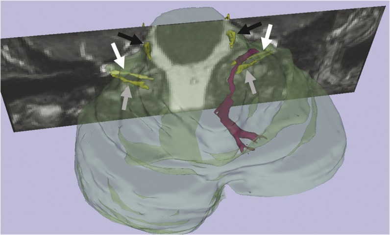
A 51-year-old man presented with 1 week of intermittent vertigo and mild left-sided dysmetria. Imaging showed contact of the left vestibular nerve by the collecting vein of a cerebellar DVA (figures 1 and 2). We speculate that the symptoms were caused by transient migration of the cisternal segment of the collecting vein. Management was conservative with spontaneous resolution.
Figure 1. MRI of cerebellar developmental venous anomaly.

Axial T1 postgadolinium MRI shows a left cerebellar developmental venous anomaly (white arrows, A, B), the collecting vein of which is seen on T2 (C) and steady-state free precession (D) images (white arrows) adjacent to the left vestibular nerve (hashed arrows).
Figure 2. 3D reconstruction of cerebellar developmental venous anomaly.

3D reconstructed image shows the developmental venous anomaly and collecting vein (purple) and cranial nerves (yellow): trigeminal, black arrows; facial, white arrows; vestibulocochlear, hashed arrows.
Acknowledgments
Acknowledgement: The authors thank Dr. Mark R. Etherton, Dr. Taha Gholipour, and Matthew W. Rondeau of the Department of Neurology, Brigham and Women's Hospital, for discussion of the case.
Footnotes
Author contributions: Analysis and interpretation of data: Dr. Narendra, Dr. Wang, Dr. Erkkinen, Dr. Jagadeesan, Dr. Lee, Dr. Zimmerman, and Dr. Klein. Drafting of the manuscript: Dr. Narendra and Dr. Klein. Critical revision of the manuscript for important intellectual content: Dr. Narendra, Dr. Wang, Dr. Erkkinen, Dr. Jagadeesan, Dr. Zimmerman, and Dr. Klein.
Study funding: No targeted funding reported.
Disclosure: The authors report no disclosures relevant to the manuscript. Go to Neurology.org for full disclosures.
REFERENCES
- 1.Sarwar M, McCormick WF. Intracerebral venous angioma. Arch Neurol 1978;35:323–325 [DOI] [PubMed] [Google Scholar]
- 2.Malinvaud M, Lecanu JB, Halimi P, Avan P, Bonfils P. Tinnitus, cerebellar developmental venous anomaly. Arch Otolaryngol Head Neck Surg 2006;132:550–553 [DOI] [PubMed] [Google Scholar]


