SUMMARY
Hematopoietic stem cells (HSCs) are the precursors of the hematopoietic system responsible for the lifelong production of blood and bone marrow. Given the emerging importance of epigenetic regulation in HSC fate decisions and malignant transformation, we investigated the role of the DNA methyltransferase Dnmt3b through genetic ablation in HSCs – either alone or in combinatorial deletion with its paralog Dnmt3a. While conditional inactivation of Dnmt3b alone in adult HSCs had minor functional impact, simultaneous deletion of Dnmt3a and Dnmt3b was synergistic resulting in a severe block in differentiation and enhanced HSC self-renewal. Dnmt3a/b-null HSCs displayed activated β-catenin signaling, partly accounting for the differentiation block. Loss of Dnmt3a in HSCs resulted in global DNA hypomethylation, but a paradoxical hypermethylation of CpG islands, most of which was eliminated in Dnmt3a/b-null HSCs. These data demonstrate distinct roles for Dnmt3b in HSC differentiation and provide unprecedented resolution into the epigenetic regulation of HSC fate decisions.
INTRODUCTION
DNA methylation of CpG dinucleotides is a major mode of epigenetic regulation in the vertebrate genome, with functions including silencing of repetitive elements, X chromosome inactivation, genomic imprinting and regulation of gene expression (Jaenisch and Jahner, 1984; Ng and Bird, 1999; Surani, 1998). Of the three catalytic DNA methyltransferase enzymes, Dnmt1 has a higher affinity for hemimethylated DNA than unmethylated DNA (Bestor, 1992) and ablation of Dnmt1 in embryonic stem (ES) cells leads to extensive but non-specific loss of DNA methylation, leading to the view that Dnmt1 is a “maintenance” methyltransferase (Lei et al., 1996; Li et al., 1992). Conversely, Dnmt3a and Dnmt3b act as de novo DNA methyltransferases responsible for establishment of DNA methylation patterns, predominantly during early development (Hata et al., 2002; Okano et al., 1999). However, Dnmt3a and Dnmt3b also appear important for the stable inheritance of some DNA methylation, as loss of both enzymes in ES cells results in progressive loss of DNA methylation in repetitive and some single-copy elements (Chen et al., 2003).
While Dnmt3a and Dnmt3b are highly homologous, their biological functions remain unclear. Dnmt3b-null mice die at mid-gestation with multiple developmental defects, and Dnmt3a-null mice die shortly after birth (Okano et al., 1999), but their cell type-specific roles have not been fully elucidated. Compound Dnmt3a/Dnmt3b double knockout (DKO) embryos arrest shortly after gastrulation (Okano et al., 1999), and DKO ES cells display inefficient differentiation which is increasingly pronounced with extended passage (Chen et al., 2003). In neural stem cells, Dnmt3a is required for neurogenesis as Dnmt3a-dependent nonproximal promoter methylation regulates expression of neurogenic genes (Wu et al., 2010).
Our group has recently reported that Dnmt3a is essential for hematopoietic stem cell (HSC) differentiation as Dnmt3a-null HSCs show a marked decline in differentiation capacity on a per-HSC basis over serial transplantation, resulting in accumulation of undifferentiated HSCs in the bone marrow (Challen et al., 2012). Moreover, mutations in DNMT3A are prevalent in myeloid malignancies (Ley et al., 2010; Walter et al., 2011; Yan et al., 2011) and lymphoid leukemias (Grossmann et al., 2013), consistent with an important function in hematopoiesis. However, no distinct role for Dnmt3b has been identified in HSCs or any other adult stem cell. Here, we conditionally ablate Dnmt3b alone, or in combination with Dnmt3a, and investigate the functional consequences for HSCs.
RESULTS
Dnmt3s co-operatively enable long-term HSC differentiation in vivo
Similar to Dnmt3a, Dnmt3b is most highly expressed in long-term HSCs relative to differentiated cells (Figure S1A). To investigate the functional role of Dnmt3b, we generated conditional KO mice by crossing Dnmt3bfl/fl and Dnmt3afl/fl mice (Dodge et al., 2005) with Mx1-cre mice to generate inducible Dnmt3b-KO mice (3bKO) and Dnmt3a/Dnmt3b double KO (DKO) mice; we also compare data from these mice in some cases with that from Dnmt3a-KO mice (3aKO; data generated here or from (Challen et al., 2012)). The function of KO HSCs was examined by competitive transplantation. Two-hundred-fifty HSCs (Side Population+ c-Kit+ Lineage− Sca-1+) were transplanted into lethally irradiated recipient mice along with 250 × 103 whole bone marrow (WBM) cells from genetically distinguishable wild-type (WT) mice (CD45 allelic differences; see methods). Floxed allele deletion was induced in donor HSCs four-weeks post-transplantation to avoid confounding effects of Dnmt3 deletion in the niche or a requirement during homing. Control mice throughout this study (unless otherwise specified) consisted of Dnmt3bfl/fl, Dnmt3afl/flDnmt3bfl/fl (or similarly genetically-matched) littermates that lacked Mx1-cre, which were otherwise treated identically, including treatment with pIpC to control for interferon-mediated effects.
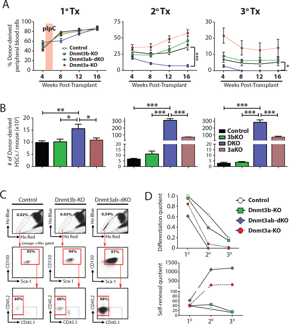
Analysis of peripheral blood chimerism in primary recipients revealed no significant differences in mice transplanted with 3bKO, or DKO HSCs compared to control HSCs (Figure 1A). As 3aKO HSCs also did not exhibit a phenotype in primary transplants (Challen et al., 2012), we performed serial transplantation. HSCs were purified from primary recipients 18-weeks post-transplant and 250 were transferred to secondary recipients along with fresh WT competitor WBM cells. While 3bKO HSCs performed similarly to control HSCs, blood production by DKO HSCs dropped precipitously (Figure 1A), with a dearth of DKO-derived cells in all peripheral blood lineages 16-weeks post-transplant (Figure S1B). In a third round of transplantation, 3bKO HSCs continued to perform like control HSCs, but with a slight increase in B-cell generation (Figure S1B). In contrast, DKO HSCs contributed to peripheral blood only transiently after the third round of transplantation (Figure 1A). These phenotypes are markedly distinct from the 3aKO HSCs, which showed enhanced blood production relative to control cells over four rounds of serial transplantation (Challen et al., 2012).
Figure 1. de novo DNA methylation is required for long-term HSC differentiation in vivo.
(A) Donor cell-derived peripheral blood chimerism in mice transplanted with control, Dnmt3b-KO (3bKO) or Dnmt3ab-dKO (DKO) HSCs. The previously determined values for Dnmt3a-KO (3aKO) HSCs (Challen et al., 2012) are included for reference in this and following panels. (B) Enumeration of donor-derived HSCs per mouse 18-weeks post-transplant over serial transplantation. (C) Bone marrow FACS analysis of tertiary transplanted mice showing accumulation of DKO HSCs. (E) Differentiation and self-renewal quotients of HSC genotypes over serial transplant. Mean ± SEM values are shown (n = 16–35 recipient mice per genotype), *P < 0.05, **P < 0.01, ***P < 0.001. See also Figure S1.
Dnmt3b enables some HSC differentiation in the absence of Dnmt3a
Self-renewal was evaluated by enumerating phenotypically-defined HSCs regenerated in the bone marrow 18-weeks after each round of transplantation. In primary mice, the numbers of HSCs generated from control and 3bKO was the same, but the number of DKO HSCs was increased significantly (Figure 1B). This behavior was exacerbated in subsequent rounds of transplantation. In secondary recipients, input of 250 control HSCs generated 6,692 ± 675 HSCs while the same input of DKO HSCs generated 305,472 ± 25,144 DKO HSCs. Similarly in tertiary transplants, 250 input control HSCs generated 3,061 ± 965 HSCs while DKO HSCs produced 295,429 ± 37,813 HSCs (Figure S1C). The bone marrow of tertiary mice transplanted with DKO HSCs harbored nearly 50-fold more HSCs than those from control or 3bKO transplanted recipients, with nearly all of the HSCs derived from DKO cells (Figure 1C; Figure S1D) even though these animals were almost devoid of DKO-derived peripheral blood cells (Figure 1A). DKO HSCs show superior self-renewal compared to 3aKO with approximately 5-fold more HSCs generated in each round of transplant (Figure 1B). However, unlike 3aKO HSCs which retain some differentiation capacity even after four rounds of transplantation (Challen et al., 2012), the differentiation of DKO cells is decimated after secondary transfer.
These data show that Dnmt3b plays a critical role in enabling differentiation in the absence of Dnmt3a. While loss of Dnmt3a alone has a more dramatic effect than loss of Dnmt3b, the effect of Dnmt3b is evidenced by the comparison between 3aKO and DKO. This concept is underscored when we examine on a per-HSC basis the output of differentiation (16-week white blood cell count per uL blood × percentage test-cell blood chimerism / number of donor-derived HSCs, the “differentiation quotient”) versus self-renewal (the number of donor-derived HSCs recovered at end of transplant per original input HSC, the “self-renewal quotient”) in each of the genotypes (Figure 1D). The DKO HSCs resemble the 3aKO HSCs in overall behavior, but the kinetics are distinct, with loss of differentiation ability and increase in self-renewal apparent in the first round of transplantation. Moreover, the self-renewal capacity of the DKO HSCs is roughly five-times that of the 3aKO HSCs on a per-cell basis.
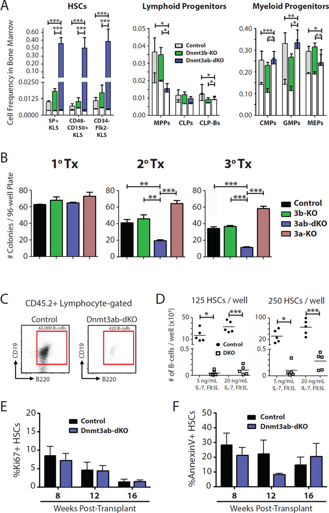
This is consistent with the idea that the Dnmt3s serve as a critical regulators at the decision point between HSC differentiation and self-renewal. Analysis of bone marrow progenitors in secondary transplants confirmed that the block in DKO differentiation occurred predominantly at the level of the HSC (Figure S2), with a dearth of all downstream progenitors (Figure 2A). Moreover, DKO HSCs were unable to differentiate well in vitro with cytokine and stromal cell support. HSCs isolated at the end of each round of serial transplantation and cultured in methylcellulose with myeloid differentiation factors revealed a deficit in the number of myeloid colonies produced from DKO HSCs compared to transplant-matched control HSCs (Figure 2B). While DKO HSCs could be serially replated (Figure S2D), the colonies derived after the second plating were comprised of an outgrowth of mast cells (Gr-1− Mac-1− c-Kit+ FcεR1+), not the normal distribution of myeloid colonies (data not shown). No difference was noted between control and 3bKO HSCs in the same assay (Figure 2B, Figure S2D). Genotyping of single HSC-derived colonies showed efficient excision of floxed alleles (Figure S2E,F,G). Co-culture of HSCs purified after secondary transplant with OP9 stromal cells revealed a severe block in B-cell differentiation of DKO HSCs (Figure 2C), regardless of the concentrations of lymphoid cytokines (IL-7, Flt3L) or the number of HSCs seeded per well (Figure 2D). These results contrast with 3aKO HSCs, which were capable of robust myeloid and lymphoid differentiation in vitro (Challen et al., 2012), and reinforce the notion that Dnmt3b plays a critical role in enabling HSC differentiation, particularly in the absence of Dnmt3a.
Figure 2. Dnmt3b confers some differentiation capacity in HSCs.
(A) Frequencies of different cell compartments in the bone marrow of secondary recipient mice. White areas indicate the proportion of CD45.1+ (competitor-derived) cells, n = 9–16. Statistical significance refers to comparisons of CD45.2+ (donor-derived) populations. (B) Number of colonies generated per plate from HSCs at the end of each round of serial transplantation, n = 4. Single HSCs were sorted into individual wells of 96-well plates in Methocult media containing SCF, IL-3, IL-6 and EPO. (C) Flow cytometric analysis of B-cell differentiation from control and DKO HSCs in vitro. (D) Numbers of B-cells generated from control and DKO HSCs after 14-days culture, n = 5. (F) Dynamic analysis of HSC proliferation during secondary hematopoietic reconstitution with Ki67 staining, n = 4. (G) Dynamic analysis of HSC apoptosis during secondary hematopoietic reconstitution with AnnexinV staining, n = 3. Mean ± SEM values are shown, *P < 0.05, **P < 0.01, ***P < 0.001. See also Figure S2.
DKO HSC expansion cannot be explained by altered proliferation or apoptosis
To further investigate the mechanisms of DKO HSC accumulation in the bone marrow of transplant recipients, a kinetic analysis of HSC proliferation and apoptosis was performed at monthly intervals during secondary hematopoietic repopulation. At no time point did DKO HSCs exhibit greater proliferation (measured by Ki67 positivity) than control HSCs (Figure 2E), and both populations increasingly entered quiescence over time. DKO and control HSCs exhibited similar levels AnnexinV staining, indicating the accumulation of DKO HSCs cannot be attributed to apoptosis resistance (Figure 2F). Together these data suggest that the accumulation of DKO HSCs is likely to be a result of cell divisions that almost exclusively result in symmetric self-renewal, rather than in differentiation.
Ablation of Dnmt3a and Dnmt3b in HSCs drive localized DNA methylation loss
To address the epigenetic differences between Dnmt3-mutant HSCs, we performed whole-genome bisulfite sequencing (WGBS) of age-matched control versus tertiary-transplanted 3aKO and DKO HSCs. It was not possible to obtain sufficient 3bKO HSCs at the equivalent stage due to their limited expansion. These studies differed from our previous comparisons of control and 3aKO HSCs (Challen et al., 2012) in (1) comprehensive nature (WGBS instead of reduced representation bisulfite sequencing (RRBS)), (2) analysis at the tertiary rather than secondary transplant stage, as that is when the DKO phenotype is most dramatic, and (3) control HSCs used here were from age-matched, untransplanted control mice due to the limited number of post-transplant control HSCs obtainable.
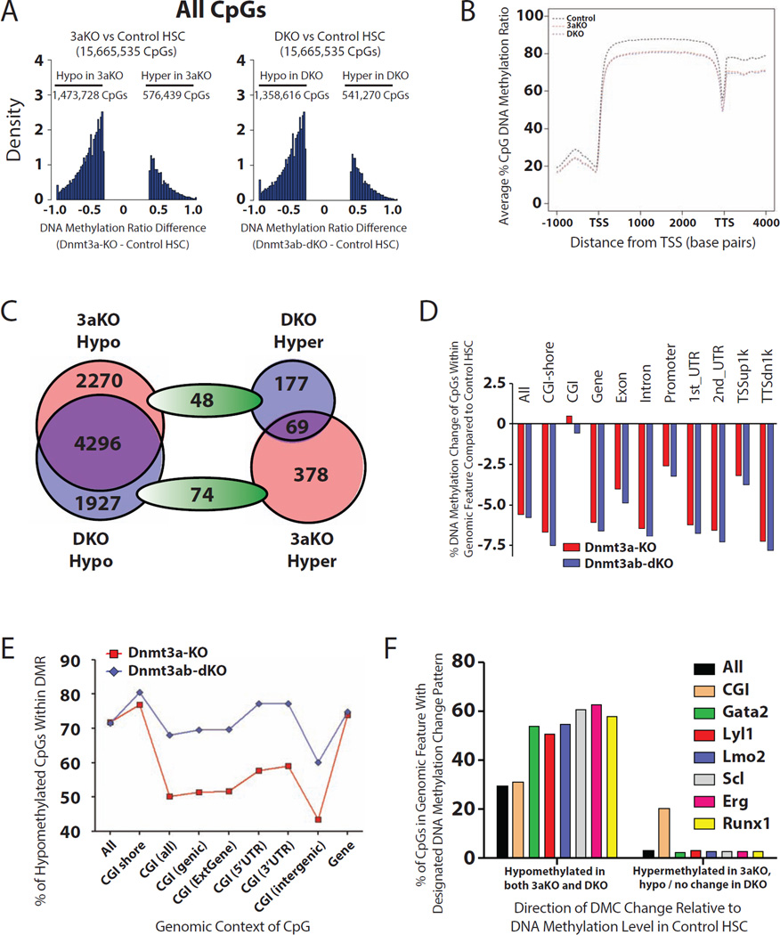
We generated between 1.1 to 1.2 billion raw reads for each of three genotypes, covering nearly all 21 million CpGs in the mouse genome (mm9) with an average depth of 40× to 47× (Table S1). Compared to control HSCs, 3aKO and DKO HSCs showed a global decrease in DNA methylation (average CpG methylation: 83.94% for control, 78.38% for 3aKO, and 78.01% for DKO; Figure 3A). Methylation was reduced across all genomic features (Table S1) and genic elements (Figure 3B). This contrast with our previous RRBS analysis, which indicated no significant difference between control and 3aKO HSCs (Challen et al., 2012), can be attributed to the bias of RRBS toward CpG-dense genomic regions. To control for possible transplantation effects on methylation (potentially masked by our use of untransplanted control HSCs), we examined the impact of transplantation as revealed by an independent study of aging HSCs (Beerman et al., 2013). Comparison of CpGs covered by both methods showed an average loss of DNA methylation of 11.8% in Dnmt3-mutant HSCs versus 6.9% due to transplantation as identified by Beerman et al. Thus, we cannot exclude some impact of transplantation itself, but the loss of Dnmt3a/b has a much greater impact.
Figure 3. Global DNA methylation defects in Dnmt3-mutant HSCs.
(A) Distribution of all DMCs in the genome of Dnmt3-mutant HSCs relative to control from WGBS data. (B) Average DNA methylation profile of CpGs across genic elements in control, 3aKO and DKO HSCs. (C) Distribution of DMCs within CGIs of Dnmt3-mutant HSCs compared to control. (D) Average DNA methylation changes across genomic elements in Dnmt3-mutant HSCs relative to control. (E) Percentages of hypomethylated DMCs within genomic elements in Dnmt3-mutant HSCs compared to control. (F) Hypomethylated DMCs are enriched in transcription factor binding sites, hypermethylated DMCs in 3aKO HSCs are enriched in CGIs. See also Figure S3 and Tables S1, S2 and S3.
This high sequencing coverage allowed quantification of differential DNA methylation throughout specific genomic features, for which we defined a change of ± 33% of DNA methylation (FDR <0.05) as a differentially methylated CpG (DMC) (Gu et al., 2010). One of the genomic loci that showed the most dramatic DNA methylation changes was repetitive elements (Table S2), with 66% and 61% of DMCs within repeats in 3aKO and DKO HSCs respectively becoming hypomethylated (Figure S3A). This included repetitive elements targeted by the de novo DNA methyltransferases in ES cells (Figure S3B). This hypomethylation was often associated with increases in repeat expression (Figure S3C), reinforcing the notion that one role of the Dnmt3s is to silence these repetitive elements to safeguard the genome.
To examine whether ablation of the Dnmt3s affected DNA methylation in specific gene sets, DMCs were grouped into differentially methylated regions (DMRs), defined as at least three neighboring DMCs of the same class (see methods). Because comparison of control versus mutant methylomes identified over 70,000 DMRs across the genome (Table S3), we initially focused on gene-associated DMRs (defined as being within a gene or 3kB upstream of transcription start site or 3kB downstream of transcription termination site). 92.5% (9,353 / 10,110) and 95.6% (8,765 / 9,171) of DMRs in 3aKO and DKO HSCs respectively became hypomethylated compared to control HSCs (Table S3). Comparison of the DKO with the 3aKO revealed fewer DMRs (3,495), of which the majority showed a further loss of DNA methylation after additional loss of Dnmt3b. Most of the strongly hypomethylated DMRs were conserved between the two mutant HSC genotypes (Figure 3C).
The most striking difference between 3aKO and DKO HSCs was within CpG islands (CGIs). Compared to control HSCs, 3aKO HSCs show a net gain of DNA methylation in CGIs while DKO HSCs experience a net loss of DNA methylation as in other genomic features (Figure 3D). CGIs are the only definable class of elements in the genome that exhibit this dichotomy (Figure 3D). While CpGs within loci groups such as “All” and “Gene” contain similar proportions of hypomethylated DMCs in 3aKO and DKO HSCs, there is a strikingly less hypomethylation within CGIs from 3aKO HSC, regardless of the genomic context of where the CGI is located (Figure 3E). Of the DMCs that were hypomethylated in both 3aKO and DKO HSCs, there was enrichment for the distribution of these CpGs within binding sites for transcription factors (TF) important for HSC regulation (Wilson et al., 2010)(Figure 3F). This may provide an indirect mechanism for transcriptional changes in Dnmt3-mutant HSCs, as lower DNA methylation may influencing TF binding, although how these DNA methylation changes alter physical association of these sites with TFs remains to be confirmed. The DMCs that were hypermethylated in 3aKO HSCs compared to both control and DKO HSCs were specifically enriched within CGIs (Figure 3F). These WGBS data suggest that while the additional loss of Dnmt3b augments the global net loss of DNA methylation resulting from inactivation of Dnmt3a, Dnmt3b contributes to inappropriate hypermethylation of CGIs in in the absence of Dnmt3a.
We have recently shown Dnmt3a maintains distinct subsets of large DNA methylation lacunae we termed canyons (Jeong et al., 2013). To examine the impact of additional loss of Dnmt3b on canyons, we compared the sizes of the 3aKO canyons with DKO canyons. Of the canyons that previously contracted (largely harboring unexpressed genes), 78% now expand in DKO HSCs (Figure S3D). Similarly, 27% of the canyons that expanded in absence of Dnmt3a (harboring highly expressed genes) are further enlarged in the DKO (Figure S3D). These data demonstrate that like Dnmt3a, Dnmt3b is involved in canyon size maintenance. Canyon views also demonstrate the remarkable specificity of Dnmt3a and Dnmt3b action, exemplified by a distant perspective of the Meis1 and HoxA loci, in which the associated canyon regions are eroded in 3aKO and DKO HSCs, but methylation loss outside these regions is minimal (Figure S3E,F).
Dnmt3a and Dnmt3b combine to epigenetically repress the stem cell gene network during HSC differentiation
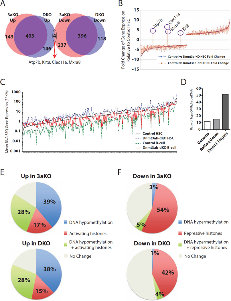
To examine the effects of DNA methylation changes in 3aKO and DKO HSCs on gene expression, we performed RNA-SEQ on the same cell populations used for WGBS. With an FDR 5% cutoff, there are 546 and 633 genes significantly upregulated and downregulated respectively in 3aKO HSCs compared to control HSCs (Table S4). Similarly, 549 and 514 genes were upregulated and downregulated respectively in DKO HSCs compared to control HSCs (Table S4). The majority of upregulated genes in 3aKO HSCs are also upregulated in DKO HSCs, and vice versa (Figure 4A). Furthermore, KEGG pathway analysis showed many of the same functional categories enriched in both mutant genotypes, such as upregulation of TGFβ signaling. One divergence was that MAPK signaling was enriched in 3aKO HSCs whereas Hedgehog signaling was enriched in DKO HSCs (Figure S4A). The genes downregulated in both mutant genotypes involved cell cycle, niche interactions and hematopoietic lineage specification programs (Figure S4B). Gene set enrichment analysis (GSEA) on the genes significantly differentially expressed in mutant HSCs correlated the genes upregulated in mutant HSCs with genes increased in cells after exposure to the demethylating agent 5-azacytidine (Figure S4C). There was no overlap between the genes upregulated in 3aKO HSCs and downregulated in DKO HSCs, and only four genes (Atp7b, Krt8, Clec11a and Mxra8) that were downregulated in 3aKO HSCs but upregulated in DKO HSCs (Figure 4B). Some differences in the outlier genes can be explained by differential DNA methylation, such as Krt8, which in 3aKO HSCs has a hypermethylated DMR in exon 1 that is not present in control or DKO HSCs (Figure S4D).
Figure 4. The Dnmt3s repress HSC-specific genes and gene expression changes are explained by epigenetic dynamics.
(A) Overlap of differentially expressed genes between Dnmt3-mutant HSCs and control HSCs. (B) Plotting fold change in transcript expression of genes differentially expressed between 3aKO and control HSCs as the baseline, overlay of fold change in DKO HSCs relative to controls show a highly similar pattern. (C) Identification of stem cell genes repressed by Dnmt3s during HSC differentiation. The baseline is RNA-SEQ expression of genes in control HSCs. Plot shows increased expression in DKO HSCs, repression in control B-cells, and intermediate expression (incomplete repression) in DKO B-cells. (D) Ratio of hypoDMRs / hyperDMRs shows enrichment for hypomethylation of Dnmt3-regulated genes relative to the rest of the genome. (E,F) Categorical breakdown of genes differentially expressed in 3aKO and DKO HSCs compared to controls according to their epigenetic status. Increased gene expression in 3aKO and DKO HSCs is dominated by hypomethylation (E) while decreased gene expression is underpinned moreso by loss of activating chromatin (F). Activating histone = increased H3K4me3 and/or reduced H3K27me3. Repressive histones = decreased H3K4me3 and/or increased H3K27me3. See also Figure S4, and Tables S4 and S5.
We have previously shown that a major function of Dnmt3a in HSCs was to epigenetically repress the stem cell genetic network to downregulate HSC self-renewal genes and allow differentiation (Challen et al., 2012). To examine this model in an unbiased way in the DKO situation, we performed RNA-SEQ on control and DKO-derived B-cells after secondary transplantation when some differentiation capacity remained. To identify epigenetically regulated genes, we selected genes that were upregulated >1.50-fold in DKO HSCs compared to control HSCs, in control HSCs compared to control B-cells, and in DKO B-cells compared to control B-cells; this defined a cohort of 254 genes (Table S4). Setting the expression level of each gene in control HSCs as a baseline, all genes have increased expression in DKO HSCs and dramatically decreased expression in control B-cells (Figure 4C). The role of the Dnmt3s in epigenetic repression of these stem cell-specific genes is clear as the expression patterns for DKO B-cells falls in between control HSCs and control B-cells. Independent analysis of DNA methylation patterns of this cohort of 254 genes in DKO HSCs revealed that the ratio of hypomethylated to hypermethylated DMRs (compared to control HSCs) was 50-fold (Figure 4D), significantly greater than the average ratio throughout the genome or the average of all genes. Thus, in normal hematopoiesis, these genes are predominantly expressed in HSCs and are silenced by Dnmt3s during differentiation. In the absence of the Dnmt3s this process is inefficient, leading to enforced HSC self-renewal and incomplete repression of stem cell-specific genes in differentiated progeny.
Differential gene expression can largely be explained by epigenetic dynamics
To gain insight into the influence of altered DNA methylation on other epigenetic marks, ChIP-SEQ was performed for the activating histone mark H3K4me3 and the repressive mark H3K27me3. We identified 14,494, 13,898 and 13,807 H3K4me3 peaks for control, 3aKO and DKO HSCs respectively, and 5,606, 4,358 and 6,531 H3K27me3 peaks for control, 3aKO and DKO HSCs respectively (Table S5) that were associated with gene promoters (+/− 3kb of transcription start site - TSS). By overlaying WGBS and ChIP-SEQ data, the majority of gene expression changes in mutant HSCs can be explained by epigenetic dynamics. 84% and 81% of the genes with significantly increased expression in 3aKO and DKO HSCs respectively contained a hypomethylated DMR, increased H3K4me3, decreased H3K27me3 (all epigenetic marks associated with increased gene expression) or some combination of the three epigenetic marks (Figure 4E, Figure S4E). Conversely, 62% of the genes significantly downregulated in 3aKO HSCs were associated with an epigenetic mark more indicative of transcriptional repression – either a hypermethylated DMR, decreased H3K4me3, increased H3K27me3 or some combination (Figure 4F, Figure S4E). However, only 47% of the downregulated genes could be explained by the same phenomena in DKO HSCs (Figure 4F). While DNA hypomethylation was the major driver of increased gene expression in Dnmt3-mutant HSCs, loss of the H3K4me3 histone modification was more predictive of transcriptional repression than changes in DNA methylation. These altered epigenetic patterns had consequences for alternative promoter utilization, an example being Tmcc3, for which the longer isoform was exclusively observed in the mutant HSCs (Figure S4F). This suggests that the Dnmt3s / DNA methylation influences chromatin patterns to co-operatively control HSC self-renewal and differentiation cell fate decisions.
β-catenin Signaling Contributes to the Block in DKO HSC Differentiation
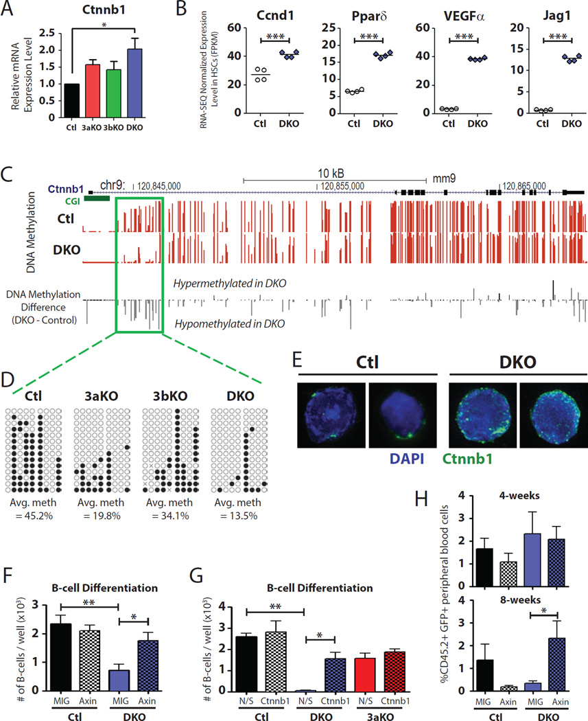
Ingenuity Pathway Analysis (IPA) was used to highlight potential gene regulatory differences between 3aKO and DKO HSCs. IPA discriminated the β-catenin (Ctnnb1) pathway to be dramatically activated in DKO HSCs (the same pathway was slightly below the IPA threshold for being considered activated in 3aKO HSCs). Ctnnb1 transcript expression was significantly increased in DKO HSCs (Figure 5A), and RNA-SEQ expression of validated β-catenin target genes such as Ccnd1, Pparδ, Vegfα and Jag1 was also significantly increased (Figure 5B). Mouse models in which HSCs constitutively express activated β-catenin show similar phenotypes to DKO HSCs, such as an 8- to 15-fold increase in the HSC pool and impaired differentiation in reconstitution assays (Scheller et al., 2006). We postulated that enhanced activity of this pathway may be responsible for some of the differentiation arrest of DKO HSCs.
Figure 5. Increased β-catenin signaling contributes to the differentiation block of DKO HSCs.
(A) Real-time PCR analysis of Ctnnb1 transcript expression in HSCs, n = 3. (B) RNA-SEQ expression of Ctnnb1 target genes in control (Ctl) and DKO HSCs. (C) DNA methylation profile of Ctnnb1 locus determined by WGBS. Green box indicates hypomethylated region in DKO HSCs. (D) Bisulfite sequencing of hypomethylated Ctnnb1 region in HSC genotypes (open circle = unmethylated CpG, closed circle = methylated CpG). (E) Immunofluorescent staining for Ctnnb1 (green) in control and DKO HSCs, with DAPI staining (blue) to define the nucleus. (F) Ectopic expression of Axin in DKO HSCs was able to partially rescue the B-cell differentiation defect compared to DKO HSCs transduced with control retrovirus (MIG). Ectopic expression of Axin in Ctl HSCs did not affect B-cell differentiation compared to MIG control cells, n = 4. (G) miRNA-mediated knockdown of Ctnnb1 was also able to restore B-cell in DKO HSCs, but had no effect on B-cell differentiation of 3aKO HSCs (N/S = scrambled non-silencing control miRNA, Ctnnb1 = Ctnnb1 miRNA#1), n = 2. (H) Peripheral blood chimerism following transduction and bone marrow transplant of control and DKO HSCs. Ectopic expression of Axin in DKO HSCs was able to significantly increase peripheral blood differentiation compared to the control MIG vector, n = 9–10 mice / group. Mean ± SEM values are shown, *P < 0.05, **P < 0.01. See also Figure S5.
DNA methylation analysis of the Ctnnb1 locus identified a DKO hypomethylated region in the shore of the promoter CGI (Figure 5C). This created a new DNA methylation canyon (Figure 5D, Figure S5A). Immunofluorescent staining of post-secondary transplant HSCs (Figure 5E) showed an increase in nuclear β-catenin in DKO HSCs (Figure S5B), as well as more total β-catenin protein (Figure S5C). Based on an arbitrary cutoff of 0.2 mean fluorescence units (MFU), 81% of DKO HSCs showed nuclear β-catenin, compared to only 48% of control HSCs. To explore whether this contributed to the differentiation deficit of DKO HSCs, we ectopically expressed Axin in post-transplant HSCs. Expression of the Wnt pathway inhibitor Axin in HSCs reduces function by degrading β-catenin (Reya et al., 2003). Control and DKO bone marrow progenitors were transduced with control empty-vector (MIG) or Axin-expressing (MIG-Axin) retrovirus and cultured for two days. Viable GFP+ HSCs were then purified (Lineage− Sca-1+ c-Kit+ CD150+) and co-cultured on OP9 stromal cells in lymphoid cytokines. Enumeration of B220+CD19+ cells after 14-days of co-culture showed that ectopic Axin expression was able to partially rescue the in vitro B-cell differentiation defect of DKO HSCs, but did not affect the number of B-cells generated from control HSCs (Figure 5F). We were able to elicit a similar response through knockdown of Ctnnb1 transcript levels (Figure S5D), although this had no effect on the B-cell potential of 3aKO HSCs (Figure 5G). As over-expression of Axin produced a similar response to Ctnnb1 knockdown in terms of β-catenin target gene response (Figure S5E), we tested whether this strategy could also restore DKO HSC differentiation potential in vivo. Post-transplant HSCs were transduced with MIG and MIG-Axin and transplanted into lethally irradiated mice. Ectopic expression of Axin significantly enhanced peripheral blood differentiation of DKO HSCs eight-weeks post-transplant (Figure 5H). Together, these data strongly support the notion that the diminished differentiation activity of the DKO HSCs is due at least in part to persistent β-catenin activation.
Abnormal Dnmt3b activity in the absence of Dnmt3a results in aberrant CGI hypermethylation
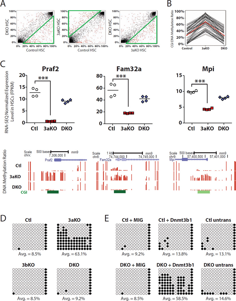
One puzzling observation was the finding that a specific subset of CGIs in HSCs actually showed increased DNA methylation levels following ablation of Dnmt3a. Promoter CGI hypermethylation is a well-established cancer methylome phenotype (Figueroa et al., 2009; Shen et al., 2010; Shen et al., 2007), so this may be an important mechanism in the early stages of HSC transformation. Compared to the control HSC methylome, 132 CGIs were significantly hypermethylated in 3aKO HSCs (Figure 6A, Table S6). The additional deletion of Dnmt3b abridged this effect, resulting in only 60 hypermethylated CGIs (Figure 6A). 63 CGIs were significantly hypermethylated in 3aKO HSCs compared to both control and DKO HSCs (Figure 6B). For genes with hypermethylated CGIs in the promoter region such as Fam32a, Mpi and Praf2, this DNA hypermethylation was associated with gene repression (Figure 6C). One possibility was that this CGI hypermethylation was actually increased 5-hydroxymethylcytosine (5-hmC), and not 5-methylcytosine (5-mC) as bisulfite sequencing cannot discriminate between these epigenetic marks. We examined the 5-hmC pattern of the 63 hypermethylated CGIs using 5-hmC profiles of control and 3aKO HSCs (Jeong et al., 2013). Only two of the 63 hypermethylated CGIs contained 5-hmC peaks in 3aKO HSCs (Figure S6A), suggesting that the majority of this hypermethylation was due to increased 5-mC. A recent epigenetic survey of ES cells deduced that CpG-dense promoters (such as those containing CGIs) are more likely to be repressed by the chromatin mark H3K27me3 while CpG-poor promoters are more likely to be epigenetically-regulated by DNA methylation (Xie et al., 2013). For the 63 CGIs that gain DNA methylation in 3aKO HSCs, 34 are marked by H3K27me3 in control HSCs but only nine in 3aKO HSCs (Figure S6B), suggesting that either of the two repressive marks was sufficient for silencing of the locus.
Figure 6. CGI hypermethylation in Dnmt3a-KO HSCs is mediated by Dnmt3b.
(A) Comparison of differentially-methylated CGIs across genotypes. Green triangles indicate the genotype with increased DNA methylation. Red dots indicate CGIs with statistically significant differences in DNA methylation. (B) Identification of CGIs that are hypermethylated in 3aKO HSCs relative to control (Ctl) and DKO HSCs. Red line is the average DNA methylation profile. (C) Gene expression and DNA methylation profiles of genes with hypermethylated promoter CGIs in 3aKO HSCs. Top panel is RNA-SEQ gene expression data, bottom panel is WGBS DNA methylation profiles. The height of each bar represents the DNA methylation level of an individual CpG. (D) Promoter CGI DNA methylation profile of Praf2 across HSC genotypes. Open circles represent unmethylated CpGs, closed circles represent methylated CpGs. (E) DNA methylation analysis of Praf2 promoter by bisulfite PCR following transduction of Ctl or DKO HSCs with a control (MIG) or Dnmt3b-expressing (Dnmt3b1) retrovirus, compared to untransduced (untrans) HSCs. ***P < 0.001. See also Figure S6, Table S6.
An alternative explanation suggested by these data is that in the absence of Dnmt3a, abnormal function of Dnmt3b may lead to aberrant CGI hypermethylation as the hypermethylation was lost when both enzymes were inactivated. To explore this in HSCs, we analyzed DNA methylation patterns of tertiary-transplant control, 3aKO, and 3bKO HSCs by RRBS (Table S6), which enriches for CGI-containing loci (Meissner et al., 2008). RRBS and WGBS showed strong concordance, with all 63 CGIs in 3aKO HSCs exhibiting hypermethylation with both methods (Figure S6C). All of these CGIs were unmethylated in the control, 3bKO and DKO HSCs (Figure S6D), strongly suggesting that absence of Dnmt3a permits inappropriate Dnmt3b action.
To confirm this mechanism, post-transplant control and DKO bone marrow progenitors were transduced with a retroviral vector encoding full-length Dnmt3b (MIG-Dnmt3b1) or control (MIG). After two-days, HSCs (GFP+ Lineage− Sca-1+ c-Kit+ CD150+ CD45.2+) were purified and assessed for DNA methylation by bisulfite sequencing. Using the promoter CGI of Praf2 as an example, hypermethylation is restricted to the 3aKO HSCs compared to control, 3bKO and DKO genotypes (Figure 6D). Enforced expression of Dnmt3b1 in DKO HSCs resulted in increased DNA methylation at this locus compared to DKO HSCs transduced with MIG control, control HSCs transduced with either MIG or MIG-Dnmt3b1, and untransduced HSCs (Figure 6E). This indicates that at least for some loci, in the absence of Dnmt3a, Dnmt3b functions abnormally in HSCs leading to aberrant CGI hypermethylation and anomalous gene silencing.
Dnmt3b isoforms in HSC function and cancer
DNMT3A mutations are prevalent in a range of hematopoietic malignancies (Grossmann et al., 2013; Ley et al., 2010; Walter et al., 2011; Yan et al., 2011), a finding congruent with the differentiation arrest of mouse 3aKO HSCs. As we show Dnmt3b also plays a role in enabling differentiation, it is surprising only two DNMT3B point mutations have been reported in hematologic cancers to date (Cancer Genome Atlas Research, 2013). Over thirty different Dnmt3b isoforms resulting from alternative promoter usage or alternative splicing have been reported (Gopalakrishnan et al., 2009; Ostler et al., 2007; Xie et al., 1999). Spatio-temporal patterns of isoform expression are largely conserved between humans and mice (Okano et al., 1998), suggesting that these isoforms carry biological significance even where splicing produces catalytically-inactive proteins, such as mouse Dnmt3b3 which lacks the catalytic motifs encoded by exons 21 and 22. Moreover, transformation is known to change the complement of DNMT3B isoforms, with aberrant forms dominating in a number of malignancies (Ostler et al., 2007).
To consider these possibilities here, we examined the expression of Dnmt3b isoforms in RNA-SEQ data, and found only two Dnmt3b isoforms are expressed in control and 3aKO HSCs - Dnmt3b1 and Dnmt3b3 (Figure 7A). The predominant isoform was Dnmt3b3, which generates a catalytically inactive protein. This expression pattern is very similar to AML patients harboring DNMT3A mutations (Cancer Genome Atlas Research, 2013). While expression of both isoforms was slightly decreased in 3aKO HSCs compared to control (Figure 7B), there was a larger proportional decrease in Dnmt3b1 levels in 3aKO HSCs. This suggests that there may be relatively little catalytically active Dnmt3b in normal HSCs, which is decreased even further following loss of Dnmt3a function. This likely explains why 3bKO alone has a relatively minor effect, and why the phenotypes of the 3aKO and DKO HSCs are similar at both the phenotypic and molecular levels. Although the importance of Dnmt3b in HSCs is clear as even residual Dnmt3b1 activity is sufficient to maintain some differentiation capacity in 3aKO HSCs, similar to a recent study showing that in the absence of Dnmt3a, Dnmt3b functions as a tumor suppressor in chronic lymphocytic leukemia (CLL) and T-cell malignancies (Peters et al., 2014).
Figure 7. Dnmt3b isoform expression and tumor-suppressor role in HSCs.
(A) RNA-SEQ expression of Dnmt3b isoforms in control (Ctl) and 3aKO HSCs. Exons 21 and 22 encoding the methyltransferase domain of Dnmt3b1 are reduced in 3aKO HSCs. (B) Ratio of Dnmt3b isoform expression in Ctl and 3aKO HSCs. (C) Frequencies of genetic mutations in DNMT3A and DNMT3B in a range of tumors extracted from the COSMIC database. (D) Overlap of DNMT3 mutations in patients with endometrial and colon cancer in COSMIC database.
As Dnmt3a and -3b are broadly expressed during embryonic development (Okano et al., 1999), we considered the possibility that DNMT3A and -3B mutations may play similar roles in other tissues. In the COSMIC database (Bamford et al., 2004), DNMT3B mutations are detected in a range of solid tumors (Figure 7C) and often overlap with mutations in DNMT3A (Figure 7D). This suggests that in some tissues, compound inactivation of de novo DNA methylation facilitates transformation, possibly reflective of a different spectrum of DNMT3B isoforms expressed in these tissues. Although it is not possible to determine at this level of analysis whether DNMT3A and -3B are drivers of these malignancies, given the data we present here, further investigation is clearly warranted.
DISCUSSION
We have demonstrated here a specific role for Dnmt3b in permitting differentiation of HSCs. Although ablation of Dnmt3b alone in HSCs produces only a modest phenotype, the function of Dnmt3b is highlighted when inactivated in combination with Dnmt3a; the synergistic impact of the DKO would not be predicted from factorial enhancement of the individual KOs. A previous study reported de novo DNA methyltransferase activity was essential for self-renewal, as Dnmt3afl/flDnmt3bfl/fl HSCs transduced with a CRE retrovirus could not sustain peripheral blood generation after bone marrow transplant (Tadokoro et al., 2007). We observe a similar effect of DKO on differentiation, but also report extensive HSC expansion (self-renewal), which may have been missed previously due to the different experimental models (retroviral Cre and no serial transplantation).
When coupled with histone ChIP-SEQ analysis, a clear pattern for epigenetic regulation of gene expression in Dnmt3-mutant HSCs emerged. The genes upregulated in Dnmt3-mutant HSCs were underpinned by some combination of loss of DNA methylation (defined by the presence of a hypomethylated DMR) with a chromatin state more permissive for gene transcription (decreased H3K27me3 and/or increased H3K4me3). The genes downregulated in Dnmt3-mutant HSCs were not generally associated with changes in DNA methylation, but rather dominated by changes in the histone code. Exactly how the chromatin architecture is instructed by the Dnmt3s remains to be determined. Possibilities include direct regulation of the writers and erasers of these marks by the Dnmt3s, recruitment of polycomb group proteins to epigenetic hotspots by the Dnmt3s, or changes in DNA methylation patterns causing conformational changes to the chromatin that permits / inhibits access to other epigenetic regulators or transcription factors.
The Wnt/β-catenin pathway drives phenotypic HSC expansion by inducing proliferation while simultaneously inhibiting apoptosis and blocking differentiation (Perry et al., 2011), and sustained canonical Wnt signaling in HSCs through constitutively activated β-catenin causes a multilineage differentiation block (Kirstetter et al., 2006; Scheller et al., 2006) reminiscent of the Dnmt3-mutant HSC phenotype. Over-expression of Axin was able to partly restore the differentiation capacity of DKO HSCs, suggesting this pathway is a mechanism of HSC fate decisions that is under epigenetic control. However, in addition to Ctnnb1, there are surely other causative targets that contribute to the enhanced self-renewal and differentiation arrest of Dnmt3-mutant HSCs. For example, NF-κB signaling regulates HSC self-renewal (Stein and Baldwin, 2013; Zhao et al., 2012), and Nfkb2 is over-expressed ~4-fold in Dnmt3-mutant HSCs. Furthermore, loss of DNA methylation in transcription-factor binding sites following ablation of the Dnmt3s in HSCs (Figure 3F) may alter binding of master regulators such as Gata2, Runx1, Lmo2 and Scl, presenting an indirect mechanism with substantial downstream consequences.
With the mild in vivo phenotype of 3bKO HSCs, our data suggests Dnmt3a can compensate almost completely for Dnmt3b loss, but Dnmt3b is only partially able to reciprocate in the reverse situation. It is possible that the target specificity of the remaining catalytically functional Dnmt3b is distinct, leading to aberrant DNA methylation patterns such as CGI hypermethylation. On a gross level, the phenotypes of 3aKO and DKO HSCs are broadly similar. We propose this is because the predominant Dnmt3b isoform expressed in 3aKO HSCs is the catalytically-inactive Dnmt3b3, resulting in quite minimal levels of de novo DNA methylation activity in 3aKO HSCs. Perhaps this explains the paucity of DNMT3B mutations in hematopoietic cancers because the relatively low level of catalytically competent DNMT3B means there is no selective pressure to genetically inactivate this gene following mutation of DNMT3A, with the DNMT3A-mutant cancer cells functionally operating as a DNMT3A/B compound null. However, although the potency of Dnmt3b is less than Dnmt3a in HSCs, even residual levels of catalytic Dnmt3b clearly enable significant HSC differentiation, as even after three rounds of serial transplantation the self-renewal quotient of 3aKO HSCs is five-fold less than that of DKO HSCs. The findings here build on the growing appreciation for epigenetic regulation in stem cell function. We show Dnmt3b has subtle, yet significant, roles in adult HSCs. Understanding the fundamental roles of Dnmt3s and DNA methylation in normal hematopoiesis is essential for deducing the impact of DNMT3 mutations and altered DNA methylation patterns in cancer.
EXPERIMENTAL PROCEDURES
Animals
All animal procedures were IACUC-approved and conducted in accordance the Baylor College of Medicine and Washington University in St. Louis institutional guidelines. All mice were C57Bl/6 background distinguished by CD45.1 or CD45.2 alleles. Dnmt3afl/fl and Dnmt3bfl/fl mice were obtained from the Beaudet lab at Baylor College of Medicine (with the consent of En Li) and crossed to Mx1-cre mice. For bone marrow transplantation, recipient C57Bl/6 CD45.1 mice were transplanted by retroorbital injection following a split dose of 10.5 Gy of lethal irradiation. 250 donor HSCs (CD45.2) were competed against 2.5 × 10⁵ WBM cells with the opposite CD45 allele (matched to the recipient). In competitive transplantation experiments, deletion of floxed alleles in donor HSCs was mediated 5–6 weeks post-transplantation in primary recipients. Conditional deletion was mediated by six intraperitoneal injections (300µg / mouse) of polyinosinic-polycytidylic acid (pIpC; Sigma, St Louis, MO) in PBS every other. For serial HSC transplantation, WBM from transplanted recipients was isolated 18-weeks post-transplant and donor HSCs were re-purified using CD45.2+SPKLS gating. 250 of these re-purified donor HSCs were competed against 2.5 × 10⁵ fresh CD45.1 WBM. PCR screening of Dnmt3a and Dnmt3b floxed allele deletion was performed as previously described (Tadokoro et al., 2007).
Hematopoietic Stem Cell Purification and Flow Cytometry
For transplantation, HSCs were purified from the bone marrow using side population (SP) plus KLS (c-kit+, Lineage−, Sca1+) gating (see also supplemental experimental procedures). Bone marrow cells were stained for 90 minutes with Hoechst, magnetically enriched for c-Kit+ cells, stained with additional antibodies, then purified by flow cytometric cell sorting.
Whole Genome Bisulfite Sequencing (WGBS)
300ng genomic DNA was isolated from and fragmented using a Covaris sonication system (Covaris S2). Libraries were constructed using the Illumina TruSeq DNA sample preparation kit. After ligation, libraries were bisulfite-treated using the EpiTech Bisulfite Kit (Qiagen, Valencia, CA). Ligation efficiency tested by PCR using TrueSeq primers and Pfu TurboCx hotstart DNA polymerase (Stratagene). After determining the optimized PCR cycle number for each samples, a large scale PCR reaction (100ul) were performed as described previously (Gu et al., 2011). PCR products were sequenced with Illumina HiSeq sequencing systems. WGBS data analyses were based on MOABS: MOdel based Analysis of Bisulfite Sequencing.
RNA-sequencing
RNA was isolated from ~70,000 HSCs with the RNeasy Micro column (Qiagen, Valencia, CA). Paired end libraries were generated by using Illumina TruSeq RNA sample preparation kit. Illumina HiSeq was used for sequencing with a paired-end sequencing length of 100bp. The alignment was performed by RUM, which first mapped reads to the genome and transcriptome by Bowtie, and then used blat to re-map those initially unmapped reads to the genome. The information from the two rounds of mappings was merged. The multiply mapped reads were discarded. The gene annotations used for transcriptome alignment include RefSeq, UCSC knownGene and ensemble gene models. The gene expression, FPKM value, was calculated by counting the reads matching the exons of each gene. Differential expression was performed using edgeR.
ChIP-sequencing (ChIP-SEQ)
Chromatin Immunoprecipitation (ChIP) was performed with 50,000 HSCs. ChIPed DNA was purified by MinElute Purification Kit (Qiagen) and prepared for library construction using ThruPLEX-FD preparation kit without extra amplification (Rubicon, Ann Arbor, MI). Sequencing was performed on a HiSeq 2000 (Illumina). Sequenced reads were mapped to the mm9 mouse genome and peaks were identified by model-based analysis of ChIP-SEQ data (MACS).
Statistical and Bioinformatic Analysis
See Supplemental Experimental Procedures. All data has been uploaded into GEO under accession number GSE50793. All the RNA-SEQ, DNA methylation, and histone ChIP-SEQ data can be visualized on the UCSC Genome Browser through the URL http://dldcc-web.brc.bcm.edu/lilab/deqiangs/dko/tracks.txt.Visit http://genome.ucsc.edu/cgi-bin/hgTracks?db=mm9&hubUrl=http://dldcc-web.brc.bcm.edu/lilab/deqiangs/dko/tracks.txt to visualize the trackhub.
Supplementary Material
HIGHLIGHTS.
de novo DNA methyltransferase enzymes synergize to enable HSC differentiation
β-catenin activation is partly responsible for Dnmt3a/b-null HSC differentiation arrest
Abnormal Dnmt3b function in the absence of Dnmt3a leads to DNA hypermethylation
The predominant Dnmt3b isoforms expressed in HSCs are catalytically inactive
ACKNOWLEDGEMENTS
We thank members of the Challen, Li and Goodell Labs for helpful discussions. This work was supported by NIH grants DK084259, AG036562, CA126752, DK092883, CA125123, DK084259, AI07495, the Ellison Medical Foundation, CPRIT grant RP110028 and the Samuel Waxman foundation; to W.L.: CPRIT RP110471 and NIH R01HG007538.
Footnotes
Publisher's Disclaimer: This is a PDF file of an unedited manuscript that has been accepted for publication. As a service to our customers we are providing this early version of the manuscript. The manuscript will undergo copyediting, typesetting, and review of the resulting proof before it is published in its final citable form. Please note that during the production process errors may be discovered which could affect the content, and all legal disclaimers that apply to the journal pertain.
AUTHOR CONTRIBUTIONS
Designed and performed experiments: GAC, AM, ML, MJ, CM, LY, SC, GJD, MAG.
Analyzed data: GAC, DS, BR, ZX, WL, MAG.
Wrote and edited the paper: GAC, MAG.
COMPETING FINANCIAL INTERESTS
The authors declare no competing financial interests.
REFERENCES
- Bamford S, Dawson E, Forbes S, Clements J, Pettett R, Dogan A, Flanagan A, Teague J, Futreal PA, Stratton MR, et al. The COSMIC (Catalogue of Somatic Mutations in Cancer) database and website. British journal of cancer. 2004;91:355–358. doi: 10.1038/sj.bjc.6601894. [DOI] [PMC free article] [PubMed] [Google Scholar]
- Beerman I, Bock C, Garrison BS, Smith ZD, Gu H, Meissner A, Rossi DJ. Proliferation-dependent alterations of the DNA methylation landscape underlie hematopoietic stem cell aging. Cell Stem Cell. 2013;12:413–425. doi: 10.1016/j.stem.2013.01.017. [DOI] [PubMed] [Google Scholar]
- Bestor TH. Activation of mammalian DNA methyltransferase by cleavage of a Zn binding regulatory domain. Embo J. 1992;11:2611–2617. doi: 10.1002/j.1460-2075.1992.tb05326.x. [DOI] [PMC free article] [PubMed] [Google Scholar]
- Cancer Genome Atlas Research, N. Genomic and epigenomic landscapes of adult de novo acute myeloid leukemia. N Engl J Med. 2013;368:2059–2074. doi: 10.1056/NEJMoa1301689. [DOI] [PMC free article] [PubMed] [Google Scholar]
- Challen GA, Sun D, Jeong M, Luo M, Jelinek J, Berg JS, Bock C, Vasanthakumar A, Gu H, Xi Y, et al. Dnmt3a is essential for hematopoietic stem cell differentiation. Nat Genet. 2012;44:23–31. doi: 10.1038/ng.1009. [DOI] [PMC free article] [PubMed] [Google Scholar]
- Chen T, Ueda Y, Dodge JE, Wang Z, Li E. Establishment and maintenance of genomic methylation patterns in mouse embryonic stem cells by Dnmt3a and Dnmt3b. Mol Cell Biol. 2003;23:5594–5605. doi: 10.1128/MCB.23.16.5594-5605.2003. [DOI] [PMC free article] [PubMed] [Google Scholar]
- Dodge JE, Okano M, Dick F, Tsujimoto N, Chen T, Wang S, Ueda Y, Dyson N, Li E. Inactivation of Dnmt3b in mouse embryonic fibroblasts results in DNA hypomethylation, chromosomal instability, and spontaneous immortalization. J Biol Chem. 2005;280:17986–17991. doi: 10.1074/jbc.M413246200. [DOI] [PubMed] [Google Scholar]
- Figueroa ME, Skrabanek L, Li Y, Jiemjit A, Fandy TE, Paietta E, Fernandez H, Tallman MS, Greally JM, Carraway H, et al. MDS and secondary AML display unique patterns and abundance of aberrant DNA methylation. Blood. 2009;114:3448–3458. doi: 10.1182/blood-2009-01-200519. [DOI] [PMC free article] [PubMed] [Google Scholar]
- Gopalakrishnan S, Van Emburgh BO, Shan J, Su Z, Fields CR, Vieweg J, Hamazaki T, Schwartz PH, Terada N, Robertson KD. A novel DNMT3B splice variant expressed in tumor and pluripotent cells modulates genomic DNA methylation patterns and displays altered DNA binding. Molecular cancer research : MCR. 2009;7:1622–1634. doi: 10.1158/1541-7786.MCR-09-0018. [DOI] [PMC free article] [PubMed] [Google Scholar]
- Grossmann V, Haferlach C, Weissmann S, Roller A, Schindela S, Poetzinger F, Stadler K, Bellos F, Kern W, Haferlach T, et al. The molecular profile of adult T-cell acute lymphoblastic leukemia: mutations in RUNX1 and DNMT3A are associated with poor prognosis in T-ALL. Genes Chromosomes Cancer. 2013;52:410–422. doi: 10.1002/gcc.22039. [DOI] [PubMed] [Google Scholar]
- Gu C, Bock C, Mikkelsen TS, Jager N, Smith ZD, Tomazou E, Gnirke A, Lander ES, Meissner A. Genome-scale DNA methylation mapping of clinical samples at single-nucleotide resolution. Nat Methods. 2010;7:133–136. doi: 10.1038/nmeth.1414. [DOI] [PMC free article] [PubMed] [Google Scholar]
- Gu H, Smith ZD, Bock C, Boyle P, Gnirke A, Meissner A. Preparation of reduced representation bisulfite sequencing libraries for genome-scale DNA methylation profiling. Nat Protoc. 2011;6:468–481. doi: 10.1038/nprot.2010.190. [DOI] [PubMed] [Google Scholar]
- Hata K, Okano M, Lei H, Li E. Dnmt3L cooperates with the Dnmt3 family of de novo DNA methyltransferases to establish maternal imprints in mice. Development. 2002;129:1983–1993. doi: 10.1242/dev.129.8.1983. [DOI] [PubMed] [Google Scholar]
- Jaenisch R, Jahner D. Methylation, expression and chromosomal position of genes in mammals. Biochim Biophys Acta. 1984;782:1–9. doi: 10.1016/0167-4781(84)90099-x. [DOI] [PubMed] [Google Scholar]
- Jeong M, Sun D, Luo M, Huang Y, Challen GA, Rodriguez B, Zhang X, Chavez L, Wang H, Hannah R, et al. Large conserved domains of low DNA methylation maintained by Dnmt3a. Nat Genet. 2013 doi: 10.1038/ng.2836. [DOI] [PMC free article] [PubMed] [Google Scholar]
- Kirstetter P, Anderson K, Porse BT, Jacobsen SE, Nerlov C. Activation of the canonical Wnt pathway leads to loss of hematopoietic stem cell repopulation and multilineage differentiation block. Nat Immunol. 2006;7:1048–1056. doi: 10.1038/ni1381. [DOI] [PubMed] [Google Scholar]
- Lei H, Oh SP, Okano M, Juttermann R, Goss KA, Jaenisch R, Li E. De novo DNA cytosine methyltransferase activities in mouse embryonic stem cells. Development. 1996;122:3195–3205. doi: 10.1242/dev.122.10.3195. [DOI] [PubMed] [Google Scholar]
- Ley TJ, Ding L, Walter MJ, McLellan MD, Lamprecht T, Larson DE, Kandoth C, Payton JE, Baty J, Welch J, et al. DNMT3A mutations in acute myeloid leukemia. N Engl J Med. 2010;363:2424–2433. doi: 10.1056/NEJMoa1005143. [DOI] [PMC free article] [PubMed] [Google Scholar]
- Li E, Bestor TH, Jaenisch R. Targeted mutation of the DNA methyltransferase gene results in embryonic lethality. Cell. 1992;69:915–926. doi: 10.1016/0092-8674(92)90611-f. [DOI] [PubMed] [Google Scholar]
- Meissner A, Mikkelsen TS, Gu H, Wernig M, Hanna J, Sivachenko A, Zhang X, Bernstein BE, Nusbaum C, Jaffe DB, et al. Genome-scale DNA methylation maps of pluripotent and differentiated cells. Nature. 2008;454:766–770. doi: 10.1038/nature07107. [DOI] [PMC free article] [PubMed] [Google Scholar]
- Ng HH, Bird A. DNA methylation and chromatin modification. Curr Opin Genet Dev. 1999;9:158–163. doi: 10.1016/s0959-437x(99)80024-0. [DOI] [PubMed] [Google Scholar]
- Okano M, Bell DW, Haber DA, Li E. DNA methyltransferases Dnmt3a and Dnmt3b are essential for de novo methylation and mammalian development. Cell. 1999;99:247–257. doi: 10.1016/s0092-8674(00)81656-6. [DOI] [PubMed] [Google Scholar]
- Okano M, Xie S, Li E. Cloning and characterization of a family of novel mammalian DNA (cytosine-5) methyltransferases. Nat Genet. 1998;19:219–220. doi: 10.1038/890. [DOI] [PubMed] [Google Scholar]
- Ostler KR, Davis EM, Payne SL, Gosalia BB, Exposito-Cespedes J, Le Beau MM, Godley LA. Cancer cells express aberrant DNMT3B transcripts encoding truncated proteins. Oncogene. 2007;26:5553–5563. doi: 10.1038/sj.onc.1210351. [DOI] [PMC free article] [PubMed] [Google Scholar]
- Perry JM, He XC, Sugimura R, Grindley JC, Haug JS, Ding S, Li L. Cooperation between both Wnt/{beta}-catenin and PTEN/PI3K/Akt signaling promotes primitive hematopoietic stem cell self-renewal and expansion. Genes Dev. 2011;25:1928–1942. doi: 10.1101/gad.17421911. [DOI] [PMC free article] [PubMed] [Google Scholar]
- Peters SL, Hlady RA, Opavska J, Klinkebiel D, Pirruccello SJ, Talmon GA, Sharp JG, Wu L, Jaenisch R, Simpson MA, et al. Tumor suppressor functions of Dnmt3a and Dnmt3b in the prevention of malignant mouse lymphopoiesis. Leukemia. 2014;28:1138–1142. doi: 10.1038/leu.2013.364. [DOI] [PubMed] [Google Scholar]
- Reya T, Duncan AW, Ailles L, Domen J, Scherer DC, Willert K, Hintz L, Nusse R, Weissman IL. A role for Wnt signalling in self-renewal of haematopoietic stem cells. Nature. 2003;423:409–414. doi: 10.1038/nature01593. [DOI] [PubMed] [Google Scholar]
- Scheller M, Huelsken J, Rosenbauer F, Taketo MM, Birchmeier W, Tenen DG, Leutz A. Hematopoietic stem cell and multilineage defects generated by constitutive beta-catenin activation. Nat Immunol. 2006;7:1037–1047. doi: 10.1038/ni1387. [DOI] [PubMed] [Google Scholar]
- Shen L, Kantarjian H, Guo Y, Lin E, Shan J, Huang X, Berry D, Ahmed S, Zhu W, Pierce S, et al. DNA methylation predicts survival and response to therapy in patients with myelodysplastic syndromes. Journal of clinical oncology : official journal of the American Society of Clinical Oncology. 2010;28:605–613. doi: 10.1200/JCO.2009.23.4781. [DOI] [PMC free article] [PubMed] [Google Scholar]
- Shen L, Kondo Y, Guo Y, Zhang J, Zhang L, Ahmed S, Shu J, Chen X, Waterland RA, Issa JP. Genome-wide profiling of DNA methylation reveals a class of normally methylated CpG island promoters. PLoS Genet. 2007;3:2023–2036. doi: 10.1371/journal.pgen.0030181. [DOI] [PMC free article] [PubMed] [Google Scholar]
- Stein SJ, Baldwin AS. Deletion of the NF-kappaB subunit p65/RelA in the hematopoietic compartment leads to defects in hematopoietic stem cell function. Blood. 2013;121:5015–5024. doi: 10.1182/blood-2013-02-486142. [DOI] [PMC free article] [PubMed] [Google Scholar]
- Surani MA. Imprinting and the initiation of gene silencing in the germ line. Cell. 1998;93:309–312. doi: 10.1016/s0092-8674(00)81156-3. [DOI] [PubMed] [Google Scholar]
- Tadokoro Y, Ema H, Okano M, Li E, Nakauchi H. De novo DNA methyltransferase is essential for self-renewal, but not for differentiation, in hematopoietic stem cells. J Exp Med. 2007;204:715–722. doi: 10.1084/jem.20060750. [DOI] [PMC free article] [PubMed] [Google Scholar]
- Walter MJ, Ding L, Shen D, Shao J, Grillot M, McLellan M, Fulton R, Schmidt H, Kalicki-Veizer J, O'Laughlin M, et al. Recurrent DNMT3A mutations in patients with myelodysplastic syndromes. Leukemia. 2011;25:1153–1158. doi: 10.1038/leu.2011.44. [DOI] [PMC free article] [PubMed] [Google Scholar]
- Wilson NK, Foster SD, Wang X, Knezevic K, Schutte J, Kaimakis P, Chilarska PM, Kinston S, Ouwehand WH, Dzierzak E, et al. Combinatorial transcriptional control in blood stem/progenitor cells: genome-wide analysis of ten major transcriptional regulators. Cell Stem Cell. 2010;7:532–544. doi: 10.1016/j.stem.2010.07.016. [DOI] [PubMed] [Google Scholar]
- Wu H, Coskun V, Tao J, Xie W, Ge W, Yoshikawa K, Li E, Zhang Y, Sun YE. Dnmt3a-dependent nonpromoter DNA methylation facilitates transcription of neurogenic genes. Science. 2010;329:444–448. doi: 10.1126/science.1190485. [DOI] [PMC free article] [PubMed] [Google Scholar]
- Xie S, Wang Z, Okano M, Nogami M, Li Y, He WW, Okumura K, Li E. Cloning, expression and chromosome locations of the human DNMT3 gene family. Gene. 1999;236:87–95. doi: 10.1016/s0378-1119(99)00252-8. [DOI] [PubMed] [Google Scholar]
- Xie W, Schultz MD, Lister R, Hou Z, Rajagopal N, Ray P, Whitaker JW, Tian S, Hawkins RD, Leung D, et al. Epigenomic analysis of multilineage differentiation of human embryonic stem cells. Cell. 2013;153:1134–1148. doi: 10.1016/j.cell.2013.04.022. [DOI] [PMC free article] [PubMed] [Google Scholar]
- Yan XJ, Xu J, Gu ZH, Pan CM, Lu G, Shen Y, Shi JY, Zhu YM, Tang L, Zhang XW, et al. Exome sequencing identifies somatic mutations of DNA methyltransferase gene DNMT3A in acute monocytic leukemia. Nat Genet. 2011;43:309–315. doi: 10.1038/ng.788. [DOI] [PubMed] [Google Scholar]
- Zhao C, Xiu Y, Ashton J, Xing L, Morita Y, Jordan CT, Boyce BF. Noncanonical NF-kappaB signaling regulates hematopoietic stem cell self-renewal and microenvironment interactions. Stem Cells. 2012;30:709–718. doi: 10.1002/stem.1050. [DOI] [PMC free article] [PubMed] [Google Scholar]
Associated Data
This section collects any data citations, data availability statements, or supplementary materials included in this article.


