Abstract
In this study, we examined the role(s) of syndecan-4 in regulating the formation of an actin geodesic dome structure called a cross-linked actin network (CLAN) in which syndecan-4 has previously been localized. CLANs have been described in several different cell types, but they have been most widely studied in human trabecular meshwork (HTM) cells where they may play a key role in controlling intraocular pressure by regulating aqueous humor outflow from the eye. In this study we show that a loss of cell surface synedcan-4 significantly reduces CLAN formation in HTM cells. Analysis of HTM cultures treated with or without dexamethasone shows that laminin 5 deposition within the extracellular matrix is increased by glucocorticoid treatment and that a laminin 5-derived, syndecan-4-binding peptide (PEP75), induces CLAN formation in TM cells. This PEP75-induced CLAN formation was inhibited by heparin and the broad spectrum PKC inhibitor Ro-31-7549. In contrast, the more specific PKCα inhibitor Go 6976 had no effect, thus excluding PKCα as a downstream effector of syndecan-4 signaling. Analysis of PKC isozyme expression showed that HTM cells also expressed both PKCγ and PKCε. Cells treated with a PKCε agonist formed CLANs while a PKCα/γ agonist had no effect. These data suggest that syndecan-4 is essential for CLAN formation in HTM cells and that a novel PKCε-mediated signaling pathway can regulate formation of this unique actin structure.
Keywords: syndecan-4, laminin 5, protein kinase C, trabecular meshwork, CLANs, cytoskeleton
Introduction
The syndecans are a four-member family of transmembrane HSPGs. Their extracellular domains bear multiple HS glycosaminoglycan chains that bind ECM proteins and other extracellular ligands [1]. All four members of the syndecan family are found within the TM [2] with syndecans-3 and −4 being the most predominant. The TM is the primary structure within the anterior segment of the eye that regulates aqueous humor outflow and is critical for maintaining normal intraocular pressure [3]. The function of syndecans within the TM is unknown.
In other tissues, syndecans function as co-receptors for integrins such as αvβ3 integrin [4] and various heparin-binding growth factors [5]. More recently syndecan-4 has been implicated as a mechanotransducer because, like integrins, they appear capable of regulating the organization of the actin cytoskeleton in response to mechanical forces [6, 7] and may be part of the stimuli that senses mechanical changes caused by the movement or vibration of the extracellular milieu [8]. Most of the signaling activity of syndecans can be linked, either directly or indirectly, to Rho family GTPase-mediated signaling pathways and typically these pathways involve PKCα [9–11]. However other isoforms such as PKCs β and γ [10], PKCδ [12, 13] and PKCε [14] may also be associated with syndecan signaling, albeit indirectly through phosphorylation events [15].
Of the syndecans, syndecan-4 is best known for having a regulatory role in the organization of actin stress filaments and is found in focal adhesions [16–18]. However, a more recent study [19] has suggested that syndecan-4 may also play a role in the formation of CLANs. CLANs are unique actin geodesic dome structures observed in a variety of cell types [20–23] including HTM cells [19, 24]. CLANs are a specialized actin structure composed of 3–5 actin filaments radiating from a central vertex (hub) that is very reminiscent of the actin geodesic domes first described by Lazarides[25] and later by Ingber in his model of tensegrity [26]. Syndecan-4 is found within CLAN vertices along with PIP2, PDLIM1, and α-actinin [19, 27, 28]. The function of CLANs is unknown, however, they have been found with increased frequency within the TM from eyes of glaucomatous patients [29], and they form spontaneously in cultured TM cells from glaucomatous donors [30]. Hence, CLANs may be a stress response structure resulting from increased intraocular pressure that subsequently alters the contractile properties of the TM making the cells less responsive to additional changes in intraocular pressure [19, 31, 32].
CLANs also form in response to glucocorticoid treatments both in anterior segment tissue and isolated TM cell cultures [33]. In addition, CLANs can be induced in cultured TM cells by the activation of an αvβ3 integrin signaling pathway [34]. This αvβ3 signaling cascade includes Src, the Rho family GTPase Racl, the Racl GEF Trio and may include the atypical GPCR CD47 [31]. CLAN formation induced by αvβ3 signaling also requires cross-talk with a PI-3 kinase-mediated β1 integrin signaling pathway [19].
In this study we investigated the role of syndecan-4 in the formation of CLANs using siRNA to knock down expression of syndecan-4 and a syndecan-4-binding peptide called PEP75 [35, 36]. PEP75 is found in the fourth module of the LG domain of the unprocessed α3 chain in laminin 5 (laminin 332) and has been shown to bind syndecan-4 and syndecan-2, but not syndecan-1 [35, 36]. Laminin 5 is composed of α3, β3 and γ2 subunits or chains. It is typically found in basement membranes of stratified epithelia and plays an important role in maintaining epithelial-mesenchymal integrity in tissues subjected to external forces [37, 38]. Both the LG4 and LG5 modules of laminin 5 play a role in regulating deposition of the protein into the ECM. Once laminin 5 has been secreted and deposited within the ECM, however, these modules are proteolytically removed. Thus the LG4 module containing the PEP75 sequence is potentially available for binding to cells expressing syndecan-4 via the PEP75 sequence. This peptide appears to be biologically active and has been shown to regulate migration, adhesions, and MMP-1 expression [39–42].
In this paper, we show that laminin 5 is present in cultures of HTM cells and that dexamethasone appeared to increase its deposition within the ECM. We also show that CLAN formation is dependent on the expression of syndecan-4 and that the laminin 5-derived PEP75 appears to activate a novel PKCε- signaling pathway that triggers CLAN formation. Activation of this pathway appears to involve HS chains and can enhance CLAN formation induced by αvβ3 integrin activation. This is the first study to show that HTM cells express laminin 5, and, to the best of our knowledge, this is the first time that PKCε has been shown to be involved with syndecan-4 in the formation of an actin structure.
Materials and Methods
Cell culture
Four different HTM strains were used for this study. N17TM-2 HTM cells were isolated from the OD eye of a 17 year old donor. N27TM-2, N27TM-3 and N27TM-6 cells were isolated from the OD eye (TM-2) and the OS eye (TM-3 and TM-6) respectively of a 27 year old donor. Neither donor had any known history of ocular disease. The cells strains were isolated as previously described [2, 43, 44]. Cells were cultured in low glucose DMEM (Sigma, St. Louis, MO), 15% fetal bovine serum (Atlanta Biologicals, Atlanta, GA), 2 mM L-glutamine (Sigma), 1% amphoteracin B (Mediatech, Herndon, VA), 0.05% gentamicin (Mediatech) and 1 ng/mL FGF-2 (Peprotech, Rocky Hill, NJ) [43, 44]. For experiments where cells were treated with or without DEX (Sigma), cells were grown to confluence on glass coverslips and maintained as monolayers for 7 days. The cultures were then switched to 10% FBS-containing medium plus 0.1% ethanol alone or ethanol containing 500 nM DEX for an additional 7 days. Cells were re-fed with fresh medium containing ethanol or DEX every other day prior to fixation in p-formaldehyde.
Cell spreading assays
The spreading assays were performed as described previously [19, 31, 34]. Confluent HTM monolayers were serum-starved for 24 hours and then re-plated in the presence of 25 µg/ml cycloheximide onto coverslips pre-coated with 20 nM fibronectin with or without the β3 integrin activating mAb AP-5 (Blood Center of Wisconsin, Milwaukee, WI). Alternatively, glass chamber slides were coated with 20 nM fibronectin or laminin 5 (Abcam, Inc, Cambridge, MA). In some experiments cells were plated in the presence of the laminin 5-derived peptide PEP75 [35, 36]
(KNSFMALYLSKGRLVFALG) with or without increasing concentrations (0.02 µM, 0.2 µM, 2.0 µM, 20 µM or 200 µM) of porcine intestinal mucosa heparin (Sigma) or the PEPcon control peptide (LVAGAFFKRKLLLMNSSGY). Both peptides were synthesized at the University of Wisconsin Biotechnology Center (Madison, WI). Alternatively, cells were pre-incubated with the PKC inhibitors Ro-31–7549 (5µM) or Gö 6976 (10µM) (both from EMD Millipore, Billerica, MA), or the PI3-Kinase inhibitor LY294002 (20µM) (EMD Millipore) for 1 hour prior to re-plating in the presence or absence of PEP75. In some experiments, cells were pre-treated for 30 minutes with 5 µM of the PKCα/γ selective agonist ROPA (LC Laboratories, Woburn MA) [45] or with 10 µM of the PKCε selective agonist FR236924 (DCP-LA, R&D Systems, Minneapolis, MN) [46] Cells were allowed to spread for 2–3 hours and then fixed with 4% p-formaldehyde plus 0.18% TritonX-100 for 30 minutes. Human plasma fibronectin was prepared as described [47].
Immunofluorescence microscopy and quantification of CLANs
For laminin localization studies, cells were treated with ethanol alone or with 500 nM DEX, fixed in 4% p-formaldehyde and then double-labeled with either a mouse mAb against laminin 5 (clone P3H9-2, Abcam, Inc, Cambridge, MA) or a rabbit pAb against laminin 1 (EMD-Millipore Corp.). As a negative control, cells were double-labeled with mAb G-A-5 (Sigma) against GFAP and rabbit IgG. For syndecan localization, cells plated and spread on fibronectin for 3 hours were fixed in −20°C methanol and then double-labeled with either mouse mAb 150.9 [48] against syndecan-4 or mouse mAb 10H4 [49] against syndecan-2 (kindly provided by Guido David, Katholieke Universiteit, Leuven, Belgium) together with a rabbit pAb against F-actin (Sigma). As a negative control, cells were double-labeled with mAb G-A-5 together with rabbit anti-F-actin. The primary antibodies were detected using Alexa Fluor®546-conjugated goat anti-mouse IgG or Alexa Fluor®488-conjugated goat-anti-rabbit IgG, respectively (both from Invitrogen, Carlsbad, CA).
Fixed cells used in spreading assays for CLAN quantification were labeled with Alexa Fluor®488-conjugated phalloidin and Hoechst 33342 (both from Invitrogen) to visualize F-actin and nuclei respectively. Fluorescence was observed with an epifluorescence microscope (Zeiss Axioplan 2) equipped with a digital camera (Axiocam HRm) and image acquisition software (Axiovision ver. 4.8).
To quantify the number of CPCs, 5–8 low-power (200X) fluorescence images from each treatment group were captured. The minimum requirement for an actin structure to be counted as a CLAN was previously described [31]. The number of CPCs per image, along with the total number of cells, was counted to calculate the percentage of CPCs per image. Data were pooled from 2–3 experiments for each treatment and represent the mean percentage of CPC ± the standard deviation (s.d.) of the mean. Statistical analysis comparing the different treatment groups for CLAN formation was performed using ANOVA. Where pairs of treatment groups had to be compared, ANOVA analysis was used in conjunction with the Tukey HSD test.
Western blot analysis
Confluent HTM monolayers were lysed with a 25 mM Hepes, pH 7.4 buffer containing 150 mM NaCl, 1 mM EDTA, 1% NP-40, 0.25% deoxycholate, 1X Halt protease inhibitor and 1X Halt phosphatase inhibitor (both inhibitors were from Thermo Fisher Scientific). Twenty micrograms of whole cell lysate per lane was electrophoresed and transferred to Immobilon-FL (EMD-Millipore). All primary antibodies were from Abcam and used at the following dilutions: rabbit monoclonal anti-PKCα clone Y124 at 1:5000, rabbit monoclonal anti-PKCβII clone Y125 at 1:500, rabbit monoclonal anti-PKCγ (phosphotyrosine T515) at 1:1000 and mouse anti-PKCε clone 1B7 at 1:1000. Secondary antibodies were from LI-COR, Inc (Lincoln, NE). Rabbit antibodies were detected with IRDye 800CW-conjugated goat anti-rabbit IgG and the mouse antibodies were detected with IRDye 800CW-conjugated goat anti-mouse IgG. Blots were read on an Odyssey CLx system (LI-COR, Inc).
siRNA transfection
Confluent HTM monolayers were transfected with siRNA in conjunction with Lipofectamine 2000 (Invitrogen). Transfections were performed according to the manufacturer’s instructions. The siRNA used were ON-TARGETplus siCONTROL Non-Targeting Pool (D-001810-10-05, Dharmacon) and syndecan4 siRNA ON-TARGETplus SMARTpool (L-003706-00-0005, Dharmacon) at a final concentration of 125 nM. Subsequent experiments were performed 72 hours post-transfection which included a 24 hour serum starvation period.
RNA isolation, reverse transcription and real-time PCR
Total RNA was isolated from confluent monolayers treated with or without siRNA using the QIAshredder and RNeasy Plus Mini Kits (Qiagen Inc.). Total RNA was reverse transcribed with the RETROscript reverse transcription kit (Life Technologies, Inc.) or AffinityScript QPCR cDNA Synthesis Kit (Agilent Technologies) using random primers according to the manufacturer’s instructions. Real-time PCR experiments using the synthesized cDNA were performed using an Applied Biosystems 7300 real-time PCR system (Life Technologies., Inc.) with SYBR Green PCR Master Mix (Life Technologies, Inc.). The PCR profile used was 2 min at 50°C and 10 min at 95°C followed by 40 cycles of 15 sec at 95°C and 1 min at 60°C. Data were normalized to GAPDH and the fold change compared to untreated cells was determined. The following primers were used for the PKC isozyme analysis: PKCα: (forward primer) GTCCACAAGAGGTGCCATGAA, (reverse primer) AAGGTGGGGCTTCCGTAAGT; PKCβII: (forward) GGATTGGGATTTGACCAGCAG; (reverse) TGGCACAGGCACATTGAAGT; PKCγ: (forward) GAAGACCCGAACGGTGAAAG; (reverse) GTCCCAGTCCCACACCTCCA; PKCε: (forward) TCGGGTGAAGCCCCTAAAGA; (reverse) GGCTGCCGAAGATAGGTGG.
FACScan analysis
FACS analysis was performed on HTM cells as previously described [34, 50]. Cells were lifted from tissue culture dishes non-enzymatically using Cell Dissociation Solution™ (Sigma) prior to blocking with 1% goat serum. Cells were incubated with either mAb 8G3 against human syndecan-4 [51], kindly provided by Guido David, (Katholieke Universiteit Leuven) or purified mouse IgG1fibronectin formed CLANs in response (EMD-Millipore Corp) at 10 µg/ml in phosphate-buffered saline (PBS) +1% BSA for 30 min, on ice. Primary antibodies were detected with 5 µg/mL Alexa 488-conjugated goat anti-mouse IgG at in PBS + 1% BSA for 30 min, on ice. Analysis was performed with the FACSCalibur System (BD Biosciences, San Jose, CA). Percent changes in syndecan-4 levels in response to siRNA treatment were assessed by comparing the normalized geometric means of the untreated, negative control siRNA, and syndecan-4 siRNA treatment peaks.
Results
Loss of syndecan-4 inhibits CLAN formation
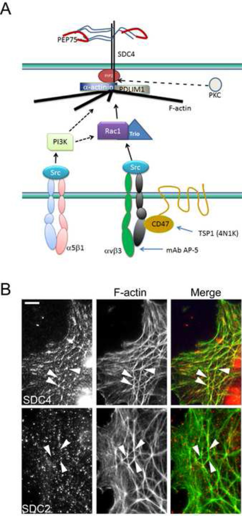
Our earlier studies [19, 28, 31, 34], summarized in Figure 1A, sought to characterize both the structural components responsible for the formation of CLANs and the signaling pathways regulating their formation. Consistent with our earlier studies [19, 34], syndecan-4 was found in CLAN vertices (Fig. 1B). In contrast syndecan-2, which is closely related to syndecan-4 [52], fails to localize within this structure. Negative control cells labeled for GFAP failed to show any staining (not shown). Likewise, the other two members of the family, syndecans 1 and 3, have not been localized within CLANs (not shown). Thus, it appears that localization to CLANs is specific to syndecan-4.
Figure 1. Schematic of CLAN components and integrin-mediated signaling pathways involved.
A) Syndecan-4 (SDC4), α-actinin, PIP2 and PDLIM1 have previously been shown to localize within the vertices of interconnecting F-actin bundles in CLANs where we propose they may form a molecular signaling complex or “vertisome” [19, 28]. Other known CLAN components not depicted include filamins A and B, myosin and tropomysosin [19, 20, 22, 28].The formation of this structure has been shown to involve distinct β1 and β3 integrin pathways that utilize PI3-kinase (PI3K) and CD47/Rac1/Trio respectively [31]. PI3K may activate Trio, or it may converge with the Rac/Trio pathway downstream of Rac1 (dotted line). The formation of this CLAN structure can be triggered by PEP75 (red curved line) which is a peptide from laminin 5 that can bind syndecan-4. It can also be triggered by an activating antibody to αvβ3 integrin (mAb AP-5) or a peptide from thrombospondin-1 (TSP1) called 4N1K [31]. PKCs may also activate other signaling molecules that regulate CLAN formation. B) Syndecan-4 (SDC4) is an integral part of the CLAN structure while syndecan-2 (SDC2) is not. The merged image shows that syndecan-4 localizes specifically to the vertices of the CLAN structure. This localization is not observed for syndecan-2. Arrowheads show location of CLAN vertices obvserved with F-actin labeling. Scale bar = 5 µm.
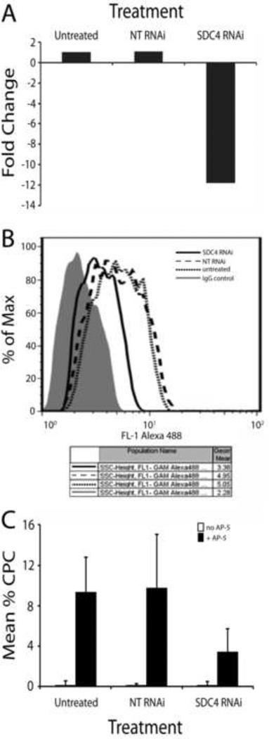
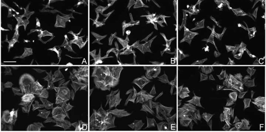
To determine if syndecan-4 was necessary for the formation of CLANs induced by αvβ3 integrin signaling, syndecan-4 siRNA was used to knockdown expression of syndecan-4 in HTM cells. Figure 2A shows that a twelve-fold reduction in syndecan-4 RNA resulted in a 32–33% decrease in the cell surface expression of syndecan-4 relative to cells treated with the non-targeting siRNA or untreated cells respectively (Fig. 2B). CLAN formation induced by activation of αvβ3 integrin using mAb AP-5 was also significantly impaired by the knockdown of syndecan-4 expression and resulted in a corresponding 64% decrease (p < 0.01) in the percentage of cells forming CLANs (Fig. 2C). Knockdown of syndecan-4 did not impair attachment and cell spreading (Fig. 3). This suggests that syndecan-4 plays an important structural role in CLAN formation.
Figure 2. Loss of syndecan-4 reduces β3 integrin-mediated CLAN formation.
A) N27TM-3 HTM cells were left untreated or transfected with a non-targeting (NT) RNAi or syndecan-4 (SDC4) RNAi. 72 hours post-transfection, RNA was extracted and analyzed for loss of RNA expression using real-time PCR. Cells were processed as described in Materials and Methods. Data were normalized to GAPDH and the fold change compared to untreated cells was determined. B) FACS analysis was performed on HTM cells transfected with NT or SDC4 RNAi 72 hours post-transfection to determine cell surface expression of SDC4. Cells were processed as described in Materials and Methods. C) Untreated cells or cells treated with NT or SDC4 RNAi were re-plated onto fibronectin-coated coverslips 72 hours post-transfection in the absence or presence of the β3 integrin-activating antibody AP-5. Three hours later the cells were fixed and labeled with phalloidin. The percentage of CLAN-positive cells (CPCs) is shown as the mean ± the SD; n ranged from 1295-1582 cells for each treatment group. For all 3 treatment groups, the cells treated with mAb AP-5 showed increased CLAN formation over the corresponding untreated cells (P < 0.01). For AP-5-treated cells, SDC4 RNAi significantly reduced CLAN formation over untransfected cells or cells transfected with non-targeting RNAi (P < 0.01).
Figure 3. Loss of syndecan-4 does not impair cell spreading of HTM cells on fibronectin.
N27TM-3 HTM cells were left untreated (A, D) or transfected with a non-targeting RNAi (B, E) or a syndecan-4 RNAi (C, F). Seventy two hours post-transfection, the cells were re-plated onto fibronectin-coated coverslips in the absence (A–C) or presence (D–F) of the β3 integrin-activating antibody AP-5. Three hours later the cells were fixed and labeled with phalloidin. Scale bar =100 µm.
Syndecan-4-binding peptide induces CLAN formation
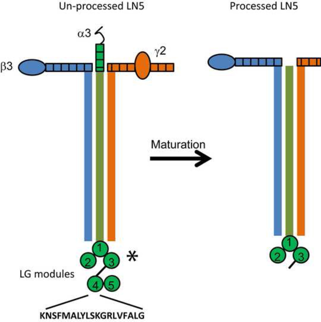
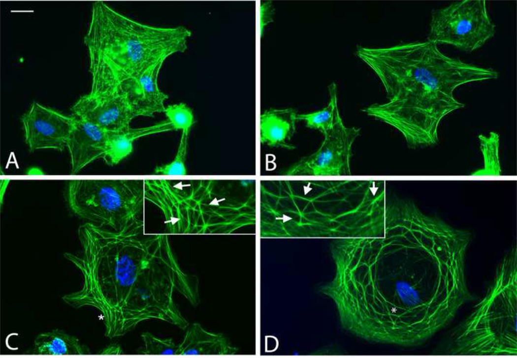
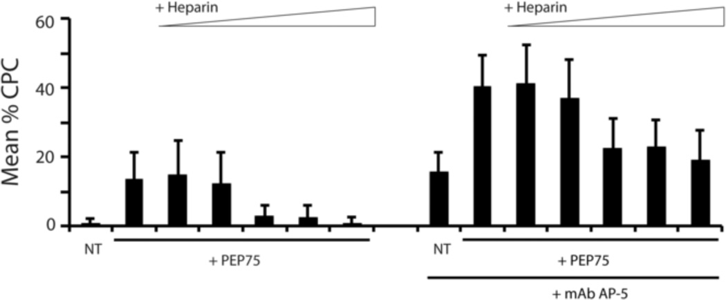
To further verify that syndecan-4 played a role in CLAN formation, HTM cells were incubated with a laminin 5-derived peptide, PEP75, which has been shown to bind to syndecan-4 in a HS-dependent manner [36]. This is a biologically active peptide that is found within the LG4 module that is proteolytically released from laminin 5 (Fig. 4). Untreated cells or cells treated with a control peptide, typically failed to form CLANs when plated for 3 hours on fibronectin-coated coverslips (Figs. 5A, B). In contrast, HTM cells plated on fibronectin formed CLANs in response to PEP75 (Figs. 5C) which were similar to those induced by the β3 integrin-activating antibody AP-5 (Fig. 5D). As shown in Figure 6, treatment of cells with PEP75 alone significantly induced CLAN formation relative to untreated cells (p < 0.01) at a level comparable to that induced by mAb AP-5.
Figure 4. Schematic of laminin 5.
Laminin 5 (laminin 332) is a heterotrimeric, multi-domain-containing ECM protein that is composed of α3, β3 and γ2 subunits. The unprocessed form of the protein contains a large globular domain at the C-terminal end of the α3 chain which is composed of five LG modules (LG1-5). The binding sites for α3β1, a6β1 and α6β4 integrins are located within the first three LG modules (asterisk) while the syndecan 4 binding PEP75 sequence (KNSFMALYLSKGRLVFALG) is located within the LG4 module. After secretion and deposition into the ECM, the protein undergoes proteolytic processing that results in the loss of N-terminal portions of both the α3 and γ2 chains as well as cleavage of the LG4 and LG5 modules from the C-terminus of the α3 chain. Thus the PEP75-containing module of laminin 5 is potentially free to interact with cell surface receptors such as syndecan 4 independent of the rest of the protein.
Figure 5. Comparison of HTM cells treated with PepCon, PEP75 or mAb AP-5.
N27TM-2 HTM cells were plated onto fibronectin-coated coverslips and allowed to spread for 3 hours prior to fixation and labeling with Alexa 488-conjugated phalloidin and Hoechst 33342. A) untreated cells; B) cells treated with 60 µg/mL PEPcon; C) cells treated with 60 µg/mL PEP75 and D) cells treated with mAb AP-5. Insets = enlarged areas of cells with CLANs; asterisks = areas in panels C & D enlarged in insets. Scale bar = 20µm.
Figure 6. PEP75 induces CLAN formation which is sensitive to competition with heparin.
N27TM-2 HTM cells were plated onto fibronectin-coated coverslips in the absence or presence of 60 µg/mL (28.5 µM) PEP75 together with increasing concentrations of heparin (0.02 µM – 200µM in 10-fold increments). The treatment groups on the right half of the graph included mAb AP-5. After 3 hours the cells were fixed and labeled with phalloidin. The percentage of CPCs is shown as the mean ± the SD; n ranged from 1134–2149 cells for each treatment group. Both PEP75 and mAb AP-5 alone increased CLAN formation over untreated contols (P < 0.01). 2.0 µM, 20 µM and 200 µM concentrations of heparin decreased PEP75-induced CLAN formation to near control levels (P > 0.01) while 0.02 µM and 0.2 µM concentrations had no effect. PEP75 and AP-5 in combination induced significantly greater CLAN formation than PEP75 alone or AP-5 alone, respectively (P < 0.01). 2.0 µM, 20 µM and 200 µM heparin significantly decreased CLAN formation in cells treated with PEP75 and AP-5 to levels seen in cells treated with AP-5 alone (P < 0.01) while 0.02 µM and 0.2 µM, again, had no effect.
CLAN formation induced by PEP75 could be blocked, in a dose dependent manner (p < 0.01), by the addition of heparin (Fig. 6) suggesting that PEP75 was inducing CLAN formation through a heparan sulfate proteoglycan. Syndecan-4 and αvβ3 integrin appeared to be working together in the formation of CLANs, since the addition of PEP75 in the presence of mAb AP-5 caused an additional 2.5-fold increase (p < 0.01) in the number of cells forming CLANs. Heparin also inhibited this increase in a dose dependent manner. At 200 µM heparin the level of CLAN returned back to the level observed in the presence of mAb AP-5 alone.
Dexamethasone increases deposition of laminin 5 into the ECM of HTM cultures
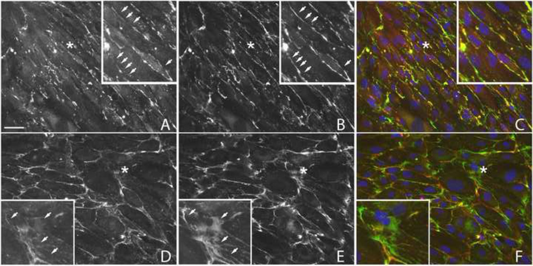
Given that PEP75 is derived from laminin 5 which is generally considered to be specific to the basement membranes of stratified epithelia [37] we sought to determine whether or not HTM cells made this protein and whether treatment with DEX increased its deposition within the ECM of HTM cultures. Figure 7A shows that in the absence of DEX, laminin 5 and laminin 1 are both detectable within the ECM of HTM cells. The labeling patterns were similar, but not identical (compare panels 7A and 7B). Control cells double-labeled with monoclonal anti-GFAP and rabbit IgG failed to show any staining (not shown). Treatment with DEX for 7 days resulted in heavier labeling patterns for both laminin 5 (panel 7D) and laminin 1 (panel 7E) suggesting that the expression of both laminins is up-regulated by steroid treatment. As in the absence of DEX, the labeling patterns for the two ECM proteins were still not completely identical. This suggests that the LG4 domain containing the PEP75 sequence could be available to bind to syndecan-4 expressed by HTM cells and that this interaction would be enhanced in DEX treated cultures when CLANs are more prevalent.
Figure 7. Localization of laminin 5 and laminin 1 in HTM cells treated with or without dexamethasone.
Confluent monolayers of N27TM-2 cells were treated for 7 days with 0.1% ethanol (A–C) or 500 nM DEX (D–F) prior to fixation and labeling with either mouse mAb P3H9-2 against laminin 5 (A, D; red channel) or a rabbit pAb against laminin 1 (B, E; green channel). DEX increased the labeling for both laminins, however, the labeling patterns for laminin 5 and laminin 1 did not completely overlap. Merged images (C, F) are also shown. Similar results were obtained with the N17TM-2 HTM cell strain (data not shown). Asterisks = areas enlarged within insets; arrows = regions where laminin 1 and laminin 5 labeling show incomplete co-localization. Scale bar = 50 µm.
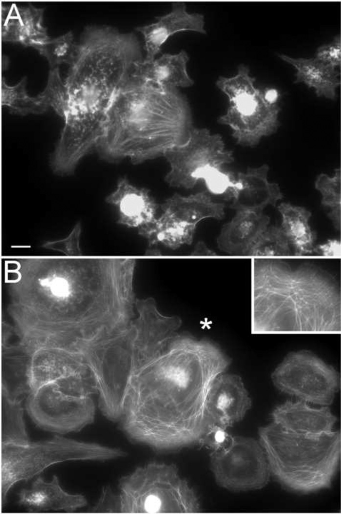
To determine if laminin alone was sufficient to induce CLAN formation, HTM cells were plated on a purified mixture of processed and unprocessed laminin 5. As shown in Figure 8, these cells failed to demonstrate any appreciable CLAN formation (≪ 1% CLAN-positive cells, not shown) in the absence (Fig. 8A) or presence (not shown) of PEP75. This was not unexpected, since our earlier studies showed that CLAN formation is a co-signaling event with β1 and αvβ3 integrins [31]. When we treated HTM cells plated on laminin 5 with mAb AP-5, however, the HTM cells formed CLANs (Fig 8B) indicating that αvβ3 integrin signaling was essential for CLAN formation.
Figure 8. HTM cells plated on laminin 5 only form CLANs in the presence of mAb AP-5.
N27TM-2 HTM cells were plated on laminin 5 for 3 hrs in the absence (A) or presence (B) of the αvβ3 integrin-activating antibody AP-5 prior to fixation and labeling with phalloidin. CLANs did not form on laminin 5 alone but did so in the presence of soluble mAb AP-5. Spreading also appeared to be enhanced when cells were spread on laminin 5 in the presence of AP-5. Bar = 20 µm.
PEP75 induces PKC-dependent CLAN formation
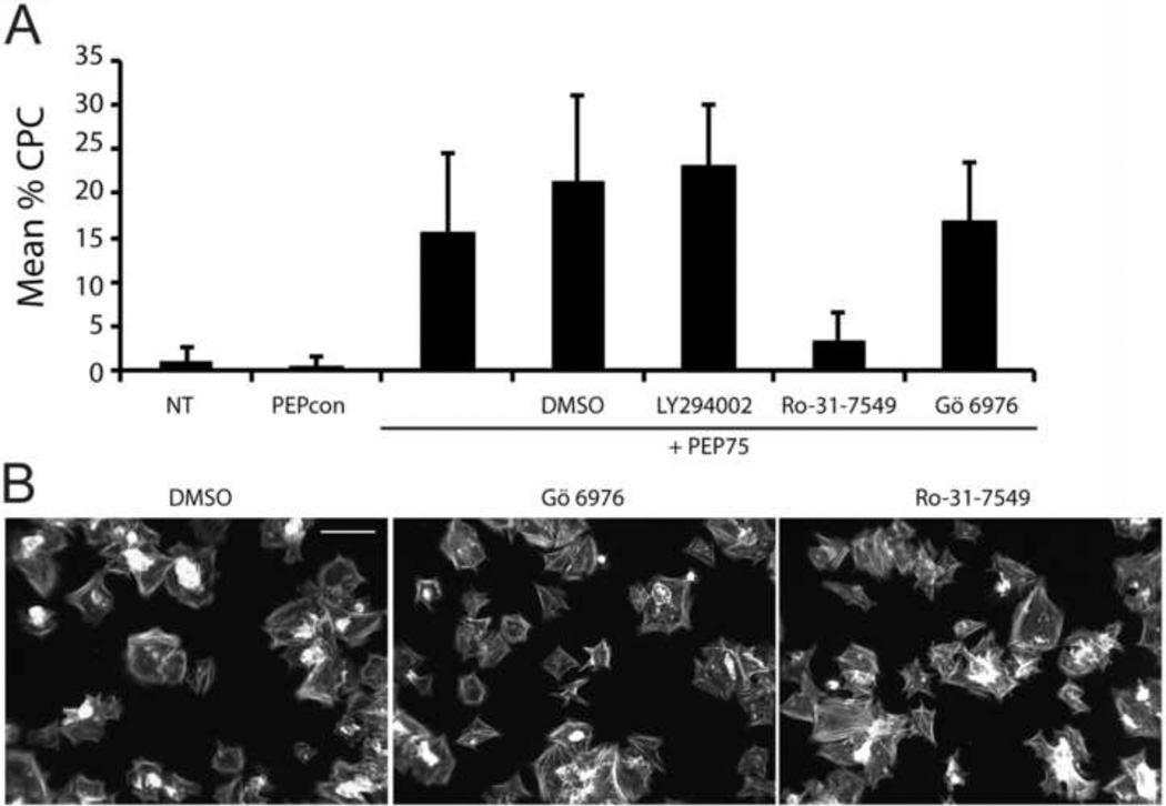
Since syndecan-4 has been shown to promote signaling events such as focal adhesion and stress fiber formation via PKCα [16–18], we then examined if syndecan-4 was using a PKC-dependent signaling pathway to form CLANs. Figure 9A shows the effects of two different PKC inhibitors on PEP75-induced CLAN formation. Gö 6976 is a relatively selective PKC inhibitor that targets PKCα, PKCβ1 and PKCµ [53]. In contrast, the PKC inhibitor Ro-31–7549 is a broad spectrum inhibitor [54]. Gö 6976 had no effect on PEP75-induced CLAN formation, while Ro-31–7549 reduced CLAN formation nearly five-fold relative to cells treated with PEP75 alone (p < 0.01) and six-fold relative to vehicle-treated cells (p < 0.1). This suggested that PKCγ, PKCβII, or PKCε may by mediating CLAN formation induced by PEP75 while PKCα was not involved. Relative to the DMSO control group, neither of the PKC inhibitors had any obvious effect on overall cell spreading (Fig. 9B)
Figure 9. PEP75-induced CLAN formation is dependent upon PKC, but independent of PI3-Kinase.
A) N27TM-2 HTM cells were plated onto fibronectin-coated coverslips in the absence or presence of 60 µg/mL PEP75 or 60 µg/mL PEPcon. PEP75-treated cells were also treated with 0.1% DMSO alone, 20 µM LY294002, 5 µM Ro-31-7549 or 10 µM Go 6976. After 3 hours the cells were fixed and labeled with phalloidin. The percentage of CPCs is shown as the mean ± the SD; n ranged from 1211–2624 cells for each treatment group. There was no difference between untreated control cells and cells treated with PEPcon. PEP75 alone significantly increased CLAN formation over untreated controls and PEPcon-treated cells, respectively (P < 0.01). Ro-31-7549 significantly decreased PEP75-induced CLAN formation over cells treated with PEP75 alone (P < 0.01). DMSO alone, LY294002 and Gö 6976 all failed to reduce PEP75-induced CLAN formation relative to cells treated with PEP75 alone. B) Cells were treated with PEP75 together with DMSO only, Gö 6976 or Ro-31-7549. Neither of the inhibitors had any significant effect upon cell spreading relative to the DMSO control-treated cells.
We also examined the potential role of PI-3 kinase in PEP75-mediated CLAN formation. A recent study by Araki, et al. [35] found that PEP75 induced clustering of syndecan-4 and β1 integrins, and we had previously found that CLAN formation was regulated, in part, by a PI-3 kinase-dependent β1 integrin signaling pathway [31]. However, the PI-3 kinase inhibitor LY294002 had no effect on PEP75-induced CLAN formation (Fig. 9A) indicating that PEP75 was not utilizing the syndecan-4/ β1 integrin pathway previously described by Araki, et al.
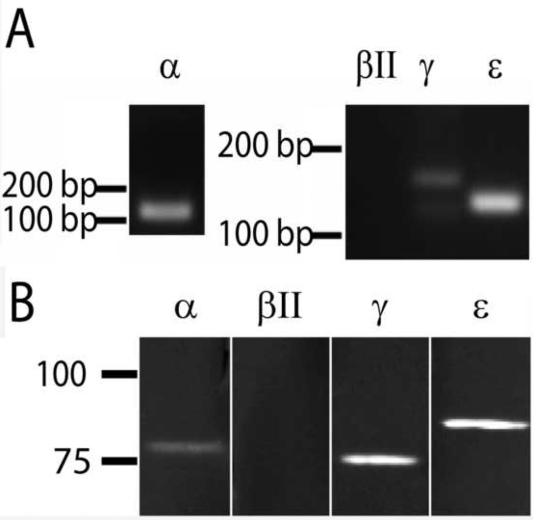
To help determine which PKC isozyme might be involved, we performed PCR and western blot analysis to determine which PKCs were present in the HTM cells. As shown in Figure 10, PKCβII was undetectable by either PCR or western blotting. In contrast, the α, γ and ε PKC isozymes were all found at both the RNA and protein level. With the exception of PKCβII, these results are consistent with an earlier report [55]. Given the absence of any inhibition by Gö 6976 on PEP75-induced CLAN formation, PKCα is unlikely to mediate the effects of the peptide on HTM cells. However, PKCγ and PKCε are both inhibited by Ro-31-7549 suggesting that one or both of these PKCs could be utilized by syndecan-4 to regulate PEP75-induced CLAN formation.
Figure 10. PKCα, γ and ε, but not PKCβII, are expressed in HTM cells.
A) mRNA was extracted from N27TM-2 HTM monolayers and qPCR performed using primers for PKCα, βII, γ and ε. The location of molecular weight markers are indicated. B) Western blot analysis of protein extracts from HTM monolayers. 20 µg was loaded per lane and the samples were probed using antibodies for for PKCα, βII, γ or ε respectively. The location of molecular weight markers are indicated.
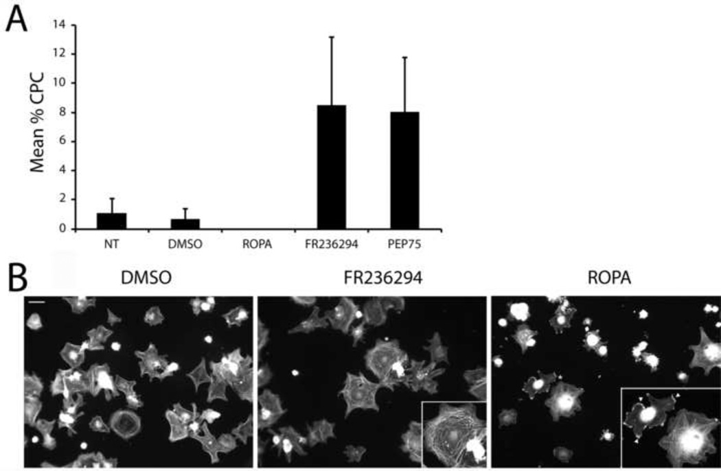
In order to try to identify specifically which PKC was regulating PEP75-mediated CLAN formation, two PKC agonists were used that have been reported to demonstrate a high degree of selectivity with regards to which isozymes they activate. ROPA preferentially activates PKCs α and γ [45] while FR236924 (DCP-LA) is a highly selective PKCε agonist [46]. As shown in Figure 11A, the PKCε agonist FR236924 increased the percentage of CLAN-positive cells seven-to twelve-fold over the percentage seen in control cells (P > 0.01). In fact the level of CLAN formation with FR236924 was virtually identical to that observed in PEP75-treated cells. In contrast, the PKCα/γ agonist ROPA completely failed to induce CLAN formation in HTM cells. The morphology of FR236924-treated cells appeared similar to control cells while ROPA-treated cells often exhibited reduced spreading relative to control cells (Fig. 11B). ROPA-treated cells also often exhibited pronounced membrane ruffling which was absent in the other treatment groups. These data suggest that the PKC isozyme involved in mediating PEP75-induced CLAN formation is PKCε.
Figure 11. A PKCε agonist induces CLAN formation in HTM cells while a PKCγ agonist does not.
A) N27TM-3 HTM cells were plated onto fibronectin-coated coverslips in the absence or presence of 0.1% DMSO, 5 µM ROPA (PKCα/γ agonist), 10 µM FR236924 (PKCε agonist) or 60 µg/mL PEP75. After 2 hours the cells were fixed and labeled with phalloidin. The percentage of CPCs is shown as the mean ± the SD; n ranged from 988–1591 cells for each treatment group. Cells treated with the PKCε agonist (P < 0.01) or PEP75 (P < 0.01) showed a higher percentage of CLAN-positive cells compared to untreated cells or cells treated with DMSO. Cells treated with the PKCα/γ agonist ROPA did not form CLANs. B) Representative images of N27TM-3 HTM cells treated with either DMSO, FR236924 or ROPA. FR236924 induced CLAN formation (see inset) while ROPA did not. ROPA-treated cells, however, frequently demonstrated pronounced membrane ruffling (see inset, arrowheads) which was absent in the other treatment groups.
Attempts to use siRNA-based procedures to verify that PKCε was involved were unsuccessful due to reagent-induced toxicity. We also attempted to overexpress dominant negative versions of both PKCγ and PKCε in order to abrogate PEP75-induced CLAN formation. However, we were unable to achieve high enough transfection efficiencies in our quiescent cell strains necessary to make any statistically significant conclusions.
Discussion
In this study we show that syndecan-4 plays a key role in the formation of atypical actin structures known as CLANs since the knockdown of syndecan-4 expression reduced CLAN formation. Whether syndecan-4 is playing a strictly structural role as a membrane docking receptor that mediates the assembly of this actin structure or whether it has a signaling role as well is unknown [15]. Formation of syndecan-4-containing CLANs appeared to use a novel PKCε pathway rather than the PKCα pathway previously shown to be involved with syndecan-4-in focal adhesion and stress fiber formation [16–18]. This suggests that varying the PKC-syndecan signaling interaction could impact the organization of the actin cytoskeleton.
The fact that data pointed to PKCε rather than PKCα as mediating PEP75-induced CLAN formation was not completely unexpected. There is precedent for finding syndecan-4 interacting with other PKCs within discreet signaling structures other than focal adhesions. A study by Vanwinkle, et al. [14] localized syndecan-4 within costameres of cardiomyocytes and suggested that, based upon immunolocalization data, PKCε could be a downstream effector of syndecan-4.
PKCε is a member of the novel subclass of PKC isozymes while PKCα, γ, βI and βII all belong to the conventional subclass of PKCs [56]. Thus, the interaction(s) between PKCε and syndecan-4 likely differ from that reported for syndecan-4 and PKCα. PKCα binds the cytoplasmic tail of syndecan-4 in the presence of PIP2, [10] and this interaction involves the PKCα C2 regulatory domain [57]. Since the C2 domains of the conventional PKCs are structurally distinct from those of the novel PKCs [56], the PKCε C2 domain is unlikely to interact with the syndecan-4 cytoplasmic tail. This is consistent with the yeast two-hybrid studies of Lim, et al. [58] that failed to demonstrate a direct interaction between syndecan-4 and PKCε. Nevertheless, PKCε could be regulating syndecan-4 activity in connection with PEP75-mediated CLAN formation given that novel PKCs have been shown to regulate serine phosphorylation of syndecan-4 [59, 60]. Whether a PKCε- mediated phosphorylation event regulates the incorporation of syndecan-4 into CLANs or the formation of the actin CLAN structure is unknown.
It is interesting that PKCε is involved in the formation of CLANs. Previous reports have shown that PKCs in general are involved in regulating aqueous humor outflow from the eye and that they do so in part by regulating the contractility of the TM [61–64]. However, those studies were concerned with showing that PKC regulated stress fiber formation and did not address any role for PKCs in CLAN formation. Interestingly, PKCε is known to be part of a mechanical stress response [65, 66] and CLANs may be a stress response structure [26] Thus, it seems reasonable that PKCε could be involved in regulating the rearrangement of stress fibers to form CLANs in response to mechanical stress.
It was not entirely unexpected that a matrix fragment like laminin 5-derived PEP75 could induce CLAN formation. A recent study [31] found that a thrombospondin-1-derived peptide, 4N1K, also induced CLAN formation in HTM cells. This peptide binds to the αvβ3 integrin co-receptor CD47 (Figure 1A). In addition to these matrix fragments, various ECM proteins have been found to induce CLAN formation [19]. Fibronectin, in particular, elicited the strongest CLAN response. However, upon activation of an αvβ3 signaling pathway, other ECM proteins such as type I and type IV collagen also induced significant levels of CLAN formation indicating that several β1 integrins can co-signal with αvβ3 integrins to regulate CLAN formation.
These co-signaling events could be the result of glucocorticoid-induced up-regulation and activation of αvβ3 integrins [67] and ECM proteins [28, 34]. Glucocorticoids have previously been shown to increase production and matrix deposition of unidentified laminins as well as thrombospondin-1, fibronectin and type IV collagen in HTM cells [68–70]. Here we have shown dexamethasone specifically increases matrix deposition of laminin 1 and laminin 5 which contains the PEP75 peptide used in this study. PEP75 is derived from the LG4 module of laminin 5 that is normally released from the mature protein once it has been deposited into the ECM. Therefore, this domain could potentially contribute to the increased CLAN formation observed following steroid treatment [33] by binding to syndecan-4 expressed on HTM cells. In addition, if the LG4 domain remains as a component of the ECM, this domain also has the potential to stabilize CLANs.
It is interesting that PEP75 also enhanced CLAN formation induced by αvβ3 activation. This indicates that there is more than one signaling pathway capable of regulating CLAN formation. In addition to the β1/β3 integrin co-signaling pathway previously reported [19], there appears to be a syndecan-4/αvβ3 integrin-mediated pathway possibly triggered by the LG4 domain of laminin 5. Other signaling pathways shown to trigger CLAN formation include those involving TGFβ2 [71], Wnt5a [72] and GPCRs [73]. Whether these other pathways represent distinct pathways from the syndecan-4/αvβ3 integrin or β1/β3 integrin pathways is unclear. TGFβ2- and Wnt5a–mediated CLAN formation requires prolonged treatments with these agents that can often result in the modulation of integrin mediated events and their ECM ligands. CLAN formation induced by GPCR signaling involved Schwann cells plated on laminin 1 [73] following short term treatments with either lysophosphatidic acid or sphingosine 1-phosphate. Activation of this pathway would target members of the Rho GTPase family which have previously been shown to be involved in the β1/β3 integrin co-signaling [19]. Clearly, understanding how these separate pathways trigger CLAN formation and determining if there is any commonality among these pathways could lead to a better understanding of steroid-induced glaucoma.
Several questions concerning syndecan-4 and this signaling pathway remain open. In particular, it is unknown whether syndecan-4 acts as a transmembrane structural component that mediates the formation of CLANs or if it also plays a role in meditating downstream signaling events in CLAN formation. There is also the question of whether an integrin is directly associated with syndecan-4 as a co-receptor in CLANs. Integrins have been shown to physically associate with syndecans and act as their co-receptors [4]. However, an earlier study found that neither β1 nor β3 integrins could be detected with syndecan-4 in CLAN vertisomes [19]. Yet combining PEP75 and AP-5 treatments resulted in greater CLAN formation than either treatment by itself. This suggests at least an indirect association between the syndecan-4, PKCε and the αvβ3 integrin signaling pathways. It is also worth noting that a number of the same signaling molecules that are associated with integrins in focal adhesions and stress fiber formation are also seen in CLAN verstisomes and appear to be involved in regulating CLAN formation despite them being completely different structures found at opposite aspects of cells [21, 74]. Thus there probably are some shared signaling components between the syndecan-4 and αvβ3 integrin triggered pathways.
In summary, the data demonstrate that the HSPG syndecan-4 plays a critical role in regulating the formation of a unique actin structure distinct from stress fibers. That role could be structural, but it may also have a signaling role as well since activation of a novel syndecan-4/PKC pathway induces CLAN formation.
Highlights.
Knockdown of syndecan-4 expression inhibits CLAN formation.
Dexamethasone increases deposition of laminin 5 into the ECM.
The syndecan-4 binding peptide PEP75, derived from laminin 5, induces CLAN formation.
PKCε regulates PEP75-mediated CLAN formation.
Acknowledgments
Grant Support: EY017006, EY0020490 (DMP), and a Core grant to the Department of Ophthalmology and Visual Sciences (P30 EY016665).
Abbreviations
- HSPG
heparan sulfate proteoglycan
- HS
heparan sulfate
- ECM
extracellular matrix
- TM
trabecular meshwork
- PKC
protein kinase C
- CLANs
cross-linked actin networks
- HTM
human trabecular meshwork
- PIP2
phosphatidylinositol 4,5-bisphosphate
- GEF
guanine nucleotide exchange factor
- GPCR
G-protein-coupled receptor
- LG
large globular
- DEX
dexamethasone
- OD
oculus dexter
- OS
oculus sinister
- ROPA
resiniferonol 9,13,14-orthophenylacetate
- FR236924
2-[(2-pentylcyclopropyl)methyl]cyclopropaneoctanoic acid
- mAb
monoclonal antibody
- pAb
polyclonal antibody
- GFAP
glial fibrillary acidic protein
- CPCs
CLAN-positive cells
- FACS
fluorescence activated cell sorting
Footnotes
This is a PDF file of an unedited manuscript that has been accepted for publication. As a service to our customers we are providing this early version of the manuscript. The manuscript will undergo copyediting, typesetting, and review of the resulting proof before it is published in its final citable form. Please note that during the production process errors may be discovered which could affect the content, and all legal disclaimers that apply to the journal pertain.
Contributor Information
Mark S. Filla, Email: msfilla@wisc.edu.
Ross Clark, Email: rclark2@wisc.edu.
Donna M. Peters, Email: dmpeter2@wiscmail.wisc.edu.
REFERENCES
- 1.Choi Y, Chung H, Jung H, et al. Syndecans as cell surface receptors: unique structure equates with functional diversity. Matrix Biol. 2011;30:93–99. doi: 10.1016/j.matbio.2010.10.006. [DOI] [PubMed] [Google Scholar]
- 2.Filla MS, David G, Weinreb RN, et al. Distribution of syndecans 1–4 within the anterior segment of the human eye: expression of a variant syndecan-3 and matrix associated syndecan-2. Exp Eye Res. 2004;79:61–74. doi: 10.1016/j.exer.2004.02.010. [DOI] [PubMed] [Google Scholar]
- 3.Tamm E. The trabecular meshwork outflow pathways: structural and functional aspects. Exp. Eye Res. 2009;88:648–655. doi: 10.1016/j.exer.2009.02.007. [DOI] [PubMed] [Google Scholar]
- 4.Beauvais DM, Burbach BJ, Rapraeger AC. The syndecan-1 ectodomain regulates αvβ3 integrin activity in human mammary carcinoma cells. J Cell Biol. 2004;167:171–181. doi: 10.1083/jcb.200404171. [DOI] [PMC free article] [PubMed] [Google Scholar]
- 5.Bernfield M, Gotte M, Park PW, et al. Functions of cell surface heparan sulfate proteoglycans. Ann. Rev. Biochem. 1999;68:729–777. doi: 10.1146/annurev.biochem.68.1.729. [DOI] [PubMed] [Google Scholar]
- 6.Bass MD, Roach KA, Morgan MR, et al. Syndecan-4-dependent Rac1 regulation determines directional migration in response to the extracellular matrix. J. Cell Biol. 2007;177:527–538. doi: 10.1083/jcb.200610076. [DOI] [PMC free article] [PubMed] [Google Scholar]
- 7.Bellin RM, Kubicek JD, Frigault MJ, et al. Defining the role of syndecan-4 in mechanotransduction using surfacemodification approaches. Proc. Natl. Acad. Sci. U.S.A. 2009;106:22102–22107. doi: 10.1073/pnas.0902639106. [DOI] [PMC free article] [PubMed] [Google Scholar]
- 8.Mahoney CM, Morgan MR, Harrison A, et al. Therapeutic ultrasound bypasses canonical syndecan-4 signaling to activate Rac1. J. Biol. Chem. 2009;284:8898–8909. doi: 10.1074/jbc.M804281200. [DOI] [PMC free article] [PubMed] [Google Scholar]
- 9.Dovas A, Yoneda A, Couchman JR. PKCα-dependent activation of RhoA by syndecan-4 during focal adhesion formation. J. Cell Sci. 2006;119:2837–2846. doi: 10.1242/jcs.03020. [DOI] [PubMed] [Google Scholar]
- 10.Oh ES, Woods A, Lim ST, et al. Syndecan-4 proteoglycan cytoplasmic domain and phosphatidylinositol 4,5-bisphosphate coordinately regulate protein kinase C activity. J. Biol. Chem. 1998;273:10624–10629. doi: 10.1074/jbc.273.17.10624. [DOI] [PubMed] [Google Scholar]
- 11.Okina E, Manon-Jensen T, Whiteford JR, et al. Syndecan proteoglycan contributions to cytoskeletal organization and contractility. Scand. J. Med. Sci. Sports. 2009;19:479–489. doi: 10.1111/j.1600-0838.2009.00941.x. [DOI] [PubMed] [Google Scholar]
- 12.Jung SY, Kim J-M, Kang HK, et al. A biologically active sequence of the laminin α2 large globular 1 domain promotes cell adhesion through syndecan-1 by inducing phosphorylation and membrane localization of protein kinase Cδ. J. Biol. Chem. 2009;284:31764–31775. doi: 10.1074/jbc.M109.038547. [DOI] [PMC free article] [PubMed] [Google Scholar]
- 13.Orosco A, Fromigue’ O, Hay” E, et al. Dual involvement of protein kinase C δ in apoptosis induced by syndecan-2 in osteoblasts. J. Cell. Biochem. 2006;98:838–850. doi: 10.1002/jcb.20826. [DOI] [PubMed] [Google Scholar]
- 14.Vanwinkle WB, Snuggs MB, De Hostos EL, et al. Localization of the transmembrane proteoglycan syndecan-4 and its regulatory kinases in costameres of rat cardiomyocytes: a deconvolution microscopic study. Anatom. Rec. 2002;268:38–46. doi: 10.1002/ar.10130. [DOI] [PubMed] [Google Scholar]
- 15.Roper JA, Williamson RC, Bass MD. Syndecan and integrin interactomes: large complexes in small spaces. Curr. Opin. Struct. Biol. 2012;22:583–590. doi: 10.1016/j.sbi.2012.07.003. [DOI] [PMC free article] [PubMed] [Google Scholar]
- 16.Oh ES, Woods A, Couchman JR. Syndecan-4 proteoglycan regulates the distribution and activity of protein kinase C. J. Biol. Chem. 1997;272:8133–8136. doi: 10.1074/jbc.272.13.8133. [DOI] [PubMed] [Google Scholar]
- 17.Woods A, Couchman JR. Protein kinase C involvement in focal adhesion formation. J. Cell Sci. 1992;101:277–290. doi: 10.1242/jcs.101.2.277. [DOI] [PubMed] [Google Scholar]
- 18.Woods A, Couchman JR. Heparan sulfate proteoglycans and signalling in cell adhesion. Adv. Exp. Med. Biol. 1992;313:87–96. doi: 10.1007/978-1-4899-2444-5_9. [DOI] [PubMed] [Google Scholar]
- 19.Filla MS, Woods A, Kaufman PL, et al. β1 and β3 integrins cooperate to induce syndecan-4 containing cross-linked actin networks (CLANs) in human trabecular meshwork (HTM) cells. Invest. Ophthalmol. Vis. Sci. 2006;47:1956–1967. doi: 10.1167/iovs.05-0626. [DOI] [PMC free article] [PubMed] [Google Scholar]
- 20.Gordon WE, 3rd, Bushnell A. Immunofluorescent and ultrastructural studies of polygonal microfilament networks in respreading non-muscle cells. Exp. Cell Res. 1979;120:335–348. doi: 10.1016/0014-4827(79)90393-8. [DOI] [PubMed] [Google Scholar]
- 21.Ireland GW, Voon FC. Polygonal networks in living chick embryonic cells. J. Cell Sci. 1981;52:55–69. doi: 10.1242/jcs.52.1.55. [DOI] [PubMed] [Google Scholar]
- 22.Lazarides E. Actin, alpha-actinin, and tropomyosin interaction in the structural organization of actin filaments in nonmuscle cells. J. Cell Biol. 1976;68:202–219. doi: 10.1083/jcb.68.2.202. [DOI] [PMC free article] [PubMed] [Google Scholar]
- 23.Mochizuki Y, Furukawa K, Mitaka T, et al. Polygonal networks, "geodomes", of adult rat hepatocytes in primary culture. Cell Biol. Int. Rep. 1988;12:1–7. doi: 10.1016/0309-1651(88)90105-1. [DOI] [PubMed] [Google Scholar]
- 24.Clark AF, Wilson K, McCartney MD, et al. Glucocorticoid-induced formation of cross-linked actin networks in cultured human trabecular meshwork cells. Invest. Ophthalmol. Vis. Sci. 1994;35:281–294. [PubMed] [Google Scholar]
- 25.Lazarides E, Burridge K. α-actinin: immunofluorescent localization of a muscle structural protein in nonmuscle cells. Cell. 1975;6:289–298. doi: 10.1016/0092-8674(75)90180-4. [DOI] [PubMed] [Google Scholar]
- 26.Ingber DE, Tensegrity I. Cell structure and hierarchical systems biology. J. Cell Sci. 2003;116:1157–1173. doi: 10.1242/jcs.00359. [DOI] [PubMed] [Google Scholar]
- 27.Clark AF, Brotchie D, Read AT, et al. Dexamethasone alters F-actin architecture and promotes cross-linked actin network formation in human trabecular meshwork tissue. Cell Motil. Cytoskel. 2005;60:83–95. doi: 10.1002/cm.20049. [DOI] [PubMed] [Google Scholar]
- 28.Clark R, Nosie A, Walker T, et al. Comparative genomic and proteomic analysis of cytoskeletal changes in dexamethasone-treated trabecular meshwork cells. Mol. Cell Proteomics. 2013;12:194–206. doi: 10.1074/mcp.M112.019745. [DOI] [PMC free article] [PubMed] [Google Scholar]
- 29.Hoare M-J, Grierson I, Brotchie D, et al. Cross-linked actin Networks (CLANs) in the trabecular meshwork of the normal and glaucomatous human eye in situ. Invest. Ophthalmol. Vis. Sci. 2008;50:1255–1263. doi: 10.1167/iovs.08-2706. [DOI] [PubMed] [Google Scholar]
- 30.Clark AF, Miggans ST, Wilson K, et al. Cytoskeletal changes in cultured human glaucoma trabecular meshwork cells. J. Glaucoma. 1995;4:183–188. [PubMed] [Google Scholar]
- 31.Filla MS, Schwinn MK, Sheibani N, et al. Regulation of cross-linked actin network (CLAN) formation in human trabecular meshwork (HTM) cells by convergence of distinct β1 and β3 integrin pathways. Invest. Ophthalmol. Vis. Sci. 2009;50:5723–5731. doi: 10.1167/iovs.08-3215. [DOI] [PMC free article] [PubMed] [Google Scholar]
- 32.Clark AF, Wordinger RJ. The role of steroids in outflow resistance. Exp. Eye Res. 2009;88:752–759. doi: 10.1016/j.exer.2008.10.004. [DOI] [PubMed] [Google Scholar]
- 33.Clark AF, Wilson K, de Kater AW, et al. Dexamethasone-induced ocular hypertension in perfusion-cultured human eyes. Invest. Ophthalmol. Vis. Sci. 1995;36:478–489. [PubMed] [Google Scholar]
- 34.Filla MS, Schwinn MK, Nosie AK, et al. Dexamethasone-associated cross-linked actin network (CLAN) formation in human trabecular meshwork (HTM) cells involves β3 integrin signaling. Invest. Ophthalmol. Vis. Sci. 2011;52:2952–2959. doi: 10.1167/iovs.10-6618. [DOI] [PMC free article] [PubMed] [Google Scholar]
- 35.Araki E, Momota Y, Togo T, et al. Clustering of syndecan-4 and integrin β1 by Laminin α3 chain-derived peptide promotes keratinocyte migration. Molec. Biol. Cell. 2009;20:3012–3024. doi: 10.1091/mbc.E08-09-0977. [DOI] [PMC free article] [PubMed] [Google Scholar]
- 36.Utani A, Nomizu M, Matsuura H, et al. A unique sequence of the laminin alpha 3 G domain binds to heparin and promotes cell adhesion through syndecan-2 and −4. J. Biol. Chem. 2001;276:28779–28788. doi: 10.1074/jbc.M101420200. [DOI] [PubMed] [Google Scholar]
- 37.Rousselle P, Beck K. Laminin 332 processing impacts cellular behavior. Cell Adhes. Migrat. 2013;7:122–134. doi: 10.4161/cam.23132. [DOI] [PMC free article] [PubMed] [Google Scholar]
- 38.Schéele S, Nyström A, Durbeej M, et al. Laminin isoforms in development and disease. J. Mol. Med. 2007;85:825–836. doi: 10.1007/s00109-007-0182-5. [DOI] [PubMed] [Google Scholar]
- 39.Utani A, Momota Y, Endo H, et al. Laminin α3 LG4 module induces matrix metalloproteinase-1 through mitogen-activated protein kinase signaling. J. Biol. Chem. 2003;278:34483–34490. doi: 10.1074/jbc.M304827200. [DOI] [PubMed] [Google Scholar]
- 40.Momota Y, Suzuki N, Kasuya Y, et al. Laminin α3 LG4 module induces keratinocyte migration: involvement of matrix metalloproteinase-9. J. Recept. Signal Transduct. Res. 2005;25:1–17. doi: 10.1081/rrs-200047870. [DOI] [PubMed] [Google Scholar]
- 41.Tran M, Rousselle P, Nokelainen P. Targeting a tumor-specific laminin domain critical for human carciongenesis. Canc. Res. 2008;68:2885–2894. doi: 10.1158/0008-5472.CAN-07-6160. [DOI] [PubMed] [Google Scholar]
- 42.Carulli S, Beck K, Dayan G, et al. Cell surface proteoglycans syndecan-1 and −4 bind overlapping but distinct sites in laminin α3 LG45 protein domain. J. Biol. Chem. 2012;287:12204–12216. doi: 10.1074/jbc.M111.300061. [DOI] [PMC free article] [PubMed] [Google Scholar]
- 43.Polansky JR, Weinreb RN, Baxter JD, et al. Human trabecular cells. I. establishment in tissue culture and growth characteristics. Invest. Ophthalmol. Vis. Sci. 1979;18:1043–1049. [PubMed] [Google Scholar]
- 44.Polansky JR, Weinreb R, Alvarado JA. Studies on human trabecular cells propagated in vitro. Vis. Res. 1981;21:155–160. doi: 10.1016/0042-6989(81)90151-6. [DOI] [PubMed] [Google Scholar]
- 45.Geiges D, Meyer T, Marte B, et al. Activation of protein kinase C subtypes α, γ, δ, ε, ζ αvδ η by tumor-promoting and nontumor-promoting agents. Biochem. Pharmacol. 1997;53:865–875. doi: 10.1016/s0006-2952(96)00885-4. [DOI] [PubMed] [Google Scholar]
- 46.Kanno T, Yamamoto H, Yaguchi T, et al. The linoleic acid derivative DCP-LA selectively activates PKC-ε, possibly binding to the phosphatidylserine binding site. J. Lipid Res. 2006;47:1146–1156. doi: 10.1194/jlr.M500329-JLR200. [DOI] [PubMed] [Google Scholar]
- 47.Mosher DF, Johnson RB. In vitro formation of disulfide-bonded fibronectin multimers. J. Biol. Chem. 1983;258:6595–6560. [PubMed] [Google Scholar]
- 48.Longley RL, Woods A, Fleetwood A, et al. Control of morphology, cytoskeleton and migration by syndecan-4. J. Cell Sci. 1999;112:3421–3431. doi: 10.1242/jcs.112.20.3421. [DOI] [PubMed] [Google Scholar]
- 49.Lories V, Cassiman JJ, Van den Berghe H, et al. Multiple distinct membrane heparan sulfate proteoglycans in human lung fibroblasts. J. Biol. Chem. 1989;264:7009–7016. [PubMed] [Google Scholar]
- 50.Peterson JA, Sheibani N, David G, et al. Heparin II domain of fibronectin uses α4β1 integrin to control focal adhesion and stress fiber formation, independent of syndecan-4. J. Biol. Chem. 2005;280:6915–6922. doi: 10.1074/jbc.M406625200. [DOI] [PubMed] [Google Scholar]
- 51.David G, van der Schueren B, Marynen P, et al. Molecular cloning of amphiglycan, a novel integral membrane heparan sulfate proteoglycan expressed by epithelial and fibroblastic cells. J. Cell Biol. 1992;118:961–969. doi: 10.1083/jcb.118.4.961. [DOI] [PMC free article] [PubMed] [Google Scholar]
- 52.Couchman JR. Syndecans: proteoglycan regulators of cell-surface microdomains? Nat. Rev. Molec. Cell Biol. 2003;4:926–937. doi: 10.1038/nrm1257. [DOI] [PubMed] [Google Scholar]
- 53.Martiny-Baron G, Kazanietzs MG, Mischak H, et al. Selective inhibition of protein kinase C isozymes by the indolocarbazole Gö 6976. J. Biol. Chem. 1993;268:9194–9197. [PubMed] [Google Scholar]
- 54.Wilkinson SE, Parker PJ, Nixon JS. Isoenzyme specificity of bisindolylmaleimides, selective inhibitors of protein kinase C. Biochem. J. 1993;294:335–337. doi: 10.1042/bj2940335. [DOI] [PMC free article] [PubMed] [Google Scholar]
- 55.Alexander JP, Acott TS. Involvement of protein kinase C in TNFalpha regulation of trabecular matrix metalloproteinases and TIMPs. Invest. Ophthalmol. Vis. Sci. 2001;42:2831–2838. [PubMed] [Google Scholar]
- 56.Newton AC. Regulation of protein kinase C. Curr. Opin. Cell Biol. 1997;9:161–167. doi: 10.1016/s0955-0674(97)80058-0. [DOI] [PubMed] [Google Scholar]
- 57.Horowitz A, Murakami M, Gao YH, et al. Phosphatidylinositol-4,5-bisphosphate mediates the interaction of syndecan-4 with protein kinase C. Biochem. 1999;38:15871–15877. doi: 10.1021/bi991363i. [DOI] [PubMed] [Google Scholar]
- 58.Lim ST, Longley RL, Couchman JR, et al. Direct binding of syndecan-4 cytoplasmic domain to the catalytic domain of protein kinase C alpha (PKC alpha) increases focal adhesion localization of PKC alpha. J. Biol. Chem. 2003;278:13795–13802. doi: 10.1074/jbc.M208300200. [DOI] [PubMed] [Google Scholar]
- 59.Chaudhuri P, Colles SM, Fox PL, et al. Protein Kinase Cδ-dependent phosphorylation of syndecan-4 regulates cell migration. Circ. Res. 2005;97:671–684. doi: 10.1161/01.RES.0000184667.82354.b1. [DOI] [PubMed] [Google Scholar]
- 60.Horowitz A, Simons M. Regulation of syndecan-4 phosphorylation in vivo. J. Biol. Chem. 1998;273:10914–10918. doi: 10.1074/jbc.273.18.10914. [DOI] [PubMed] [Google Scholar]
- 61.Khurana RN, Deng PF, Epstein DL. The role of protein kinase C in modulation of aqueous humor outflow facility. Exp. Eye Res. 2003;76:39–47. doi: 10.1016/s0014-4835(02)00255-5. [DOI] [PubMed] [Google Scholar]
- 62.Tian B, Gabelt BT, Kaufman PL. Effect of staurosporine on outflow facility in monkeys. Invest. Ophthalmol. Vis. Sci. 1999;40:1009–1011. [PubMed] [Google Scholar]
- 63.Tian B, Brumback LC, Kaufman PL. ML-7, chelerythrine and phorbol ester increase outflow facility in the monkey eye. Exp. Eye Res. 2000;71:551–566. doi: 10.1006/exer.2000.0919. [DOI] [PubMed] [Google Scholar]
- 64.Thieme H, Nass JU, Nuskovski M, et al. The effects of protein kinase C on trabecular meshwork and ciliary muscle contractility. Invest. Ophthalmol. Vis. Sci. 1999;40:3254–3261. [PubMed] [Google Scholar]
- 65.Cheng J-J, Wung B-S, Chao Y-J, et al. Sequential activation of protein kinase C (PKC)-α and PKC-ε contributes to sustained Raf/ERK1/2 activation in endothelial cells under mechanical strain. J. Biol. Chem. 2001;276:31368–31375. doi: 10.1074/jbc.M011317200. [DOI] [PubMed] [Google Scholar]
- 66.Traub O, Monia BP, Dean NM, Berk BC. PKC-e is required for mechano-sensitive activation of ERK1/2 in endothelial cells. J. Biol. Chem. 1997;272:31251–31257. doi: 10.1074/jbc.272.50.31251. [DOI] [PubMed] [Google Scholar]
- 67.Faralli JA, Gagen D, Filla MS, et al. Dexamethasone increases αvβ3 integrin expression and affinity through a calcineurin/NFAT pathway. Biochim. Biophys. Acta. 2013;1833:3306–3313. doi: 10.1016/j.bbamcr.2013.09.020. [DOI] [PMC free article] [PubMed] [Google Scholar]
- 68.Dickerson JE, Jr, Steely HT, Jr, English-Wright SL, et al. The effect of dexamethasone on integrin and laminin expression in cultured human trabecular meshwork cells. Exp. Eye Res. 1998;66:731–738. doi: 10.1006/exer.1997.0470. [DOI] [PubMed] [Google Scholar]
- 69.Filla MS, Lui X, Nguyen TD, et al. In vitro localization of TIGR/MYOC in trabecular meshwork extracellular matrix and binding to fibronectin. Invest. Ophthalmol. Vis. Sci. 2002;43:151–161. [PubMed] [Google Scholar]
- 70.Flugel-Koch C, Ohlmann A, Fuchshofer R, et al. Thrombospondin-1 in the trabecular meshwork: localization in normal and glaucomatous eyes, and induction by TGF-betal and dexamethasone in vitro. Exp. Eye Res. 2004;79:649–663. doi: 10.1016/j.exer.2004.07.005. [DOI] [PubMed] [Google Scholar]
- 71.O’Reilly S, Pollock N, Currie L, et al. Inducers of cross-linked actin networks in trabecular meshwork cells. Invest. Ophthalmol. Vis. Sci. 2011;52:7316–7324. doi: 10.1167/iovs.10-6692. [DOI] [PubMed] [Google Scholar]
- 72.Yuan Y, Call MK, Yuan Y, et al. Trabecular meshwork cells through noncanonical Wnt signaling. Invest. Ophthalmol. Vis. Sci. 2013;54:6502–6509. doi: 10.1167/iovs.13-12447. [DOI] [PMC free article] [PubMed] [Google Scholar]
- 73.Barber SC, Mellor H, Gampel A, et al. S1P and LPA trigger Schwann cell actin changes and migration. Eur. J. Neurosci. 2004;19:3142–3150. doi: 10.1111/j.0953-816X.2004.03424.x. [DOI] [PubMed] [Google Scholar]
- 74.Ireland GW, Sanders EJ, Voon FC, et al. The ultrastructure of polygonal networks in chick embryonic cells in vitro. Cell Biol. Int. Rep. 1983;7:679–688. doi: 10.1016/0309-1651(83)90196-0. [DOI] [PubMed] [Google Scholar]


