Short abstract
Two recent studies of yeast prions have now formally demonstrated that multiple stable protein conformations are the basis of strain variation.
Abstract
Studies of mammalian prion diseases such as bovine spongiform encephalopathy have suggested that different strains consist of prion proteins with different conformations. Two recent studies of yeast prions have now formally demonstrated that multiple stable protein conformations are the basis of strain variation.
Transmissible spongiform encephalopathies (TSEs) are a group of closely related neurodegenerative conditions of animals and humans that includes sheep scrapie, chronic wasting disease of deer and elk, bovine spongiform encephalopathy (BSE) and human Creutzfeldt-Jakob disease (CJD). TSEs attracted interest and considerable controversy well before the epidemic of BSE and the subsequent appearance of a new variant of human CJD, because of their extraordinary features. It was once widely believed that TSEs were caused by infectious agents containing a nucleic-acid genome, but the prevailing view now attributes these diseases to subcellular pathogens called 'prions', which are defined as small proteinaceous infectious particles that lack informational nucleic acid [1]. Although the precise molecular structure of the infectious agent has still not been definitively identified, considerable evidence supports the unorthodox hypothesis that prions are composed largely, if not entirely, of a pathogenic conformation of the prion protein (PrP), referred to as PrPSc, and that during the disease process, PrPSc imposes its conformation on the normal, host-encoded version of PrP (PrPC), resulting in the exponential accumulation of PrPSc.
One of the biggest challenges for this theory, called the prion hypothesis, has been to explain the existence of multiple strains of the infectious agent in the absence of informational nucleic acid; this characteristic convinced some investigators that the scrapie agent must be a virus [2-4]. Mammalian prion strains are classically defined in terms of their differing incubation times and the different profiles of pathological lesions that they produce in the central nervous system of recipient animals. More recently, attempts have been made to use biochemical and/or immunological properties of PrPSc as markers of prion strain differences [5,6]. Discussion of different strains in the context of the prion hypothesis generally refers to the different conformational states of mammalian or yeast prion proteins. Two recent studies in yeast [7,8] confirm the predictions from earlier studies of mammalian prions [9-14], which suggested that strain specificity is linked to conformational differences in PrPSc.
The discovery in the mid-1990s that certain phenotypic traits in yeast were propagated by a mechanism similar to TSEs [15,16] suggested that information transfer by such epigenetic mechanisms was more widespread in nature than was once appreciated, and the discovery did much to bolster the prion hypothesis. Moreover, while progress on experimental verification of the 'protein-only' prion hypothesis in the past 20 years has been considerable, the long experimental incubation times of TSEs and the difficulties associated with characterization of PrPSc, including its extreme hydrophobicity and tendency to aggregate, have provided formidable challenges for studies of mammalian prion diseases. In contrast, although the study of prion-like agents in yeast has not been straightforward, yeast readily lends itself to genetic, cell biological and biochemical analysis, meaning that progress on the study of yeast prions has been relatively rapid.
The two most widely studied yeast prions are [URE3] [15], the prion form of the protein Ure2p which is involved in the regulation of nitrogen metabolism, and [PSI+] [16], which is the prion isoform of the essential protein Sup35p, the yeast counterpart of the animal translational termination factor eRF3. As predicted for a prion-like mode of replication, Sup35p is soluble inside [psi-] cells (and is thus equivalent to PrPC) but forms insoluble fibrillar amyloid aggregates in [PSI+] cells (equivalent to PrPSc). [PSI+] yeast cells are partially defective in translation termination because accumulation of self-replicating aggregates of Sup35p in [PSI+] cells leads to depletion of the cellular pool of the termination factor, resulting in an enhanced tendency of ribosomes to read through nonsense mutations. The [PSI+] state is readily assessed in a genetic background containing a nonsense mutation in the ade1 gene. In the [psi-] state, yeast containing such a mutant allele do not grow on medium without adenine and accumulate a red pigment on complete medium, whereas the presence of the [PSI+] prion in ade1 cells leads to read-through of UGA codons (nonsense suppression), which partially restores growth on adenine-deficient medium and results in white or pink colonies.
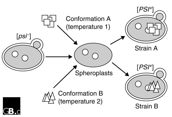
[PSI+] shows a range of phenotypic states - reminiscent of mammalian prion strains - which differ from each other in their levels of nonsense suppression, the involvement of chaperone proteins, and in the solubility and activity of Sup35p. Such [PSI+] variants can be identified on the basis of the ade1 color phenotype. In previous studies, Weissman and colleagues [17] impressively demonstrated induction of the [PSI+] state by the introduction into [psi-] cells of a bacterially produced recombinant fragment of Sup35p, referred to as Sup-NM, made up of residues 1-254. This fragment consists of the amino-terminal glutamine- and asparagine-rich region of Sup35p that is required for [PSI+] propagation, plus the highly charged middle region. In a study recently published in Nature by the same group, Tanaka and colleagues [7] used a new, highly efficient method for infecting yeast with preformed Sup-NM amyloid fibers, combined with genetic selection to identify the small numbers of yeast cells converted to the [PSI+] state, to demonstrate that infection of yeast with different conformations of yeast prion proteins results in the manifestation of different prion strains. Previous studies [18] showed that overexpression of the amino-terminal fragment Sup-NM leads to aggregation of Sup35p and the appearance of a range of phenotypic [PSI+] variants. Using a heterogenous preparation of Sup-NM fibers preformed in vitro, Tanaka and coworkers [7] were able to obtain a range of [PSI+] strains following introduction into [psi-] yeast. When extracts or partially purified Sup35p proteins were prepared from the yeast cells containing the resulting [PSI+] strains and used to transform [psi-] yeast, the different [PSI+] strains were faithfully propagated, suggesting that the different strains of prion observed in yeast transformed with pure protein arose from intrinsic heterogeneity in the Sup-NM prions formed in vitro. In a key experiment, Sup-NM amyloid fibers with different conformations were prepared in vitro at different temperatures, allowing the investigators to directly test the role of protein conformation in determining [PSI+] strain properties in vivo (Figure 1). Tanaka and colleagues [7] found that the different conformations of Sup-NM formed in vitro generated different [PSI+] strains and, once formed, the conformation-dependent strain characteristics were stably propagated in successive generations of yeast cells.
Figure 1.

A schematic representation of common aspects of the procedure that Tanaka et al. [7] and King and Diaz-Avalos [8] used to generate multiple [PSI+] strains by converting Sup35p protein to different aggregating conformations in vitro. The [psi-] budding yeast cells (left) containing normal Sup35p (circles) were made into spheroplasts (lacking some of the cell wall; middle) into which preformed conformations of a recombinant amino-terminal fragment of Sup35p (squares and triangles) were introduced. This leads to a [PSI+] state (right), as assessed by plating on a rich medium containing trace amounts of adenine; [PSI+] cells produce white colonies on this medium whereas [psi-] cells produce red colonies (not shown). Different conformations of Sup35p gave rise to phenotypically distinct strains of [PSI+] cells.
In a related paper appearing in the same issue of Nature, Chih-Yen King and Ruben Diaz-Avalos [8] used a fusion construct, referred to as Sup35(1-61)-GFP, consisting of the green fluorescent protein (GFP) fused carboxy-terminally to the first 61 amino-acid residues of Sup35p, to determine whether diluted cell extracts prepared from yeast propagating various [PSI+] strains could be used to seed the assembly of aggregates of recombinant Sup35(1-61)-GFP. When exposed to recombinant Sup35(1-61)-GFP, the initial short rod-shaped aggregates from the cell extracts elongated and the fiber morphology of the original seeding strain was maintained. Following incubation and sonication, the aggregates were found to retain strain-specific infectivity upon reintroduction into yeast. In an important final series of experiments analogous to those described by Tanaka and colleagues [7], King and Diaz-Avalos [8] demonstrated that strain-specific [PSI+] infectivity could arise from self-assembly of pure recombinant Sup35(1-61)-GFP prepared under different buffering and temperature conditions in the absence of yeast seeds. In agreement with the results of Tanaka and colleagues [7], transformation of yeast cells with these different amyloid preparations induced the formation of distinct [PSI+] strains [8].
These two studies clearly demonstrate that Sup-NM or Sup35(1-61)-GFP can be induced to adopt multiple, stable conformations before entry into the cell and that these conformational differences are the basis of [PSI+] strain variation. This demonstration satisfies a core prediction of the prion hypothesis and validates earlier studies of mammalian prion diseases. Seminal studies linking the conformation of PrPSc with prion strain arose from investigations of mink prions that, upon transmission, produced different clinical symptoms and produced PrPSc with different resistances to proteinase-K digestion and altered amino-terminal proteinase-K cleavage sites [9]. Such strain-specific conformational differences were also reproduced in cell-free conversion systems [10,11]. Evidence supporting the hypothesis that strain diversity is encoded in the tertiary structure of PrPSc emerged from studies of the transmission of inherited and sporadic human prion diseases in transgenic mice [12-14]. Banding patterns of PrPSc forms with different glycosylation patterns and sizes of PrPSc fragments following proteinase-K treatment have also been used to determine the strain of CJD cases [19,20]. In particular, a characteristic type of glycosylated PrPSc observed in patients with variant CJD and BSE-infected animals appears to distinguish vCJD PrPSc from the patterns observed in classical CJD [21].
Notwithstanding criticisms that studies of phenotypic states in yeast do not accurately reflect TSE infection [4], yeast prions provide a powerful model for understanding the general principles of protein-based inheritance with relevance to the molecular mechanisms of mammalian amyloid diseases. The challenge for investigators studying mammalian prions will be to corroborate these studies in yeast by creating new infectious material from pure recombinant PrP or from material synthesized in vitro. Disappointingly, attempts to generate infectivity from such approaches, let alone different prion strains, have so far had uniformly negative results.
References
- Prusiner SB. Novel proteinaceous infectious particles cause scrapie. Science. 1982;216:136–144. doi: 10.1126/science.6801762. [DOI] [PubMed] [Google Scholar]
- Dickinson AG, Meikle VMH, Fraser H. Identification of a gene which controls the incubation period of some strains of scrapie agent in mice. J Comp Pathol. 1968;78:293–299. doi: 10.1016/0021-9975(68)90005-4. [DOI] [PubMed] [Google Scholar]
- Bruce ME, Dickinson AG. Biological evidence that the scrapie agent has an independent genome. J Gen Virol. 1987;68:79–89. doi: 10.1099/0022-1317-68-1-79. [DOI] [PubMed] [Google Scholar]
- Manuelidis L. Transmissible encephalopathies: speculations and realities. Viral Immunol. 2003;16:123–139. doi: 10.1089/088282403322017875. [DOI] [PubMed] [Google Scholar]
- Peretz D, Williamson RA, Legname G, Matsunaga Y, Vergara J, Burton DR, DeArmond SJ, Prusiner SB, Scott MR. A change in the conformation of prions accompanies the emergence of a new prion strain. Neuron. 2002;34:921–932. doi: 10.1016/S0896-6273(02)00726-2. [DOI] [PubMed] [Google Scholar]
- Safar J, Wille H, Itri V, Groth D, Serban H, Torchia M, Cohen FE, Prusiner SB. Eight prion strains have PrPSc molecules with different conformations. Nat Med. 1998;4:1157–1165. doi: 10.1038/2654. [DOI] [PubMed] [Google Scholar]
- Tanaka M, Chien P, Naber N, Cooke R, Weissman JS. Conformational variations in an infectious protein determine prion strain differences. Nature. 2004;428:323–328. doi: 10.1038/nature02392. [DOI] [PubMed] [Google Scholar]
- King CY, Diaz-Avalos R. Protein-only transmission of three yeast prion strains. Nature. 2004;428:319–323. doi: 10.1038/nature02391. [DOI] [PubMed] [Google Scholar]
- Bessen RA, Marsh RF. Distinct PrP properties suggest the molecular basis of strain variation in transmissible mink encephalopathy. J Virol. 1994;68:7859–7868. doi: 10.1128/jvi.68.12.7859-7868.1994. [DOI] [PMC free article] [PubMed] [Google Scholar]
- Bessen RA, Kocisko DA, Raymond GJ, Nandan S, Lansbury PT, Caughey B. Non-genetic propagation of strain-specific properties of scrapie prion protein. Nature. 1995;375:698–700. doi: 10.1038/375698a0. [DOI] [PubMed] [Google Scholar]
- Lucassen R, Nishina K, Supattapone S. In vitro amplification of protease-resistant prion protein requires free sulfhydryl groups. Biochemistry. 2003;42:4127–4135. doi: 10.1021/bi027218d. [DOI] [PubMed] [Google Scholar]
- Telling GC, Parchi P, DeArmond SJ, Cortelli P, Montagna P, Gabizon R, Mastrianni J, Lugaresi E, Gambetti P, Prusiner SB. Evidence for the conformation of the pathologic isoform of the prion protein enciphering and propagating prion diversity. Science. 1996;274:2079–2082. doi: 10.1126/science.274.5295.2079. [DOI] [PubMed] [Google Scholar]
- Mastrianni J, Nixon F, Layzer R, DeArmond SJ, Prusiner SB. Fatal sporadic insomnia: fatal familial insomnia phenotype without a mutation of the prion protein gene. Neurology. 1997;48(Suppl):A296. [Google Scholar]
- Korth C, Kaneko K, Groth D, Heye N, Telling G, Mastrianni J, Parchi P, Gambetti P, Will R, Ironside J, et al. Abbreviated incubation times for human prions in mice expressing a chimeric mouse-human prion protein transgene. Proc Natl Acad Sci USA. 2003;100:4784–4789. doi: 10.1073/pnas.2627989100. [DOI] [PMC free article] [PubMed] [Google Scholar]
- Wickner RB. [URE3] as an altered URE2 protein: evidence for a prion analog in Saccharomyces cerevisiae. Science. 1994;264:566–569. doi: 10.1126/science.7909170. [DOI] [PubMed] [Google Scholar]
- Lindquist S. Mad cow meet psi-chotic yeast: the expansion of the prion hypothesis. Cell. 1997;89:495–498. doi: 10.1016/S0092-8674(00)80231-7. [DOI] [PubMed] [Google Scholar]
- Sparrer HE, Santoso A, Szoka FC, Jr, Weissman JS. Evidence for the prion hypothesis: induction of the yeast [PSI+] factor by in vitro-converted Sup35 protein. Science. 2000;289:595–599. doi: 10.1126/science.289.5479.595. [DOI] [PubMed] [Google Scholar]
- Derkatch IL, Chernoff YO, Kushnirov VV, Inge-Vechtomov SG, Liebman SW. Genesis and variability of [PSI] prion factors in Saccharomyces cerevisiae. Genetics. 1996;144:1375–1386. doi: 10.1093/genetics/144.4.1375. [DOI] [PMC free article] [PubMed] [Google Scholar]
- Collinge J, Sidle KCL, Meads J, Ironside J, Hill AF. Molecular analysis of prion strain variation and the aetiology of "new variant" CJD. Nature. 1996;383:685–690. doi: 10.1038/383685a0. [DOI] [PubMed] [Google Scholar]
- Parchi P, Castellani R, Capellari S, Ghetti B, Young K, Chen SG, Farlow M, Dickson DW, Sima AAF, Trojanowski JQ, et al. Molecular basis of phenotypic variability in sporadic Creutzfeldt-Jakob disease. Ann Neurol. 1996;39:767–778. doi: 10.1002/ana.410390613. [DOI] [PubMed] [Google Scholar]
- Hill AF, Desbruslais M, Joiner S, Sidle KCL, Gowland I, Collinge J, Doey LJ, Lantos P. The same prion strain causes vCJD and BSE. Nature. 1997;389:448–450. doi: 10.1038/38925. [DOI] [PubMed] [Google Scholar]


