Abstract
Spinal epidural lipomatosis (SEL) is a rare disorder, regarded in literature as a consequence of administration of exogenous steroids, associated with a variety of systemic diseases, endocrinopathies and the Cushing's syndrome. Occasionally, SEL may occur in patients not exposed to steroids or suffering from endocrinopathies, namely, idiopathic SEL. Thus far, case studies of SEL among Korean have been published rather sporadically. We reviewed the clinical features of SEL cases, among Koreans with journal review, including this report of three operated cases. According to this study, there were some differences between Korean and western cases. Koreans had higher incidences of idiopathic SEL, predominant involvement in the lumbar segments, very few thoracic involvement and lower MBI, as opposed to westerners.
Keywords: Spinal epidural lipomatosis, Korean, Idiopathic
INTRODUCTION
Spinal epidural lipomatosis (SEL) is a disorder defined as having pathological overgrowth of normal extradural fat, which can compress the spinal canal or nerve root. It has been regarded as a consequence of long-term administration of exogenous steroids, associated with a variety of glucocorticoid-producing systemic diseases9).
Several literatures, in western population, including retrospective reports on SEL have been analyzed with respect to disease etiology, gender, age, BMI, involved segments, treatment methods and their outcome. Until now, however, no reviewed article ever dealt with Koreans except for sporadic case reports.
The SEL characteristics of Koreans and Westerners were reviewed and compared to the following scrutiny of those reported cases studies, including three cases for this report.
CASE REPORT
Case 1
A 43-year-old male without any underlying illness, suffered a neurologic deficit and neurogenic intermittent claudication (NIC) of his lower legs, which were precipitated by ambulating a distance longer than 100 meters. His symptoms were aggravated despite the conservative treatment. This patient had no history of administering any exogenous corticosteroid. He was fatty and of a short stature without cushingoid appearance. His body weight was 57 kg and his height was 148 cm.
Lumbar MRI on T2-weighted image (T2WI) implicated that the fatty lesion extended from the posterior aspect of L1 body to the body of S1 (Fig. 1A). The axial image revealed the typical "Y-sign" or "Stellate-sign" of the thecal sac (Fig. 1B). Multi-level small laminotomy was performed on the left side of L1, L2, L3, L4, and L5, while bilateral decompression was carried out with a one-side approach (Fig. 1C). The patient showed an immediate improvement after surgery. Postoperative MRI revealed a much reduced epidural fat and decreased compression of the sacral sac (Fig. 1D). Six months after follow-up, he was free from the symptoms.
Fig. 1.

A and B : Sagittal and axial T2WI MR show an abundance of epidural fat deposition with thecal compression from L1 to L5, resulting in obliteration of the thecal sac with an inverted star appearance. C : Multi-level laminotomy was performed on L1, L2, L3, L4, and L5. D : Postoperatively, the epidural fat was shown to be almost completely removed.
Case 2
A 58-year-old male had a sustained lower back pain, leg fatigue and weakness, showing neurogenic intermittent claudication (NIC). He had diabetes mellitus and hypertension. The NIC symptoms were precipitated by walking longer than 50 meters. He had no history of administering any exogenous corticosteroid. He had no definitive cushingoid appearance. His body weight was 76 kg while his height was 164 cm.
The sagittal and axial T2WI MR images showed an extensive epidural fat deposition with a thecal compression from L1 to L5-like thread beads on the string (Fig. 2A, B). Concomitant L4-5 spinal stenosis, due to a protruded disc and ligamentum flavum hypertrophy, was also seen. Right L3 hemipartial laminectomy and L4 total laminectomy with the interbody fusion on L4-5 were performed for the treatment of underlying spinal stenosis (Fig. 2C). The fat above and below the laminectomy level was scraped out with a hook. Postoperative MRI revealed much reduced epidural fat with decompression of the thecal sac (Fig. 2D). After the operation, the patient improved immediately and one year after follow-up.
Fig. 2.

A and B : Sagittal and axial T2-weighted MRI show an extensive epidural fat deposition with thecal compression from L1 to L5, showing 16 mm in the thickest lesion. C : Right L3 hemitotal laminectomy was performed and L4 total laminectomy with interbody fusion was performed on L4-5 to manage the underlying spinal stenosis. D : Postoperative MRI reveals much reduced epidural fat with decompression of the thecal sac.
Case 3
A 69-year-old male with diabetes mellitus, hypertension, and chronic renal disease, had a longstanding radiating pain, and showed symptoms of NIC predominantly in the anterior aspect of the right knee. These symptoms were precipitated upon a mere initiation of ambulation. He had no definite cushingoid appearance. His body weight was 62 kg while his height was 155 cm. This patient had numbness in the sensory dermatomal distribution of L4 in the right leg.
The Lumbar MRI revealed moderately bulged intervertebral discs at L4-5 and abnormal hypertrophy of the adipose tissue on the posterior aspect of the spinal canal extending from L3 to L5. It was especially shown in the anterior aspect of the body of right L4, which revealed compression on the dural sac and right L4 nerve root (Fig. 3A, B). Right L4 hemi-laminectomy, L4-5 discectomy and debulking of the epidural fat, especially on the right anterior aspect of the L4 body (Fig. 3C), were performed. The postoperative MRI revealed a greatly reduced epidural fat, distinctive visualization of the decompressed sacral sac, and L4 root (Fig. 3D). He also showed satisfactory improvement during the follow-up observations.
Fig. 3.

A and B : Sagittal and axial T2WI MR show an extensive epidural fat deposition with thecal compression from L2 to L4, especially in the anterior aspect of right L4 body. C : Right L4 hemi-laminectomy, L4-5 discectomy and debulking of the epidural fat, especially on the right anterior aspect of the L4 body were performed. D : The postoperative MRI reveals a greatly reduced epidural fat, distinctive visualization of the decompressed sacral sac, and L4 root.
DISCUSSION
Lee et al.20) in 1975 first described SEL that developed in a patient who had a renal transplantation. SEL is a rare disease, which may occur after long-term steroid administration for the treatment of organ transplantation, dermatomyositis, systemic lupus erythematosus, nephrotic syndrome, asthma, sarcoidosis, rheumatoid arthritis or Crohn's disease, or by systemic disease or endocrinopathies producing excessive endogenous corticosteroid1,16,20).
Occasionally, SEL occurs without any evidence of specific cause. Badami et al.2) in 1982 first reported idiopathic SEL, associated with a symptomatic deposition of spinal epidural fat without any history of exogenous steroid administration or endogenous corticosteroid producing endocrinopathies as described above. Haddad, et al.10) first hypothesized that idiopathic SEL was caused by obesity only without steroid association. A significant number of patients with idiopathic SEL in the literature were males, and more than 75% of all reported patients were obese26).
The treatment of SEL varies from conservative management to surgical treatment. Surgery may be necessary for patients who failed in weight reduction merely to improve the symptoms or for patients with intractable clinical manifestations. So far, according to the paper, the steroid associated cases commonly occurred in thoracic segments, whereas idiopathic cases were usually found in the lumbar segments1,9,13).
Kawai et al.13) review of 30 idiopathic SEL cases showed that this disorder had a strong tendency of male-predominance, and that only 3 of these cases were females. Obesity was observed in most patients and only 4 patients were of normal body weight. The mean age was 42.3 years and the average body mass index was 29.0. The most commonly involved area was the lumbosacral spine (57%)13).
Al-Khawaja et al.1) in 2008 published another review article of SEL with 111 patients where 69 were relevant to steroids, while the remaining 42 were not. Male gender was predominant in both idiopathic SEL and secondary SEL groups, taking up 80% and 70%, respectively. 65% of idiopathic SEL occurred in the lumbar segments, whereas 73% of secondary SEL occurred in the thoracic segments.
Frogel et al.9) in 2005 published a review article with 104 SEL patients. They classified SEL into four categories : the exogenous steroid use, 55.3%; obesity only, 24.5%; the endocriopathy or endogenous steroid group, 3.2%; and the non-obese idiopathic group with approximately, 17%. According to their review, thoracic involvement was associated with the steroid-administered group, while the idiopathic SEL group was found to involve the lumbar segment predominantly9).
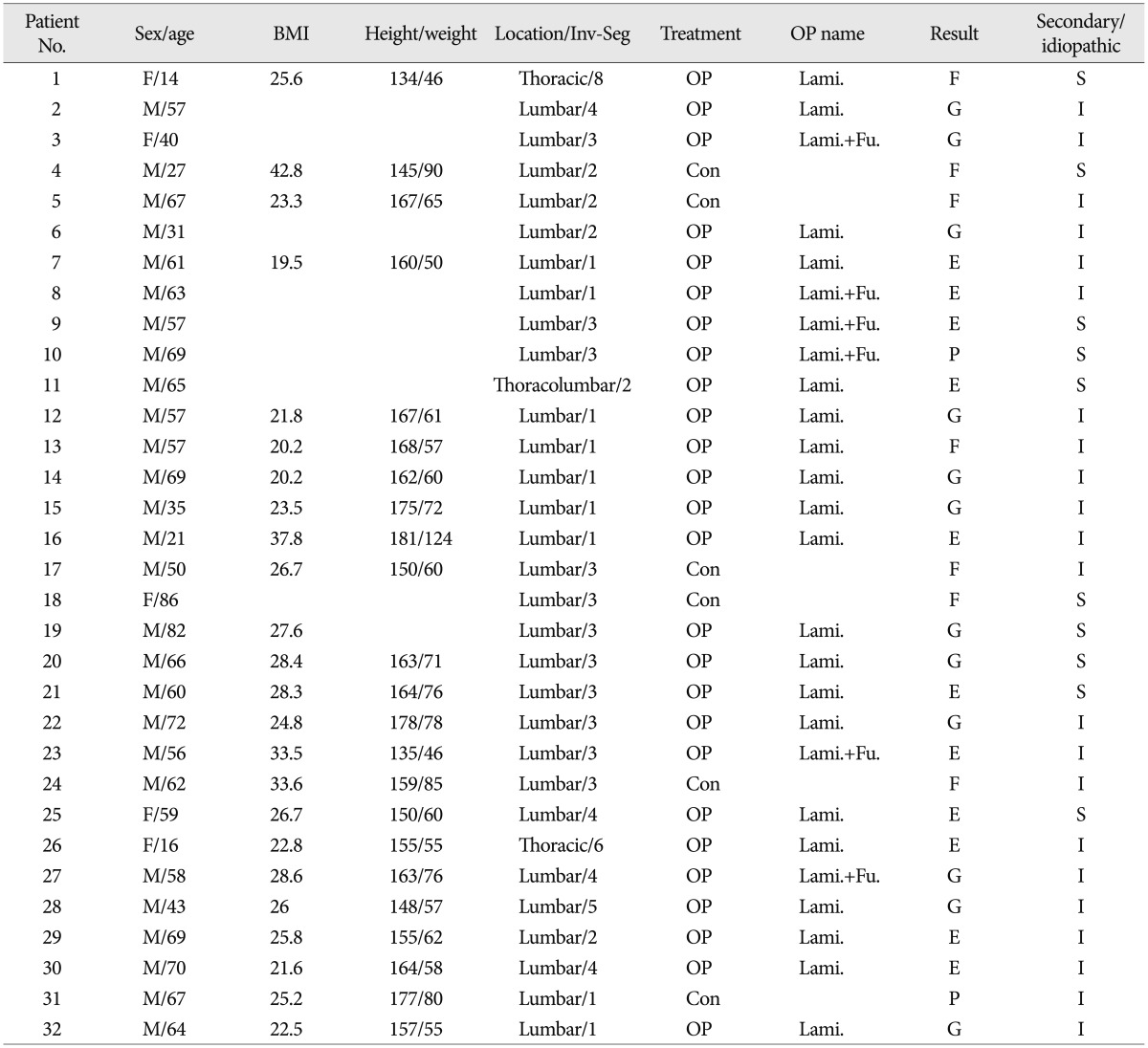
Medical Journal Search engines such as KoreaMed, KM base, and PubMed were utilized to find published articles of SEL associated with the Korean ethnicity. Kim et al.16) in 1986 first reported a case of SEL that a Korean patient with adrenocortical adenoma had developed. Up to now, 32 cases in 19 papers were found. These included 3 cases of this report. The SEL characteristics of Korean patients are shown in Table 1. Three (3) cases of this report and 29 already-published cases of Korean SEL patients were carefully examined and analyzed23). Idiopathic SEL is defined as a rare disorder that causes spinal cord compression and neurological deficits without any evidence of exogenous steroid administration, endogenous Cushing's syndrome or endocrinopathies.
Table 1.
Characteristics of Korean patients with spinal epidural lipomatosis

M : male, F : female, BMI : body mass index, Inv-Seg : involved segment, Con : conservative treatment, OP : operation, Lami. : laminectomy, Fu. : spinal fusion, E : excellent, G : good, F : fare, P : poor, S : secondary, I : idiopathic
Of those 32 cases, 22 were idiopathic and 10 were secondary SEL cases. The mean age was 55.3 years while the male-to-female ratio was 84.3 to 15.7. However, no significant difference between domestic and foreign cases was found. The mean BMI of domestic cases was 26.5, which was less than that of foreign cases. The idiopathic SEL group among Koreans took up 68.8%, which was significantly different from that of the f reign cases. The secondary SEL group of foreign cases took up 62% to 75%1,8,9,13).
In the analysis of inflicted area, only three of domestic cases involved the thoracic segment. Among those foreign cases, idiopathic SEL cases usually involved the lumbar segments while the secondary SEL group involved the thoracic spine. In our review, thoracic involvement was seen in only 3 cases. Two cases were secondary SEL and one case was idiopathic SEL. Surgical intervention was carried out in 23 cases (78.1%) and the result of surgery was excellent to good in every case except for one case of mortality which was related to medical complications3,4,5,6,7,11,12,14,15,16,17,18,19,21,22,24,25,27,28). Surgical results were similar between domestic and foreign cases. The causative factor of the racial difference was unidentified.
In summary, cases of Korean showed higher incidences of idiopathic SEL, for which the infliction (fat lesion) was manifested predominantly in the lumbar segments, and rarely developed in the thoracic segments, while Korean patients had a lower BMI, in comparison with that of western cases. The inquiry of these case studies is the first comparative analysis between Korean and western SEL cases. Nevertheless, the reason behind such racial differences has not been elucidated.
CONCLUSION
According to this study, there were differences between Korean and western cases. Koreans had higher incidences of idiopathic SEL, predominant involvement in the lumbar segments, very few thoracic involvement and lower BMI, as opposed to westerners. Owing to the lack of a large series of Korean SEL cases, direct comparisons with western reports may not be conclusive and further studies are needed in the days ahead including systemic analysis and multi-center study.
References
- 1.Al-Khawaja D, Seex K, Eslick GD. Spinal epidural lipomatosis--a brief review. J Clin Neurosci. 2008;15:1323–1326. doi: 10.1016/j.jocn.2008.03.001. [DOI] [PubMed] [Google Scholar]
- 2.Badami JP, Hinck VC. Symptomatic deposition of epidural fat in a morbidly obese woman. AJNR Am J Neuroradiol. 1982;3:664–665. [PMC free article] [PubMed] [Google Scholar]
- 3.Chang H, Bahk WJ, Lim KH. Lumbar epidural lipomatosis. J Korean Soc Spine Surg. 1996;3:256–251. [Google Scholar]
- 4.Choi KC, Kang BU, Lee CD, Lee SH. Rapid progression of spinal epidural lipomatosis. Eur Spine J. 2012;21(Suppl 4):S408–S412. doi: 10.1007/s00586-011-1855-x. [DOI] [PMC free article] [PubMed] [Google Scholar]
- 5.Choi SM, Choi G, Lee SH. Cauda equina syndrome due to spinal epidural lipomatosis. Korea J Spine. 2005;2:65–67. [Google Scholar]
- 6.Choy WS, Kim HJ, Kim KH, Ong SS, Park JH. Lumbar epidural lipomatosis. J Korean Soc Spine Surg. 1998;5:136–142. [Google Scholar]
- 7.Doo TH, Kim HJ, Shin DA. Lumbosacral epidural lipomatosis with compressive neuropathy. New Med J. 2006;49:43–47. [Google Scholar]
- 8.Fan CY, Wang ST, Liu CL, Chang MC, Chen TH. Idiopathic spinal epidural lipomatosis. J Chin Med Assoc. 2004;67:258–261. [PubMed] [Google Scholar]
- 9.Fogel GR, Cunningham PY, 3rd, Esses SI. Spinal epidural lipomatosis : case reports, literature review and meta-analysis. Spine J. 2005;5:202–211. doi: 10.1016/j.spinee.2004.05.252. [DOI] [PubMed] [Google Scholar]
- 10.Haddad SF, Hitchon PW, Godersky JC. Idiopathic and glucocorticoid-induced spinal epidural lipomatosis. J Neurosurg. 1991;74:38–42. doi: 10.3171/jns.1991.74.1.0038. [DOI] [PubMed] [Google Scholar]
- 11.Han YM, Ahn MS. Idiopathic spinal epidural lipomatosis. J Korean Neurosurg Soc. 2001;30:795–799. [Google Scholar]
- 12.Ju WI, Ahn JG, Ji C, Park SC, Cho KS, Park CK, et al. Spinal epidural lipomatosis. J Korean Neurosurg Soc. 1999;28:110–113. [Google Scholar]
- 13.Kawai M, Udaka F, Nishioka K, Houshimaru M, Koyama T, Kameyama M. A case of idiopathic spinal epidural lipomatosis presented with radicular pain caused by compression with enlarged veins surrounding nerve roots. Acta Neurol Scand. 2002;105:322–325. doi: 10.1034/j.1600-0404.2002.1c194.x. [DOI] [PubMed] [Google Scholar]
- 14.Kim DY, Kim JW, Jeong JH, Choi JH. Symptomatic spinal epidural lipomatosis secondary to obesity. Kosin Med J. 2008;23:111–113. [Google Scholar]
- 15.Kim HS, Han IH, Lee JH, Choi BK. Symptomatic spinal epidural lipomatosis induced by repeated epidural steroid injections. Korean J Spine. 2009;6:218–220. [Google Scholar]
- 16.Kim IW, Koo CH, Park JS, Baek SU, Lee SD, Lee KH, et al. Adrenal cortical adenoma associated with spinal epidural lipomatosis and paraplegia. J Korean Pediatr Soc. 1986;29:86–92. [Google Scholar]
- 17.Kim NG, Choi NC, Kwon OY, Jeon SC, Lim BH. A case of spinal epidural lipomatosis associated with phenytoin induced hypothyroidism and obesity. J Korean Neurol Assoc. 1997;15:670–676. [Google Scholar]
- 18.Kim SY. Spinal epidural lipomatosis : a case report. Korean J Pain. 2009;22:249–252. [Google Scholar]
- 19.Kim TW, Huh YS, Chi MP, Kim JO, Kim JC. Spinal epidural lipomatosis : report of four cases. J Korean Neurosurg Soc. 2000;29:1527–1532. [Google Scholar]
- 20.Lee M, Lekias J, Gubbay SS, Hurst PE. Spinal cord compression by extradural fat after renal transplantation. Med J Aust. 1975;1:201–203. doi: 10.5694/j.1326-5377.1975.tb111328.x. [DOI] [PubMed] [Google Scholar]
- 21.Lee SB, Park HK, Chang JC, Jin SY. Idiopathic thoracic epidural lipomatosis with chest pain. J Korean Neurosurg Soc. 2011;50:130–133. doi: 10.3340/jkns.2011.50.2.130. [DOI] [PMC free article] [PubMed] [Google Scholar]
- 22.Lee SS, Ha GH, Yon JH, Son JY, Hong KH, Shin DY. Epidural lipomatosis discovered during managing of lower back pain : a case report. Korean J Anesthesiol. 1998;35:381–384. [Google Scholar]
- 23.Min WK, Oh CW, Jeon IH, Kim SY, Park BC. Decompression of idiopathic symptomatic epidural lipomatosis of the lumbar spine. Joint Bone Spine. 2007;74:488–490. doi: 10.1016/j.jbspin.2006.11.021. [DOI] [PubMed] [Google Scholar]
- 24.Park GY, Cho JH, Lee SY. Lumbar spinal stenosis induced by epidural lipomatosis : a case report. J Korean Acad Rehabil Med. 2004;28:618–621. [Google Scholar]
- 25.Park SH, Rho SW, Sung KH. Idiopathic spinal epidural lipomatosis. Korean J Spine. 2006;3:99–101. [Google Scholar]
- 26.Robertson SC, Traynelis VC, Follett KA, Menezes AH. Idiopathic spinal epidural lipomatosis. Neurosurgery. 1997;41:68–74. doi: 10.1097/00006123-199707000-00015. discussion 74-75. [DOI] [PubMed] [Google Scholar]
- 27.Seong BY, Park CJ, Park SJ, Kim SW, Lee TG. Lumbar spinal epidural lipomatosis : two cases report. J Korean Soc Spine Surg. 1998;5:333–341. [Google Scholar]
- 28.Yim YB, Jo YJ, Kim DS, Jeong DS, Park KH, Song GS, et al. Idiopathic spinal epidural lipomatosis in a non-obese healthy man. J Korean Neurol Assoc. 1998;16:402–407. [Google Scholar]


