Abstract
Background:
Antioxidants from natural sources have a major role in reversing the effects of oxidative stress and promoting health, growth and productivity in animals.
Objective:
This study was undertaken to investigate the possible antioxidant activity and hepatoprotective effects of Phytocee™ Cool on carbon tetrachloride (CCl4) induced oxidative stress and liver damage in rats.
Materials and Methods:
Animals were pretreated with Phytocee™ Cool for 10 days and were challenged with CCl4 (1:1 v/v) in olive oil on the 10th day. After 24 h of CCl4 administration blood was collected and markers of hepatocellular damage aspartate aminotransferase (AST), alanine aminotransferase (ALT) were evaluated. Rats were sacrificed and oxidative stress in liver was estimated using malondialdehyde (MDA), reduced glutathione (GSH), superoxide dismutase (SOD) and catalase.
Results:
CCl4 caused a significant increase in serum AST, ALT, hepatic MDA and GSH levels, whereas the SOD and catalase activities were decreased. Phytocee™ Cool pretreatment attenuated the MDA, AST ALT levels and increased the activities of SOD and catalase.
Conclusion:
Phytocee™ Cool demonstrated antioxidant potential and hepatoprotective effects and plausibly be used in the amelioration of oxidative stress.
Keywords: Antioxidant, carbon tetrachloride, oxidative stress, Phytocee™ Cool
INTRODUCTION
Free radicals generation is an integral feature of normal cellular function or metabolism. The innate enzymatic and nonenzymatic antioxidant defense systems act against the free radicals generated and protect the organisms from radical toxicity.[1] However, despite the cellular antioxidant defense systems, endogenous or exogenous sources/stressors cause radical related damage due to the imbalance between free radicals and radical scavenging mechanisms. The imbalance so termed as oxidative stress has been implicated in development of various diseases. Although clinical manifestation of chronic pathologies related to oxidative stress in poultry is limited due to its short life span, it is considered as one of the potential causes that lead to deleterious effects on the cell structures, including lipids and membranes, proteins and DNA leading to poor performance and growth.[2,3] As the birds are exposed to stressful conditions such as high environmental temperatures, infections, immunization etc., oxidative stress results as an outcome, consequently resulting in reduced performance and productivity in broilers and layers.[4,5] Therefore, to alleviate the oxidative stress and improve the performance and productivity in poultry, supplementation of synthetic antioxidants has become a common practice.[6] Alternatively, to improve the growth performance of broiler chickens, productivity in layers, decrease stress challenges, addition of natural antioxidants to the feed may be the most welcome addition.
Numerous medicinal plants for decades have been investigated for their antioxidant potentials and the available literature provides evidence that herbs have gained considerable importance as natural antioxidants in improving the overall health aspects of the poultry industry.[6,7] The indigenous medicinal plants like Emblica officinalis, Ocimum sanctum and Withania somnifera have already been demonstrated for their antioxidant potentials.[8] E. officinalis (Euphorbiaceae) also known as Phyllanthus emblica, Amla or Indian gooseberry[9] is a rich source of tannoid principles emblicanin A, emblicanin B, punigluconin and pedunculagin, vitamin C and flavones. These phytochemical entities of amla are reported to have potential anti-oxidant activity.[10,11,12] O. sanctum (Labiatae) in Indian traditional systems of medicines has been used for adaptogenic/antistress, antioxidant and immune stimulating activities.[13] Furthermore, O. sanctum was found to be effective in the management of general stress symptoms in a clinical trial probably due to its antioxidant, antistress and adaptogenic activity.[14]
Withania somnifera popularly known as Ashwaganda is a perennial plant belonging to the family Solanaceae and has been widely reported for its antioxidant activity.[10,15] Based on the above considerations, a unique polyherbal preparation, Phytocee™ Cool intended for poultry containing E. officinalis, O. sanctum and W. somnifera has been formulated and in addition electrolytes were added. As there is no scientific evidence existing for the antioxidant activity of Phytocee™ Cool, this study was undertaken to evaluate its antioxidant potential in rats.
MATERIALS AND METHODS
Animals
Male Wistar rats (150-200 g), bred and reared at central animal facility, Natural Remedies (Bangalore, India) were used in this study. The animals were housed under standard conditions of illumination cycle set to 12 h light and 12 h dark, at a temperature of 20-24° C and 30-70% relative humidity. Standard pelleted rodent feed (M/s. Amrut Laboratory Animal Feeds, Maharashtra, India) and ultraviolet purified and filtered water was provided ad libitum.
Drugs and chemicals
Carbon tetrachloride (CCl4) (Rankem Fine Chemicals., India), vitamin C purified/ascorbic acid (Merck Specialties, India) and Refined olive oil (SOS Cuetara, S. A, Spain) were obtained. Other chemicals used were 2-thiobarbituric acid (TBA) and 5,5’- Dithio bis 2-nitro-benzoic acid (Sigma-Aldrich Co., USA), pyrogallol and potassium dihydrogen orthophosphate (Qualigens Fine Chemicals, India), tris buffer, hydrogen peroxide (H2O2) solution 30%, di-sodium hydrogen orthophosphate and trichloro acetic acid, (Ranbaxy fine chemicals, India). All other chemicals and reagents used were of analytical grade.
Plant materials
Phytocee™ Cool is a novel polyherbal formulation containing E. offcinalis fruits, O. sanctum whole plant and W. somnifera roots and electrolytes.
Experimental groups and protocol
Male Wistar rats were randomly allocated to six groups, each containing 5-6 animals. Normal control (Group I) and negative/CCl4 control (Group II) rats received vehicle (demineralized water 10 ml/kg) orally for 10 days whereas, Groups III to VI received vitamin C (20 mg/kg), or Phytocee™ Cool at three dose levels of 50, 100, 200 mg/kg respectively for 10 days by oral administration. On the 10th day, rats from Group II–VI were challenged with CCl4 (1:1 v/v in olive oil) orally to induce hepatotoxicity.[16] The animals were anesthetized 24 h after CCl4 administration, blood was collected and the serum was separated. Subsequently, animals were euthanized; liver was dissected out, blotted and processed for the biochemical estimations.
Estimation of marker enzymes and antioxidant indices
Malondialdehyde (MDA) levels in liver homogenates were estimated as per the method described by Knight et al.[17] The colored MDA-TBA adduct formed as a result of reaction of lipid peroxidation product, MDA with TBA was quantified spectrophotometrically at 532 nm and was considered as an index of lipid peroxidation. The results were expressed as nmol of MDA/g tissue using molar extinction coefficient of the chromophore (1.56 × 10⁵/M-1/cm-1).
Serum alanine aminotransferase (ALT), aspartate aminotransferase (AST) activities were determined by Reitman and Frankel[18] method. Measurement is based on the detection of the increase in absorbance due to the formation of 2,4-dinitrophenyl-hydrazones in the reaction. Superoxide dismutase (SOD) activity was estimated by the method of Marklund and Marklund[19] in the liver homogenates. This method employs the superoxide-driven auto-oxidation of pyrogallol. One unit of SOD activity was defined as the amount of the enzyme, which inhibits the pyrogallol autoxidation by 50% and results were normalized on the basis of total protein content to express the activity as SOD units/mg protein.
The method described by Aebi[20] was followed for the estimation of catalase activity in the liver homogenate. The rate of decomposition of H2O2 is proportional to the decrease in absorbance at 240 nm. The difference in absorbance per unit time was considered as a measure of catalase activity and was expressed as catalase units/mg protein.
Reduced glutathione (GSH) was estimated by using Ellman's reagent following Sedlak and Lindsay[21] method. The measurement of SH groups was based on the formation of yellow color product resulting from the reaction of 5,5'-dithiobis (2-nitrobenzoic acid) (DTNB) and GSH. The yellow color formed was measured spectrophotometrically at 412 nm and was expressed in terms of μmoles/g tissue using the molar extinction coefficient of DTNB-GSH conjugate (13.6 × 10³/M-1/cm-1).
Statistical analysis
Data were expressed as mean and standard error of the mean and were analyzed using one-way ANOVA followed by Bonferroni method as post-hoc test. In case of heterogeneous data after transformation, Dunnett T3 method was used. Statistical significance was set at P ≤ 0.05.
RESULTS
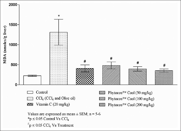
The hepatic levels of MDA were increased significantly (P ≤ 0.05) in CCl4 treated animals when compared to the normal control, while pretreatment with Phytocee™ Cool significantly decreased the CCl4 induced increase in hepatic MDA levels in a dose dependent manner [Figure 1].
Figure 1.

Effect of Phytocee™ Cool on carbon tetrachloride induced lipid peroxidation in rat liver
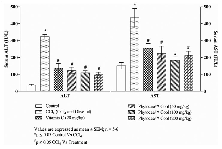
The mean serum ALT and AST levels, markers for hepatic tissue damage are depicted in Figure 2. Administration of CCl4 to rats significantly (P ≤ 0.05) increased the activity of serum hepatic marker enzymes when compared to normal control, whereas serum ALT and AST levels decreased significantly (P ≤ 0.05) in groups pretreated with Phytocee™ Cool when compared to CCl4 group.
Figure 2.

Effect of Phytocee™ Cool on carbon tetrachloride induced rise in serum alanine aminotransferase and aspartate aminotransferase in rats
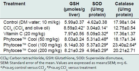
The free radical induced hepatotoxicity of CCl4 showed a significant decrease in SOD levels, nonsignificant decrease in catalase levels and significant increase in the GSH levels when compared to normal control group. However, Phytocee™ Cool pretreatment significantly increased SOD activity at all dose levels and catalase activity at a dose of 100 mg/kg, conversely hepatic levels of GSH were decreased as compared to CCl4 treated group [Table 1].
Table 1.
Effect of Phytocee™ Cool on CCl4 induced changes in reduced GSH, SOD and catalase of rat liver

DISCUSSION
Stress is of major concern for poultry production systems, as birds are exposed routinely to an array of stressors such as immunization, high and low environmental temperatures, preslaughter holding etc., As a consequence of stress, release of oxidants, imbalance between oxidants and in vivo antioxidants occurs at the cellular level leading to modification of cellular macromolecules, cell death by apoptosis or necrosis and structural tissue damage. Eventually the cellular level damage leads to reduced performance, health as well as serious economic losses. Control of oxidative stress related damage has led to a substantial increase in the use of antioxidant feed supplements in poultry diet[22,23,24] since high antioxidant status is observed as one of the most important factors positively affecting bird's performance in the poultry industry.[25,26] With the growing interest in natural feed supplements, poultry farming has been seeking sources of natural antioxidants. Considering the aforementioned facts, the present study investigated the antioxidant potential of a unique polyherbal formulation Phytocee™ Cool in rats.
In this study, oxidative stress and liver injury was induced in rats by the administration of CCl4 which is one of the well-recognized and widely used animal models to investigate the antioxidant and hepatoprotective effects of substances.[27,28,29] CCl4 induced oxidative stress is dependent on the bioactivation of CCl4 to trichloromethyl radical (CCl3) and trichloromethylperoxyl radicals. The initial step of biotransformation is the cytochrome P450 dependent reductive dechlorination of CCl4 to CCl3 that may form adducts with lipids and proteins or is trapped by molecular oxygen to form trichloromethylperoxyl radical (CCl3OO).[30,31] CCl3OO is more reactive and interact with lipids, causing lipid peroxidation along with the production of 4-hydroxyalkenals.[32,33] Earlier studies on natural products that showed protective effect against CCl4 induced hepatotoxicity attributed the protective mechanism to antioxidant property of natural products.[34,35] Hence, attenuation of CCl4 induced oxidative stress by Phytocee™ Cool directly connotes its antioxidant effect.
The active radical metabolic intermediates of CCl4 covalently bind to macromolecules such as lipids, produce hepatic lipid peroxidation. These free radicals attack polyunsaturated fatty acids (PUFA) to generate lipid peroxides, which are highly instable resulting in breakdown products such as reactive aldehydes (MDA), 4-hydroxyneonal or acrolein.[36] MDA, one of the end products of PUFA peroxidation is considered as a marker for lipid peroxidation. The level of MDA reflects extent of lipid peroxidation in hepatocytes.[37] In the present study, MDA levels in the liver homogenates of rats challenged with CCl4 were significantly increased. However, the increase in the MDA levels were significantly prevented in the rats pretreated with Phytocee™ Cool indicating the ability of Phytocee™ Cool to break the chain reaction of lipid peroxidation. Furthermore, the lipid peroxidation in CCl4 challenged rats lead to substantial increase in the serum ALT and AST concentrations. The increase in ALT and AST indicate considerable hepatocellular damage as these enzymes are normally localized in the cytoplasm and are released into the circulation after occurrence of cellular damage.[38,39] The serum enzyme levels in the groups pretreated with Phytocee™ Cool were significantly low indicating that Phytocee™ Cool was able to prevent the leakage of enzymes in the cytosol to circulation by protecting the hepatic cell membranes from CCl4 induced oxidative damage. Thus, CCl4 induced significant hepatocellular damage as evident from hepatic MDA levels coupled with marked elevation of AST and ALT enzymes, while Phytocee™ Cool pretreatment demonstrated substantial protection against oxidative stress and hepatocellular damage.
In order to further elucidate the antioxidant defense mechanisms of Phytocee™ Cool, hepatic enzymatic and nonenzymatic antioxidant defense mechanisms were evaluated. Enzymatic antioxidant defenses studied were SOD and catalase. SOD catalyzes conversion of superoxide to less toxic H2O2, while catalase converts the H2O2 into nontoxic H2O.[40] CCl4 induced generation of peroxy radicals leads to inactivation of SOD and catalase[41] a finding in corroboration with CCl4 challenged group in the present study. However, Phytocee™ Cool administration demonstrated significant increase in the activities of SOD and catalase indicating that it might have restored/activated the enzyme activities. These findings provide a supportive and convincing evidence for the antioxidant potential of Phytocee™ Cool.
Another antioxidant defense mechanism includes nonenzymatic antioxidants such as GSH. GSH in CCl4 administered rats increased and the mechanism underlying this increase is unknown, albeit such increase in hepatic GSH on CCl4 administration was also observed in several studies by Di Simplicio and Mannervik,[42] Nakagawa[43] and Lai et al.;[44] Nonetheless, Phytocee™ Cool modulated the GSH levels.
Antioxidants play a major role in delaying or inhibiting the oxidation of easily oxidizable macromolecules like lipids by protecting these molecules from actions of free radicals or reactive oxygen species[24] and accordingly lower plasma levels of antioxidants have been inversely correlated with the oxidative damage.[45,46] Phytocee™ Cool demonstrated its antioxidant activity by protecting the hepatic cells from CCl4 induced oxidative stress and by modulating the antioxidant defense systems. This protective effect of polyherbal formulation of Phytocee™ Cool can be attributed to the cumulative effect of its individual herbs, as the individual herbs (E. officinalis, O. sanctum and W. somnifera) are earlier reported for their antioxidant potential. Extracts of E. officinalis decreased the liver lipid peroxides in CCl4 treated rats and increased activities of SOD and catalase enzymes in vivo.[47,48,49] Another study revealed hepatoprotection by E. officinalis as evidenced by its ability to decrease AST and ALT levels in rats treated with ethanol.[50] Meanwhile, O. sanctum extract demonstrated significant decrease in LPO levels coupled with significant increase in SOD and catalase in liver homogenates of rats exposed to oxidative stress.[51,52] Further, earlier reports by Rajakumar and Rao (1993) have demonstrated that isoeugenol from O. sanctum with a double bond in its structure is a potent free radical scavenger.[53] Furthermore, flavonoids present in Ocimum extract protected the radiation induced cellular damage by scavenging the free radicals suggest strong evidence of antioxidant potential of Ocimum.[54] W. somnifera restored the LPO levels and enhanced the activities of catalase in stress induced rats. Moreover, Withania treatment significantly reduced the total barbituric acid reactive substances, AST and ALT when compared to ammonium chloride treated group.[55] Further, the phytoactives of W. somnifera showed antioxidant effects by modulating enzymatic and nonenzymatic defense systems.[56] The available literature on the individual ingredients of Phytocee™ Cool suggests their potent antioxidant activity and the present study also revealed similar antioxidant and protective effects that is indicative of the plausible cumulative effect of the herbs in the formulation.
Phytocee™ Cool contains several phytoactives viz., flavonoids, phenolic compounds, ascorbic acid, tannoid principles etc., Flavonoids, phenolic compounds and ascorbic acid are well-reported in earlier studies for scavenging/chelating free radicals and may be involved in chain breaking activity. Furthermore, the tannoid principles largely would have contributed for the rise in antioxidant levels.[10,49] These aforementioned mechanisms might plausibly be responsible for antioxidant activity of Phytocee™ Cool. Since, it is already established that ascorbic acid from natural sources is more potent antioxidant compared to synthetic vitamin C,[57,58] Phytocee™ Cool can be used as a better substitute for synthetic vitamin C. Overall, Phytocee™ Cool is a unique combination of herbs and electrolytes that may contribute to alleviate the heat stress in birds; although the effect of electrolytes on the heat induced stress is yet to be explored. Thus, Phytocee™ Cool revealed its antioxidant effects by ameliorating oxidative stress induced changes and by modulating the activities of antioxidant defense systems.
CONCLUSION
Our study demonstrated CCl4 induced marked oxidative stress was attenuated by Phytocee™ Cool. This protective effect of Phytocee™ Cool can be correlated to its potent antioxidant property.
Footnotes
Source of Support: Nil
Conflict of Interest: None declared
REFERENCES
- 1.Sies H. Oxidants and Antioxidants. New York: Academic Press; 1991. Oxidative Stress: pp. 2–8. [DOI] [PubMed] [Google Scholar]
- 2.Valko M, Leibfritz D, Moncol J, Cronin MT, Mazur M, Telser J. Free radicals and antioxidants in normal physiological functions and human disease. Int J Biochem Cell Biol. 2007;39:44–84. doi: 10.1016/j.biocel.2006.07.001. [DOI] [PubMed] [Google Scholar]
- 3.Christaki E. Naturally derived antioxidants in poultry nutrition. Res J Biotechnol. 2012;7:109–12. [Google Scholar]
- 4.Lykkesfeldt J, Svendsen O. Oxidants and antioxidants in disease: Oxidative stress in farm animals. Vet J. 2007;173:502–11. doi: 10.1016/j.tvjl.2006.06.005. [DOI] [PubMed] [Google Scholar]
- 5.Kucuk O, Sahin N, Sahin K, Gursu MF, Ozcelik M, Issi M. Egg production, egg quality and lipid peroxidation status in laying hens maintained at a low ambient temperature (6°C) and fed a vitamin C and vitamin E-supplemented diet. Vet Med (Praha) 2003;48:33–40. [Google Scholar]
- 6.Zhang GF, Yang ZB, Wang Y, Yang WR, Jiang SZ, Gai GS. Effects of ginger root (Zingiber officinale) processed to different particle sizes on growth performance, antioxidant status and serum metabolites of broiler chickens. Poult Sci. 2009;88:2159–66. doi: 10.3382/ps.2009-00165. [DOI] [PubMed] [Google Scholar]
- 7.Kamboh AA, Zhu WY. Effect of increasing levels of bioflavonoids in broiler feed on plasma anti-oxidative potential, lipid metabolites and fatty acid composition of meat. Poult Sci. 2013;92:454–61. doi: 10.3382/ps.2012-02584. [DOI] [PubMed] [Google Scholar]
- 8.Gupta VK, Surendra KS. Plants as natural antioxidants. Nat Prod Rad. 2006;5:326–34. [Google Scholar]
- 9.Khan KH. Roles of Emblica officinalis in medicine-A review. Bot Res Int. 2009;2:218–28. [Google Scholar]
- 10.Bhattacharya A, Ghosal S, Bhattacharya SK. Antioxidant activity of tannoid principles of Emblica officinalis (amla) in chronic stress induced changes in rat brain. Indian J Exp Biol. 2000;38:877–80. [PubMed] [Google Scholar]
- 11.Thangaraj R, Ayyappan SR, Manikandan P, Baskaran J. Antioxidant Property of Emblica officinalis during experimentally induced restrain stress in rats. J Health Sci. 2007;53:496–99. [Google Scholar]
- 12.Chatterjee A, Chatterjee S, Biswas A, Bhattacharya S, Chattopadhyay S, Bandyopadhyay SK. Gallic acid enriched fraction of Phyllanthus emblica potentiates indomethacin-induced gastric ulcer healing via e-NOS-dependent pathway. Evid Based Complement Alternat Med 2012. 2012:487380. doi: 10.1155/2012/487380. [DOI] [PMC free article] [PubMed] [Google Scholar]
- 13.Singh N, Verma P, Pandey BR, Bhalla M. Therapeutic potential of Ocimum sanctum in prevention and treatment of cancer and exposure to radiation: An overview. Int J Pharm Sci Drug Res. 2012;4:97–104. [Google Scholar]
- 14.Saxena RC, Singh R, Kumar P, Negi MP, Saxena VS, Geetharani P, et al. Efficacy of an Extract of Ocimum tenuiflorum (OciBest) in the management of general stress: A double-blind, placebo-controlled study. Evid Based Complement Alternat Med 2012. 2012:894509. doi: 10.1155/2012/894509. [DOI] [PMC free article] [PubMed] [Google Scholar]
- 15.Singh G, Sharma PK, Dudhe R, Singh S. Biological activities of Withania somnifera. Ann Biol Res. 2010;1:56–63. [Google Scholar]
- 16.Tasaduq SA, Singh K, Sethi S, Sharma SC, Bedi KL, Singh J, et al. Hepatocurative and antioxidant profile of HP-1, a polyherbal phytomedicine. Hum Exp Toxicol. 2003;22:639–45. doi: 10.1191/0960327103ht406oa. [DOI] [PubMed] [Google Scholar]
- 17.Knight JA, Pieper RK, McClellan L. Specificity of the thiobarbituric acid reaction: Its use in studies of lipid peroxidation. Clin Chem. 1988;34:2433–8. [PubMed] [Google Scholar]
- 18.Reitman S, Frankel S. A colorimetric method for the determination of serum glutamic oxalacetic and glutamic pyruvic transaminases. Am J Clin Pathol. 1957;28:56–63. doi: 10.1093/ajcp/28.1.56. [DOI] [PubMed] [Google Scholar]
- 19.Marklund S, Marklund G. Involvement of the superoxide anion radical in the autoxidation of pyrogallol and a convenient assay for superoxide dismutase. Eur J Biochem. 1974;47:469–74. doi: 10.1111/j.1432-1033.1974.tb03714.x. [DOI] [PubMed] [Google Scholar]
- 20.Aebi HE. Catalase in vitro. Methods Enzymol. 1983;105:121–6. doi: 10.1016/s0076-6879(84)05016-3. [DOI] [PubMed] [Google Scholar]
- 21.Sedlak J, Lindsay RH. Estimation of total, protein-bound and nonprotein sulfhydryl groups in tissue with Ellman's reagent. Anal Biochem. 1968;25:192–205. doi: 10.1016/0003-2697(68)90092-4. [DOI] [PubMed] [Google Scholar]
- 22.Mujahid A, Yoshiki Y, Akiba Y, Toyomizu M. Superoxide radical production in chicken skeletal muscle induced by acute heat stress. Poult Sci. 2005;84:307–14. doi: 10.1093/ps/84.2.307. [DOI] [PubMed] [Google Scholar]
- 23.Voljc M, Frankic T, Levart A, Nemec M, Salobir J. Evaluation of different vitamin E recommendations and bioactivity of α-tocopherol isomers in broiler nutrition by measuring oxidative stress in vivo and the oxidative stability of meat. Poult Sci. 2011;90:1478–88. doi: 10.3382/ps.2010-01223. [DOI] [PubMed] [Google Scholar]
- 24.Ramnath V, Rekha PS, Sujatha KS. Amelioration of heat stress induced disturbances of antioxidant defense system in chicken by brahma rasayana. Evid Based Complement Alternat Med. 2008;5:77–84. doi: 10.1093/ecam/nel116. [DOI] [PMC free article] [PubMed] [Google Scholar]
- 25.Lin H, Decuypere E, Buyse J. Acute heat stress induces oxidative stress in broiler chickens. Comp Biochem Physiol A Mol Integr Physiol. 2006;144:11–7. doi: 10.1016/j.cbpa.2006.01.032. [DOI] [PubMed] [Google Scholar]
- 26.Mujahid A, Akiba Y, Toyomizu M. Acute heat stress induces oxidative stress and decreases adaptation in young white leghorn cockerels by downregulation of avian uncoupling protein. Poult Sci. 2007;86:364–71. doi: 10.1093/ps/86.2.364. [DOI] [PubMed] [Google Scholar]
- 27.Huang Q, Zhang S, Zheng L, He M, Huang R, Lin X. Hepatoprotective effects of total saponins isolated from Taraphochlamys affinis against carbon tetrachloride induced liver injury in rats. Food Chem Toxicol. 2012;50:713–8. doi: 10.1016/j.fct.2011.12.009. [DOI] [PubMed] [Google Scholar]
- 28.Khan RA. Protective effects of Sonchus asper (L.) Hill, (Asteraceae) against CCl4-induced oxidative stress in the thyroid tissue of rats. BMC Complement Altern Med. 2012;12:181. doi: 10.1186/1472-6882-12-181. [DOI] [PMC free article] [PubMed] [Google Scholar]
- 29.Ebaid H, Bashandy SA, Alhazza IM, Rady A, El-Shehry S. Folic acid and melatonin ameliorate carbon tetrachloride-induced hepatic injury, oxidative stress and inflammation in rats. Nutr Metab (Lond) 2013;10:20. doi: 10.1186/1743-7075-10-20. [DOI] [PMC free article] [PubMed] [Google Scholar]
- 30.Fouw JD. Geneva: World Health Organization; 1999. Environmental Health Criteria 208, Carbon Tetrachloride; pp. 43–5. [Google Scholar]
- 31.Pohl LR, Schulick RD, Highet RJ, George JW. Reductive-oxygenation mechanism of metabolism of carbon tetrachloride to phosgene by cytochrome P-450. Mol Pharmacol. 1984;25:318–21. [PubMed] [Google Scholar]
- 32.Benedetti A, Fulceri R, Ferrali M, Ciccoli L, Esterbauer H, Comporti M. Detection of carbonyl functions in phospholipids of liver microsomes in CCl4- and BrCCl3-poisoned rats. Biochim Biophys Acta. 1982;712:628–38. doi: 10.1016/0005-2760(82)90292-2. [DOI] [PubMed] [Google Scholar]
- 33.Comporti M, Benedetti A, Ferrali M, Fulceri R. Reactive aldehydes (4-hydroxyalkenals) originating from the peroxidation of liver microsomal lipids: biological effects and evidence for their binding to microsomal protein in CCl 4 or BrCCl 3 intoxication. Front Gastrointest Res. 1984;8:46–62. [Google Scholar]
- 34.Jeong TC, Kim HJ, Park JI, Ha CS, Park JD, Kim SI, et al. Protective effects of red ginseng saponins against carbon tetrachloride-induced hepatotoxicity in Sprague Dawley rats. Planta Med. 1997;63:136–40. doi: 10.1055/s-2006-957630. [DOI] [PubMed] [Google Scholar]
- 35.Hsiao G, Shen MY, Lin KH, Lan MH, Wu LY, Chou DS, et al. Antioxidative and hepatoprotective effects of Antrodia camphorata extract. J Agric Food Chem. 2003;51:3302–8. doi: 10.1021/jf021159t. [DOI] [PubMed] [Google Scholar]
- 36.Weber LW, Boll M, Stampfl A. Hepatotoxicity and mechanism of action of haloalkanes: Carbon tetrachloride as a toxicological model. Crit Rev Toxicol. 2003;33:105–36. doi: 10.1080/713611034. [DOI] [PubMed] [Google Scholar]
- 37.Niki E. Lipid peroxidation: Physiological levels and dual biological effects. Free Radic Biol Med. 2009;47:469–84. doi: 10.1016/j.freeradbiomed.2009.05.032. [DOI] [PubMed] [Google Scholar]
- 38.Recknagel RO, Glende EA, Jr, Dolak JA, Waller RL. Mechanisms of carbon tetrachloride toxicity. Pharmacol Ther. 1989;43:139–54. doi: 10.1016/0163-7258(89)90050-8. [DOI] [PubMed] [Google Scholar]
- 39.Cordero-Pérez P, Torres-González L, Aguirre-Garza M, Camara-Lemarroy C, Guzmán-de la Garza F, Alarcón-Galván G, et al. Hepatoprotective effect of commercial herbal extracts on carbon tetrachloride-induced liver damage in Wistar rats. Pharmacognosy Res. 2013;5:150–6. doi: 10.4103/0974-8490.112417. [DOI] [PMC free article] [PubMed] [Google Scholar]
- 40.Celi P. The role of oxidative stress in small ruminants health and production. R Bras Zootec. 2010;39:348–63. [Google Scholar]
- 41.Tirkey N, Pilkhwal S, Kuhad A, Chopra K. Hesperidin, a citrus bioflavonoid, decreases the oxidative stress produced by carbon tetrachloride in rat liver and kidney. BMC Pharmacol. 2005;5:2. doi: 10.1186/1471-2210-5-2. [DOI] [PMC free article] [PubMed] [Google Scholar]
- 42.Di Simplicio P, Mannervik B. Enzymes involved in glutathione metabolism in rat liver and blood after carbon tetrachloride intoxication. Toxicol Lett. 1983;18:285–9. doi: 10.1016/0378-4274(83)90108-x. [DOI] [PubMed] [Google Scholar]
- 43.Nakagawa K. Carbon tetrachloride-induced alterations in hepatic glutathione and ascorbic acid contents in mice fed a diet containing ascorbate esters. Arch Toxicol. 1993;67:686–90. doi: 10.1007/BF01973692. [DOI] [PubMed] [Google Scholar]
- 44.Lai TY, Weng YJ, Kuo WW, Chen LM, Chung YT, Lin YM, et al. Taohe Chengqi Tang ameliorates acute liver injury induced by carbon tetrachloride in rats. Zhong Xi Yi Jie He Xue Bao. 2010;8:49–55. doi: 10.3736/jcim20100110. [DOI] [PubMed] [Google Scholar]
- 45.Cheng TK, Coon CN, Hamre ML. Effect of environmental stress on the ascorbic acid requirement of laying hens. Poult Sci. 1990;69:774–80. doi: 10.3382/ps.0690774. [DOI] [PubMed] [Google Scholar]
- 46.Sahin K, Sahin N, Yaralioglu S. Effects of vitamin C and vitamin E on lipid peroxidation, blood serum metabolites and mineral concentrations of laying hens reared at high ambient temperature. Biol Trace Elem Res. 2002;85:35–45. doi: 10.1385/BTER:85:1:35. [DOI] [PubMed] [Google Scholar]
- 47.Jose JK, Kuttan R. Hepatoprotective activity of Emblica officinalis and Chyavanaprash. J Ethnopharmacol. 2000;72:135–40. doi: 10.1016/s0378-8741(00)00219-1. [DOI] [PubMed] [Google Scholar]
- 48.Panda S, Kar A. Fruit extract of Emblica officinalis ameliorates hyperthyroidism and hepatic lipid peroxidation in mice. Pharmazie. 2003;58:753–5. [PubMed] [Google Scholar]
- 49.Hazra B, Sarkar R, Biswas S, Mandal N. Comparative study of the antioxidant and reactive oxygen species scavenging properties in the extracts of the fruits of Terminalia chebula, Terminalia belerica and Emblica officinalis. BMC Complement Altern Med. 2010;10:20. doi: 10.1186/1472-6882-10-20. [DOI] [PMC free article] [PubMed] [Google Scholar]
- 50.Kumar A. A review on hepatoprotective herbal drugs. Int J Res Pharm Chem. 2012;2:92–102. [Google Scholar]
- 51.Ramesh B, Satakopan VN. Antioxidant activities of hydroalcoholic extract of Ocimum sanctum against cadmium induced toxicity in rats. Indian J Clin Biochem. 2010;25:307–10. doi: 10.1007/s12291-010-0039-5. [DOI] [PMC free article] [PubMed] [Google Scholar]
- 52.Balanehru S, Nagarajan B. Intervention of adriamycin induced free radical damage. Biochem Int. 1992;28:735–44. [PubMed] [Google Scholar]
- 53.Rajakumar DV, Rao MN. Dehydrozingerone and isoeugenol as inhibitors of lipid peroxidation and as free radical scavengers. Biochem Pharmacol. 1993;46:2067–72. doi: 10.1016/0006-2952(93)90649-h. [DOI] [PubMed] [Google Scholar]
- 54.Uma Devi P. Radioprotective, anticarcinogenic and antioxidant properties of the Indian holy basil, Ocimum sanctum (Tulasi) Indian J Exp Biol. 2001;39:185–90. [PubMed] [Google Scholar]
- 55.Harikrishnan B, Subramanian P, Subash S. Effect of Withania Somnifera root powder on the levels of circulatory lipid peroxidation and liver marker enzymes in chronic hyperammonemia. J Chem. 2008;5:872–7. [Google Scholar]
- 56.Bhattacharya SK, Satyan KS, Ghosal S. Antioxidant activity of glycowithanolides from Withania somnifera. Indian J Exp Biol. 1997;35:236–9. [PubMed] [Google Scholar]
- 57.Vinson JA, Bose P. Comparative bioavailability of synthetic and natural vitamin C in guinea pigs. Nutr Rep Int. 1983;27:875–80. [Google Scholar]
- 58.Khopde SM, Indira KP, Mohan H, Gawandi VB, Satav JG, Yakhmi JV, et al. Characterizing the antioxidant activity of amla (Phyllanthus emblica) extract. Curr Sci. 2001;81:185–90. [Google Scholar]


