Abstract
Stress is an important risk factor for mood disorders. Stress also stimulates the secretion of glucocorticoids, which have been found to influence mood. To determine the role of forebrain glucocorticoid receptors (GR) in behavioral responses to chronic stress, the present experiments compared behavioral effects of repeated social defeat in mice with forebrain GR deletion and in floxed GR littermate controls. Repeated defeat produced alterations in forced swim and tail suspension immobility in floxed GR mice that did not occur in mice with forebrain GR deletion. Defeat-induced changes in immobility in floxed GR mice were prevented by chronic antidepressant treatment, indicating that these behaviors were dysphoria-related. In contrast, although mice with forebrain GR deletion exhibited antidepressant-induced decreases in tail suspension immobility in the absence of stress, this response did not occur in mice with forebrain GR deletion after defeat. There were no marked differences in plasma corticosterone between genotypes, suggesting that behavioral differences depended on forebrain GR rather than on abnormal glucocorticoid secretion. Defeat-induced gene expression of the neuronal activity marker c-fos in the ventral hippocampus, paraventricular thalamus and lateral septum correlated with genotype-related differences in behavioral effects of defeat, whereas c-fos induction in the nucleus accumbens and central and basolateral amygdala correlated with genotype-related differences in behavioral responses to antidepressant treatment. The dependence of both negative (dysphoria-related) and positive (antidepressant-induced) behaviors on forebrain GR is consistent with the contradictory effects of glucocorticoids on mood, and implicates these or other forebrain regions in these effects.
Keywords: glucocorticoid, depression, anxiety, antidepressant, defeat, stress
INTRODUCTION
A variety of studies have found that stress increases the risk of clinically significant emotional disorders (12, 23, 33). The mechanisms underlying the psychiatric risks of stress are largely undefined, although they have been linked, for example, to interaction with genetic factors such as serotonin transporter isoforms (12). However, genetic make-up is difficult to modify, indicating a need to explore other mechanisms by which stress can influence mental health.
Stress is also a major stimulus for glucocorticoid secretion by the hypothalamic-pituitary-adrenocortical (HPA) axis. Glucocorticoids have been found to have a variety of negative effects on mood ranging from anxiety and depression to psychosis, even in individuals lacking a family or personal history of psychiatric disease. Such effects have been correlated with elevated glucocorticoid levels in Cushing’s syndrome and in patients receiving glucocorticoids for immunologic disorders (10, 27). Glucocorticoids are also often elevated in depression, a finding that has been attributed to deficits in glucocorticoid receptors responsible for HPA feedback inhibition (22). Although the significance of the increase in glucocorticoids remains a matter of debate, there is limited but intriguing evidence to suggest that glucocorticoids can contribute to depression symptoms (44, 63). Consistent with the clinical literature, glucocorticoids have also been shown to have depression- and anxiety-like effects in rodents (36, 50, 53, 57, 60). However, glucocorticoids have also been found to have mood-elevating effects (10, 27), and depression has also been suggested to be due to deficits in glucocorticoids receptors mediating positive mood (51). Glucocorticoids are therefore a logical and compelling connection between stress and affective disease risk.
Glucocorticoids can bind in brain to either the higher-affinity mineralocorticoid receptor (MR) or the lower affinity glucocorticoid receptor (GR). GR is widely expressed throughout the brain and thought to mediate the effects of elevated levels of glucocorticoids, whereas MR is concentrated in but not limited to limbic structures (25) and thought to be more sensitive to low glucocorticoid levels (25). Although there is some evidence that MR can affect emotion-related behavior in rodents (32, 38, 46, 48, 64), depression- and anxiety-like symptoms are most frequently evoked in humans and animals at elevated levels of glucocorticoids (10, 27, 47), which are more likely to activate GR. Therefore, GR are the most plausible candidate to be involved in the mood effects of glucocorticoids.
GR in a variety of forebrain regions have been implicated in affective dysfunction (7, 11, 28, 30, 35, 51, 65). Mice with forebrain GR deletion have been created by transgenic expression of Cre recombinase under control of the calcium calmodulin kinase IIα (CamKIIα) promoter in floxed GR mice. The original model of forebrain GR deletion, derived on a mixed-strain background from the T50 founder line of the CamKIIα-Cre transgene, was reported to have a depression-like phenotype, consisting of increased depression-like behavior and elevated HPA activity relative to that in floxed GR mice (7). However, this depression-like phenotype was not found in mice with forebrain GR deletion derived on a pure C57BL/6 background from the T29-1 founder line of the CamKIIα-Cre transgene, even though forebrain GR deletion was at least as extensive (59). The latter mouse model, hereafter referred to as FBGRKO-T29-1 (59), offers a convenient model to test the role of forebrain GR in the behavioral effects of chronic stress, without potential confounds from baseline differences in HPA activity or depression-like behavior. The current experiments compared the effects of repeated social defeat stress or control cage exposures, with and without antidepressant treatment, on depression-related behavior in FBGRKO-T29-1 mice and their genetic controls, floxed GR mice. Although chronic stress elicited changes in behavior in floxed GR mice that were opposite to those conventionally interpreted as correlates of depression, these changes were associated with social aversion, a depression-like behavior (31), and were reversed by chronic antidepressant treatment, suggesting that behavioral alterations were dysphoria-related. FBGRKO-T29-1 mice failed to exhibit these stress-related changes in behavior and were resistant to the effects of antidepressant treatment during chronic stress, suggesting GR involvement in both negative (dysphoria-related) and positive (antidepressant-induced) effects on mood.
RESULTS
Experiment 1: Effects of forebrain GR deletion on behavioral and HPA responses to repeated social defeat
Social interaction with a novel conspecific has been used as a measure of depression-like behavior, with lower levels of interaction interpreted as greater depression-like behavior (31). Interaction with a novel mouse exhibited a significant main effect of defeat (F1,34= 8.852; P = 0.0054) but no significant main effects of genotype or genotype × defeat interaction. Defeated floxed GR mice exhibited significantly less time investigating the novel mouse than did control floxed GR mice (Table 1). There was a similar trend for defeated FBGRKO-T29-1 mice to exhibit less interaction with a novel mouse, but this effect was not significant at the post-hoc level (P = 0.097). There were no significant differences among genotypes or defeat groups in the amount of time mice spent interacting with an empty box as a neutral target (data not shown).
Table 1.
Results of the social interaction test in floxed GR and FBGRKO-T29-1mice on day 15 of control cage exposures (Control) or repeated social defeat (Defeat) from Experiment 1. Data are time (sec) mice spent interacting with a novel CD-1 male mouse that was confined in a perforated plastic box.
| Floxed GR | FBGRKO-T29-1 | ||
|---|---|---|---|
| Control | Defeat | Control | Defeat |
| 59 ± 15 N=8 |
24 ± 4* N=11 |
66 ± 19 N=8 |
35 ± 7 N=11 |
P < 0.05 vs. Control in the same genotype.
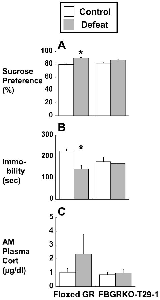
Sucrose preference, a measure of pleasure-seeking behavior (62), was similar between floxed GR and FBGRKO-T29-1 mice after control cage exposures. However, sucrose preference exhibited a significant main effect of defeat (F1,31 =19.758; P =0.0001), with defeated floxed GR but not defeated FBGRKO-T29-1 mice exhibiting significantly higher sucrose preference relative to their corresponding genotype controls (Figure 1A). There were no significant main effects of genotype or genotype × defeat interaction on sucrose preference.
Figure 1.
Effects of repeated social defeat on (A) sucrose preference, (B) forced swim immobility, and (C) basal morning plasma corticosterone in FBGRKO-T29-1 and floxed GR mice, respectively measured on days 21, 23, and 30 days of control cage exposures (Control; white bars) or repeated social defeat (Defeat; gray bars) in Experiment 1. N= 8 (control FBGRKO-T29-1 and floxed GR mice) and 11 (defeated FBGRKO-T29-1 and floxed GR mice) except for sucrose preference, in which some measurements were lost to spillage (N= 7 control FBGRKO-T29-1 and floxed GR mice, 10 defeated floxed GR mice).
*, P < 0.05 vs. Control in the same genotype.
Immobility in the forced swim test exhibited a significant main effect of defeat (F1,34 = 7.516; P = 0.0097) and a significant defeat × genotype interaction (F1,34 =5.246; P =0.0283), with no significant main effect of genotype alone. Defeated floxed GR mice displayed significantly lower levels of immobility in the forced swim test than did control floxed GR mice (Figure 1B). Control FBGRKO-T29-1 mice tended to be less immobile than were control floxed GR mice, but this difference was not significant (P > 0.1; Figure 1B). However, in contrast to floxed GR mice, FBGRKO-T29-1 mice did not exhibit any further decreases in immobility between control and defeated groups (Figure 1B).
Basal circadian nadir plasma corticosterone did not exhibit any significant main effects of defeat, genotype, or genotype × defeat interaction (Figure 1C).
Experiment 2: Effects of forebrain GR deletion on behavioral and HPA responses to antidepressant treatment during repeated social defeat
As in Experiment 1, there was a significant main effect of defeat in Experiment 2 to reduce social interaction with a novel mouse (F1,27= 5.360; P = 0.0284), but there were no significant effects of genotype, imipramine treatment or any interaction among defeat, genotype, and treatment on social interaction (Table 2). There were also no significant effects of genotype, defeat, or antidepressant treatment on interaction with the box alone (not shown) or on locomotor activity in the test arena (Table 3).
Table 2.
Results of the social interaction test in floxed GR and FBGRKO-T29-1mice on day 25 of saline or imipramine treatment during control cage exposures (Control) or repeated social defeat (Defeat) in Experiment 2. Data are time (sec) mice spent interacting with a novel CD-1 male mouse that was confined in a perforated plastic box.
| Floxed GR | FBGRKO-T29-1 | |||
|---|---|---|---|---|
| Saline | Imipramine | Saline | Imipramine | |
| Control | 35 ± 24 N=4 |
10 ± 6 N=3 |
43 ± 20 N=4 |
17 ± 4 N=4 |
| Defeat | 7 ± 2 N=4 |
10 ± 4 N=6 |
11 ± 8 N=5 |
3 ± 2 N=5 |
Table 3.
Locomotor activity in floxed GR and FBGRKO-T29-1 mice on day 25 of saline or imipramine during control cage exposures (Control) or repeated social defeat (Defeat) in Experiment 2. Data are the total distance traveled (cm) during a 2.5 min test session in a 35 × 43 cm basin.
| Floxed GR | FBGRKO-T29-1 | |||
|---|---|---|---|---|
| Saline | Imipramine | Saline | Imipramine | |
| Control | 367 ± 137 N=4 |
418 ± 158 N=3 |
528 ± 118 N=4 |
470 ± 71 N=4 |
| Defeat | 385 ± 73 N=4 |
421 ± 85 N=6 |
362 ± 43 N=5 |
372 ± 99 N=5 |
There were no significant main effects of genotype, defeat, or antidepressant treatment on sucrose preference after saline or imipramine treatment during repeated defeat or control cage exposures in Experiment 2 (Table 4).
Table 4.
Sucrose preference on day 21 of saline or imipramine treatment during control cage exposures (Control) or repeated social defeat (Defeat) in Experiment 2. Data indicate the percent of total fluid intake represented by consumption of 1% sucrose.
| Floxed GR | FBGRKO-T29-1 | |||
|---|---|---|---|---|
| Saline | Imipramine | Saline | Imipramine | |
| Control | 57 ± 7 N=4 |
51 ± 4 N=3 |
62 ± 5 N=4 |
52 ± 10 N=4 |
| Defeat | 67 ± 8 N=4 |
39 ± 9 N=6 |
46 ± 10 N=5 |
54 ± 9 N=5 |
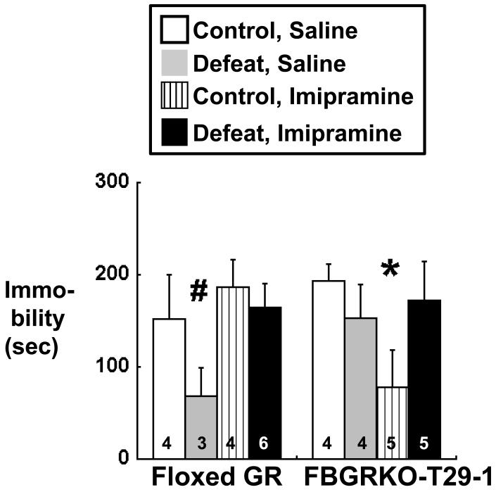
There was a significant genotype × treatment interaction on tail suspension immobility in Experiment 2 (F1,27= 4.835; P = 0.0366), although no other main effects or interactions were significant. Resembling the forced swim test results in Experiment 1, tail suspension immobility was reduced in floxed GR mice subjected to repeated defeat in Experiment 2 (Figure 2). Although chronic imipramine treatment did not affect tail suspension immobility of control floxed GR mice, imipramine treatment of defeated floxed GR mice restored their immobility to levels observed in saline-treated, control floxed GR mice (Figure 2). Also similar to the results of Experiment 1, saline-treated FBGRKO-T29-1 mice did not exhibit any changes in immobility as a result of repeated social defeat (Figure 2). Control FBGRKO-T29-1 mice did exhibit significant decreases in tail suspension immobility after chronic imipramine treatment, unlike control floxed GR mice (Figure 2). However, the lower levels of immobility observed after imipramine treatment in control FBGRKO-T29-1 mice did not occur in defeated FBGRKO-T29-1 mice treated with imipramine (Figure 2).
Figure 2.
Tail suspension immobility in floxed GR and FBGRKO-T29-1 mice from Experiment 2 on day 29 of saline or imipramine treatment during control cage exposures (Control; white or hatched bars) or repeated social defeat (Defeat; gray or black bars). Group Ns are indicated by the numbers within the data bars.
*, P < 0.05 vs. Control, Saline in the same genotype;
#, P < 0.05 vs. Defeat, Imipramine in the same genotype
Basal morning (circadian nadir) corticosterone was measured 2 d before the end of the experiment (day 28 imipramine treatment), and stress-induced corticosterone was measured in samples collected at death, 30 min after a social defeat in all groups. Basal corticosterone was not collected on the day of the final defeat in order to avoid stress effects from blood sampling on subsequent HPA responses to defeat. There were no significant effects of genotype, defeat, antidepressant treatment, or interaction among these factors on basal plasma corticosterone in Experiment 2 (Table 5). Although basal morning corticosterone appeared to be elevated in defeated floxed GR mice, this difference was not significant. There were also no significant main effects or interactions of genotype, defeat, or treatment on stress plasma corticosterone sampled 30 min after an acute social defeat in all mice (Table 6).
Table 5.
Circadian nadir (morning; AM) plasma corticosterone in floxed GR and FBGRKO-T29-1 mice in Experiment 2. Samples were collected by sub-mandibular venipuncture within 1 h of lights-on on day 28 of saline or imipramine treatment during control cage exposures (Control) or repeated social defeat (Defeat).
| AM Plasma Corticosterone (μg/dl) | ||||
|---|---|---|---|---|
| Floxed GR | FBGRKO-T29-1 | |||
| Saline | Imipramine | Saline | Imipramine | |
| Control | 0.8 ± 0.2 N=4 |
0.6 ± 0.1 N=3 |
1.0 ± 0.2 N=4 |
0.7 ± 0.1 N=4 |
| Defeat | 0.8 ± 0.3 N=4 |
2.6 ± 0.9 N=6 |
0.8 ± 0.1 N=5 |
0.9 ± 0.2 N=5 |
Table 6.
Stress-induced plasma corticosterone evoked by an acute social defeat in all floxed GR and FBGRKO-T29-1 mice in Experiment 2. Mice had been treated with saline or imipramine during the prior 29 days of control cage exposures (Control) or repeated social defeat (Defeat). Samples were collected by decapitation 30 min after the defeat. No injections were administered or basal corticosterone samples collected prior to the defeat sample in order to avoid confounding effects on subsequent responses to defeat.
| 30’ Post-defeat Plasma Corticosterone (μg/dl) | ||||
|---|---|---|---|---|
| Floxed GR | FBGRKO-T29-1 | |||
| Saline | Imipramine | Saline | Imipramine | |
| Control | 26.1 ± 3.4 N=4 |
28.4 ± 1.0 N=3 |
32.0 ± 4.5 N=4 |
26.0 ± 5.4 N=4 |
| Defeat | 23.1 ± 4.3 N=4 |
21.8 ± 3.7 N=6 |
25.6 ± 4.9 N=5 |
16.8 ± 4.3 N=5 |
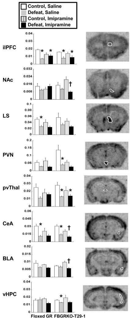
To localize GR-expressing forebrain regions potentially accounting for genotypic differences in behavioral responses to defeat or imipramine treatment, gene expression of the neuronal activity marker c-fos was analyzed by in situ hybridization in brains collected after subjecting both control and previously defeated mice in Experiment 2 to an acute defeat. Significant differences between control and repeatedly defeated mice that were present in one genotype but not the other were interpreted to indicate brain regions potentially involved in genotype-related differences in behavioral responses to defeat (saline-treated groups) or antidepressant treatment (imipramine-treated groups). In all brain regions studied, there was a significant main effect of repeated defeat to reduce levels of c-fos expression induced by a final defeat (Table 7 and Figure 3). Other ANOVA results are detailed below and in Table 7.
Table 7.
ANOVA results for c-fos gene expression data from Experiment 2. Residual degrees of freedom differ for some regions because damaged or missing sections occasionally precluded obtaining measurements from a given mouse. P values less than 0.05 are bolded for clarity.
| Region | Factor | Fdf | P |
|---|---|---|---|
| Infralimbic prefrontal cortex |
Genotype | 0.6841,25 | 0.4161 |
| Defeat | 28.2301,25 | <0.0001 | |
| Treatment | 0.6611,25 | 0.4238 | |
| Genotype × Defeat | 0.0221,25 | 0.8844 | |
| Genotype × Treatment | 0.00021,25 | 0.9892 | |
| Defeat × Treatment | 7.7551,25 | 0.0101 | |
| Genotype × Defeat × Treatment | 2.6791,25 | 0.1142 | |
| Nucleus accumbens shell |
Genotype | 1.2271,27 | 0.2779 |
| Defeat | 8.2831,27 | 0.0077 | |
| Treatment | 0.5511,27 | 0.4644 | |
| Genotype × Defeat | 5.6081, 27 | 0.0253 | |
| Genotype × Treatment | 0.1011,27 | 0.7535 | |
| Defeat × Treatment | 0.1491, 27 | 0.7023 | |
| Genotype × Defeat × Treatment | 5.4291,27 | 0.0275 | |
| Lateral septum | Genotype | 0.8531,27 | 0.3639 |
| Defeat | 16.6891,27 | 0.0004 | |
| Treatment | 2.1301,27 | 0.1560 | |
| Genotype × Defeat | 0.0121,27 | 0.9133 | |
| Genotype × Treatment | 0.0811,27 | 0.7779 | |
| Defeat × Treatment | 0.4841,27 | 0.4925 | |
| Genotype × Defeat × Treatment | 0.0071,27 | 0.9358 | |
| Paraventricular hypothalamus |
Genotype | 6.1081,26 | 0.0203 |
| Defeat | 32.0851,26 | <0.0001 | |
| Treatment | 6.3541,26 | 0.0182 | |
| Genotype × Defeat | 3.0151,26 | 0.0943 | |
| Genotype × Treatment | 8.4891,26 | 0.0073 | |
| Defeat × Treatment | 2.5861,26 | 0.1199 | |
| Genotype × Defeat × Treatment | 5.3611,26 | 0.0288 | |
| Paraventricular thalamus |
Genotype | 0.6031,27 | 0.4442 |
| Defeat | 16.3601,27 | 0.0004 | |
| Treatment | 0.9401,27 | 0.3408 | |
| Genotype × Defeat | 0.4371,27 | 0.5141 | |
| Genotype × Treatment | 0.2441,27 | 0.6250 | |
| Defeat × Treatment | 4.8011,27 | 0.0373 | |
| Genotype × Defeat × Treatment | 0.1481,27 | 0.7035 | |
| Central amygdala |
Genotype | 0.0031,27 | 0.9577 |
| Defeat | 44.9111,27 | <0.0001 | |
| Treatment | 6.1981,27 | 0.0192 | |
| Genotype × Defeat | 0.0031,27 | 0.9592 | |
| Genotype × Treatment | 0.2481,27 | 0.6224 | |
| Defeat × Treatment | 3.9551,27 | 0.0569 | |
| Genotype × Defeat × Treatment | 0.4691,27 | 0.4991 | |
| Basolateral amygdala |
Genotype | 0.00041, 27 | 0.9836 |
| Defeat | 12.5271,27 | 0.0015 | |
| Treatment | 0.0421,27 | 0.8399 | |
| Genotype × Defeat | 0.0021,27 | 0.9633 | |
| Genotype × Treatment | 0.8901,27 | 0.3540 | |
| Defeat × Treatment | 0.9991,27 | 0.3265 | |
| Genotype × Defeat × Treatment | 1.9841,27 | 0.1704 | |
| Ventral hippocampus |
Genotype | 0.0261, 27 | 0.8738 |
| Defeat | 7.9911,27 | 0.0087 | |
| Treatment | 0.0361,27 | 0.8518 | |
| Genotype × Defeat | 0.4661,27 | 0.5008 | |
| Genotype × Treatment | 1.2391,27 | 0.2755 | |
| Defeat × Treatment | 2.9691,27 | 0.0963 | |
| Genotype × Defeat × Treatment | 0.5231,27 | 0.4758 |
Figure 3.
Results of in situ hybridization analysis of c-fos gene expression in brains collected from floxed GR and FBGRKO-T29-1 mice 30 min after a social defeat in Experiment 2. Groups are the same as in Figure 2; mice had been treated with saline or imipramine for the last 29 d of a 60 d period of daily control cage exposures (Control) or social defeat (Defeat). Panels respectively depict semi-quantitative gray level readings taken from (top to bottom) the infralimbic prefrontal cortex (ilPFC), nucleus accumbens shell (NAc), lateral septum (LS), paraventricular hypothalamus (PVN), paraventricular thalamus (pvThal), central amygdala (CeA), basolateral amygdala (BLA), and ventral hippocampus (vHPC). Representative phosphorimager autoradiograms are shown to the right of each graph, with white outlines indicating the region analyzed. Ns are the same as in Figure 3 except for the PVN (N=4 for FBGRKO-T29-1 Control, Saline) and infralimbic PFC (N=4 for FBGRKO-T29-1 Defeat, Imipramine).
*, P < 0.05 vs. Control, Saline in the same genotype;
†, P < 0.05 vs. Control, Imipramine in the same genotype
In the infralimbic prefrontal cortex, where GR deletion has been shown to increase depression-like behavior (35), c-fos induction was significantly lower in saline-treated defeated mice vs. saline-treated controls in both genotypes (Figure 3, ilPFC). Imipramine did not significantly alter c-fos induction in the infralimbic prefrontal cortex in control or defeated mice of either genotype (Figure 3, ilPFC). There were no significant differences among any group in c-fos mRNA levels in the prelimbic prefrontal cortex (not shown).
In the shell of the nucleus accumbens, an area involved in motivated behavior and social interaction (1, 39), there was little difference in c-fos gene expression among groups of floxed GR mice (Figure 3, NAc). However, c-fos induction in FBGRKO-T29-1 mice was significantly lower in imipramine-treated defeated mice compared to imipramine-treated control mice (Figure 3, NAc). There was also a significant genotype × defeat × treatment interaction (Table 7) such that c-fos induction in the nucleus accumbens shell was marginally lower (P = 0.075) in imipramine-treated, defeated FBGRKO-T29-1 mice than in imipramine-treated, defeated floxed GR mice (Figure 3, NAc).
In the lateral septum, which has been implicated as a glucocorticoid target in mediating defeat-induced anxiety (11), the pattern of c-fos induction appeared to be similar between genotypes. However, significant differences occurred only in floxed GR mice, between saline-treated control mice and saline-treated defeated mice (Figure 3, LS).
The paraventricular hypothalamus was analyzed because it expresses most of the releasing factors responsible for hypothalamic-pituitary-adrenocortical axis activity (25). In the paraventricular hypothalamus, c-fos induction tended, although not significantly, to be higher in FBGRKO-T29-1 vs. floxed GR saline-treated control mice and was associated with a significant post-hoc difference between saline-treated control and saline-treated defeated FBGRKO-T29-1 mice (Figure 3, PVN). However, despite significant main effects of genotype and genotype × defeat × treatment interaction (Table 7), there were no significant differences in PVN c-fos expression between floxed GR and FBGRKO-T29-1 mice at the post-hoc level.
The posterior paraventricular thalamus, which is involved in habituation of responses to chronic stress (3, 4), did not exhibit significant differences in c-fos induction among groups of floxed GR mice (Figure 3, pvThal). However, FBGRKO-T29-1 mice treated with saline exhibited significantly lower levels of c-fos expression in defeated vs. control mice; c-fos expression in FBGRKO-T29-1 mice was not affected by imipramine treatment (Figure 3, pvThal).
The central and basolateral amygdala were investigated because of their role in fear, anxiety, and adaptation of responses to chronic stresses such as defeat (26, 28, 34, 40). In the central amygdala, both genotypes exhibited significantly lower levels of c-fos induction in saline-treated defeated mice vs. saline-treated controls (Figure 3, CeA). Imipramine treatment did not increase c-fos expression in defeated mice of either genotype. However, in FBGRKO-T29-1 mice, central amygdala c-fos expression was significantly lower in imipramine-treated defeated mice compared to that in imipramine-treated control mice (Figure 3, CeA). In the basolateral amygdala, c-fos expression was also significantly lower in imipramine-treated control vs. imipramine-treated defeated FBGRKO-T29-1 mice (Figure 3, BLA).
The CA1 field of the ventral hippocampus was analyzed because GR are prominently expressed in CA1 of floxed GR but not FBGRKO-T29-1 mice (7, 8, 59), and because the ventral hippocampus has limbic connections that are both distinct from the connections of the dorsal hippocampus and relevant to emotion-related behavior (16, 17, 34). Induction of c-fos gene expression in CA1 ventral hippocampus did not differ significantly among groups of floxed GR mice, although c-fos tended to be lower (P=0.054) in defeated vs. control floxed GR mice treated with imipramine (Figure 3, vHPC). In contrast, the ventral CA1 hippocampus of FBGRKO-T29-1 mice exhibited significantly lower c-fos expression in saline-treated defeated vs. control mice but not had similar levels of c-fos expression between imipramine-treated defeated and control mice (Figure 3, vHPC).
DISCUSSION
These experiments demonstrate that forebrain GR deletion can have state-specific effects on behavior. Forebrain GR deletion, at least in the FBGRKO-T29-1 mice used here (59), did not affect baseline behavior and attenuated behaviors, particularly the immobility response to inescapable situations, induced by the chronic stress of repeated defeat. Furthermore, although the current experiments confirm prior findings (7, 59) that forebrain GR deletion does not impair, and may even increase, behavioral sensitivity to antidepressants under baseline conditions, forebrain GR loss prevented antidepressant-induced changes in immobility behavior during chronic stress. The behavioral effects of forebrain GR deletion during chronic stress occurred in the absence of marked differences in glucocorticoid secretion between FBGRKO-T29-1 and floxed GR mice, suggesting that alterations in behavior were more likely to depend on differences in GR expression than on differences in HPA activity. Differential activation of GR-expressing forebrain regions after defeat, as assessed by c-fos gene expression, suggested that the ventral hippocampus, paraventricular thalamus and lateral septum might be involved in the behavioral resilience of FBGRKO-T29-1 mice to repeated social defeat, whereas the nucleus accumbens shell and central and basolateral amygdala might be involved in the resistance of FBGRKO-T29-1 mice to antidepressant treatment during chronic defeat stress.
Forebrain GR deletion was associated with fewer changes in behavior after chronic social defeat, even if these changes did not resemble conventional definitions of depression-like behavior (discussed below). Floxed GR but not FBGRKO-T29-1 mice exhibited increased activity in the forced swim and tail suspension tests after repeated defeat. Increased forced swim and tail suspension activity was not due to general hyperlocomotion, since overall activity was not increased. Although sucrose preference only exhibited significant effects of defeat in Experiment 1, even this behavioral change was present only in floxed GR and not in FBGRKO-T29-1 mice.
Antidepressant sensitivity was also influenced by forebrain GR. Imipramine had the expected action of an antidepressant to decrease immobility in the tail suspension test, but only in control FBGRKO-T29-1 mice. Although prior evidence of acute imipramine effects on immobility in unstressed floxed GR mice (59) appears to conflict with the lack of response to chronic imipramine treatment in control floxed GR mice, peak antidepressant levels after acute injection are likely to be higher than those at the time of testing in the present experiments, since testing occurred at least 16 h after the previous day’s injection. The greater sensitivity of unstressed FBGRKO-T29-1 mice to imipramine is consistent with the original report that mice with forebrain GR deletion respond with changes in immobility to imipramine doses that do not affect floxed GR mice (7). The sensitivity to imipramine in FBGRKO-T29-1 mice was abrogated by repeated social defeat, since imipramine-treated, defeated FBGRKO-T29-1 mice exhibited neither the decrease in immobility induced by imipramine in FBGRKO-T29-1 controls nor the increase in immobility elicited by imipramine in defeated floxed GR mice. Thus, immobility behavior in FBGRKO-T29-1 mice was responsive to antidepressants in unstressed but not chronically stressed conditions. These findings indicate that forebrain glucocorticoid receptors are involved not only in changes in immobility behavior evoked by chronic stress, but also in the response of immobility behavior to antidepressants during chronic stress. The lack of imipramine effects on social interaction or sucrose preference in the current experiments may have been due to administering antidepressant during rather than after defeat, as has been done in other studies (2, 5, 45).
There were no detectable differences in basal or stress-induced glucocorticoids between genotypes. Although the strong paraventricular hypothalamus c-fos induction in defeated FBGRKO-T29-1 mice suggested that greater HPA activity might be occurring, PVN c-fos induction is not a predictor of HPA activity (20) and did not differ between genotypes. While it cannot be excluded that more or different sampling points might have revealed divergent HPA activity between FBGRKO-T29-1 and floxed GR mice, prior measurements of adrenocorticotropin as well as corticosterone in FBGRKO-T29-1 mice have been consistently similar to those of floxed GR mice under a variety of conditions (59). Furthermore, forebrain GR have not been found to influence changes in HPA activity induced by chronic stress (19), and preliminary data in FBGRKO-T29-1 mice corroborate these findings (Jacobson, unpublished findings). Thus, it is most likely that forebrain GR loss, rather than differences in HPA activity or habituation, accounted for the behavioral differences between floxed GR and FBGRKO-T29-1 mice after defeat.
Mapping c-fos gene expression after exposing all mice to a social defeat suggested areas in which GR loss might modify behavioral responses to defeat stress or antidepressant treatment. Genotype-associated differences between saline-treated control and defeated mice, potentially indicating mechanisms for the lack of stress-induced changes in immobility behavior in FBGRKO-T29-1 mice, were found in the ventral hippocampus, the paraventricular thalamus, and the lateral septum. The paraventricular thalamus is involved in suppression of behavioral and HPA responses to chronic stress (3, 4), while the septum and ventral hippocampus are involved in anxiety-like behavior, an effect influenced by septal GR (11, 34). Thus, differential activity in these areas would be in accord with the relative lack of stress-induced changes in immobility behavior in FBGRKO-T29-1 mice. However, only the ventral hippocampus displayed differences in the overall pattern of c-fos induction between FBGRKO-T29-1 and floxed GR mice, with only FBGRKO-T29-1 mice exhibiting significant differences between saline-treated control and defeated groups. Therefore, ventral hippocampal activity may be most closely related to the resilience of FBGRKO-T29-1 mice to stress-induced abnormalities in immobility responses to inescapable situations. This interpretation is in keeping with evidence that the ventral hippocampus can regulate fear- and anxiety-related behavior (11, 17, 34, 49).
Genotype-related differences in defeat-induced c-fos expression between imipramine-treated control and imipramine-treated defeated mice, potentially indicating pathways underlying the antidepressant resistance of stress-induced immobility behavior in defeated FBGRKO-T29-1 mice, occurred in the nucleus accumbens shell, central amygdala, and basolateral amygdala. These regions have all been found to be activated by defeat (34, 40). The central and basolateral amygdala, as well as central amygdala GR, are involved in fear responses (26, 28). The more pronounced differences in basolateral and central amygdala c-fos expression between saline- and imipramine-treated FBGRKO-T29-1 mice after defeat could be connected to the impairment of imipramine responsiveness by fear-related experiences of defeat. However, the pattern of c-fos expression differed most strongly between genotypes in the nucleus accumbens shell. Gene expression of c-fos in the nucleus accumbens shell was essentially similar in all floxed GR groups but was lower in imipramine-treated FBGRKO-T29-1 defeated mice compared not only to imipramine-treated FBGRKO-T29-1 control mice, but also, marginally, to imipramine-treated, floxed GR defeated mice. Thus, the nucleus accumbens shell seems to be the best candidate of these three regions to account for the loss of imipramine effects on immobility in FBGRKO-T29-1 mice during chronic defeat stress. Involvement of the nucleus accumbens in GR-related effects of defeat is consistent with findings that dopamine release in the nucleus accumbens correlates with GR-dependent changes in defeat-induced behavior (1).
Limitations of this study include small group sizes in Experiment 2, phenotypic discrepancies between FBGRKO-T29-1 mice and the originally reported model of forebrain GR deletion, limited sampling times, and atypical effects of stress on behavior in floxed GR mice. Larger group sizes might have permitted confirmation in Experiment 2 of behavioral effects that were significant in Experiment 1, such as changes in sucrose preference, and might also have allowed more conclusive or extensive identification of brain regions exhibiting changes in c-fos expression related to stress or antidepressant response. Nevertheless, when behavioral alterations were significant in floxed GR mice, such changes were reliably missing in FBGRKO-T29-1 mice, and 6 of the 9 brain regions examined showed genotype-related differences in c-fos induction.
In these and previous experiments (59), FBGRKO-T29-1 mice did not exhibit the higher baseline immobility, lower sucrose preference, or elevated HPA activity originally reported for mixed-strain mice with forebrain GR deletion derived from the T50 CamKIIα-Cre founder line, but otherwise have GR deletion that is similar to that of the original model (7, 59). The reasons for the discrepancies in baseline behavior and endocrine function between the two lines are unclear, but may be related to differences in strain, founder, central amygdala GR loss, or mineralocorticoid receptor expression (59). Nevertheless, the current findings still indicate important influences of forebrain GR on emotion-related immobility behavior during chronic stress that are independent of, or more robust than, the effects of forebrain GR on baseline behavior.
The main effect of prior defeat to decrease defeat-induced c-fos expression in all brain regions was consistent with habituation of responses to repeated stress (29, 61). Since only one time point was studied after defeat or antidepressant treatment, it is possible that differences in c-fos or behavior between floxed GR and FBGRKO-T-29 mice are due to differential habituation to chronic stress, rather than to differences in inherent responses. This possibility seems unlikely because of the long period during which mice were stressed and because of the similar patterns in immobility behavior between control and defeated mice in Experiments 1 (30 d) and 2 (60 d), by which time habituation would be expected to be complete. Even if results are attributable to differences in habituation, these findings indicate an important impact of forebrain GR on the habituation process.
In floxed GR mice, the decreased immobility and, when observed, increased sucrose preference after repeated defeat appeared to conflict with the increases in forced swim immobility and decreases in sucrose preference that are often reported after chronic stress (5, 31, 62). Although there is no ready explanation for the contradictory direction of these stress-induced changes, there is precedent in the literature for the paradoxical behavioral changes observed in the current experiments. Chronic stress, including repeated social defeat, has been found by other investigators to decrease forced swim immobility or increase sucrose preference (6, 14, 15, 41, 45, 52, 58, 62). These atypical behavioral responses to stress, although less common, have been proposed to be part of a continuum of behavioral repertoires that is represented to varying degrees in all study populations and may account for inconsistent results in any given behavioral assay (62). There is also evidence that lower immobility in the forced swim or tail suspension test reflects an increase in anxiety-like behavior rather than a decrease in depression-like behavior (8, 56). The possibility that decreased immobility is a dysphoria-related behavior is supported by its consistent association with reductions in social interaction, an additional marker of depression– or anxiety-like behavior (2, 18, 31), and by findings, here and in other studies (37), that the decreased immobility is reversed by chronic antidepressant treatment.
The present experiments implicate forebrain GR in both the expression and the antidepressant reversal of chronic stress effects on dysphoria-related immobility. The dependence of both negative (stress-induced) and positive (antidepressant-induced) behaviors on forebrain glucocorticoid receptors is consistent with the evidence that glucocorticoids can have mood-elevating as well as depressive effects (10, 27). Defining the brain regions, cell types, and downstream targets mediating these contradictory effects of glucocorticoids will aid in reducing the risk of affective dysfunction from major life stress or immunosuppressive glucocorticoid therapy.
EXPERIMENTAL PROCEDURES
Animals
All animal use was approved by the Institutional Animal Care and Use Committee of Albany Medical College and was consistent with the standards of the National Institutes of Health Guide for the Care and Use of Laboratory Animals (21) and European Commission Directive 2010/63/EU. Mice were housed on a 12:12 light cycle (lights-on, 6:30 am) and had free access to food and water. Floxed GR mice were derived from C57BL/6 founders homozygous for a floxed exon 2 and were generously donated by Dr. Louis Muglia (University of Cincinnati; (9)). FBGRKO-T29-1 mice were bred on a pure C57 background from crosses between female floxed GR mice and male floxed GR mice hemizygous for the CamKIIα-Cre transgene (59). The latter were derived from commercially available, C57BL/6 CamKIIα-Cre transgenic mice (Jackson Laboratories stock number 005359, Bar Harbor, ME) from the T-29-1 founder line (54). Only male FBGRKO-T29-1 and floxed GR mice littermates were used for experiments and were 1.5-2.5 months old at the time of use.
Experiments
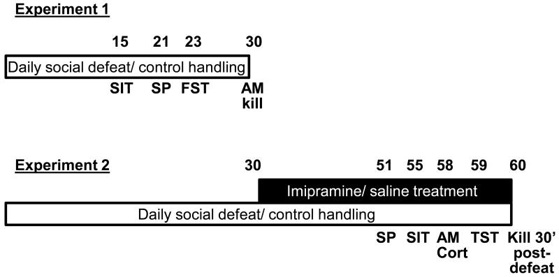
The relative timing of experimental manipulations and tests is diagrammed in Figure 4. In Experiment 1, mice were killed by decapitation in the basal state within 3h of lights-on after 30 days of defeat. The 30 day period of defeat was used to allow time to conduct tests of behaviors in addition to social defeat, changes in which were not observed until at least 21 days of defeat.
Figure 4.
Diagram of the design of Experiments 1 and 2 (top and bottom, respectively). Numbers above the bars indicate the days on which tests indicated below the bars were performed. SIT, social interaction test; SP, sucrose preference; FST, forced swim test; AM, morning; AM Cort, morning plasma corticosterone sample; TST, tail suspension test.
In Experiment 2, daily defeats were continued for another 29 d while mice were injected ip with either 30 mg/kg imipramine or saline. Injections were given once per day after each defeat or control cage exposure experience and after any behavioral testing or blood sampling was performed. Mice in Experiment 2 were killed by decapitation on d 60, 1 d after their last injection and 30 min after an acute social defeat in all control and defeat groups. The 30 min time point was previously shown to be appropriate for analysis of both HPA hormones and c-fos gene expression (6).
Social defeat was performed by placing a FBGRKO-T29-1 or floxed GR “intruder” mouse into the home cage of a singly-housed, CD-1 retired male breeder (Charles River, Wilmington, MA). Intruder mice were initially left in the resident’s cage for 5 min; however, to avoid injuries to the intruder, encounters were subsequently limited to the time the CD-1 resident first attacked and bit the intruder. Latency to the first attack was typically 30-60 sec, and all attacks elicited consistent submissive or fleeing behavior from all intruder mice, regardless of genotype. Only CD-1 males exhibiting attack latencies less than 1 min were used as resident aggressors. Intruders were returned to their home cage and housed individually after each daily attack. Preliminary experiments in which intruders were co-housed with CD-1 residents, separated by a clear, perforated partition, produced similar behavioral effects (data not shown). Intruders, designated as the “defeat” groups in the rest of text and figures, were rotated among different residents on a daily basis to minimize habituation of any behavioral interaction. Control mice of each genotype were placed for ~30 sec each day into a cage from which the resident male had been removed. Controls (designated as the “control” groups in the rest of text and figures) were also rotated among different cages each day.
Daily defeats and control cage exposures were always performed after any behavioral tests or blood sampling on the same day. Defeats and control cage exposures occurred at variable times in the light phase (31) between 1 and 10 h after lights-on, as dictated by the need to complete behavioral or hormonal testing beforehand and share access to the animal room with other users. However, defeats and control cage exposures were performed during the same 1.5 h time window on any given day for all mice in the experiment.
Behavioral tests
Behavioral tests were typically performed within the first 3 h of the light phase under quiet conditions. Social interaction tests required somewhat longer but were completed with 5 h of lights-on. All testing chambers were cleaned with MB-10 disinfectant (Quip Laboratories, Wilmington, DE) in between mice. Tests were performed on separate days in the order of least (sucrose preference or social interaction) to most stressful (forced swim or tail suspension), as diagrammed in Figure 4. Tests in Experiment 2 were timed to occur after at least 3 weeks of imipramine treatment; other differences in the sequence of tests between Experiments 1 and 2 resulted from the need to fit experimental procedures around caretaker schedules in the animal room.
Social interaction test
Social interaction was measured on day 15 of social defeat in Experiment 1 and on day 25 of imipramine treatment (55 d defeat) in Experiment 2 as a positive control for the effects of defeat to induce social aversion, a measure of depression-like behavior (2, 31). Mice were placed in a 35 × 43 cm basin with a clear, perforated box (6.5 × 8 × 12 cm) in the opposite corner. For the first 2.5 min of the trial, the box was empty, providing a neutral interaction target. During the second 2.5 min, an identical perforated box containing an unfamiliar CD-1 male mouse as a novel social target was placed in the corner, allowing mice to have olfactory, auditory, and visual contact without physical interaction. The time mice spent in the 10 cm interaction zone surrounding the box, without and with the unfamiliar mouse, was recorded with Ethovision 8.0 software (Noldus, Leesburg, VA). The total distance traveled in the trial with the empty box was also recorded as measure of overall locomotor activity.
Sucrose preference
Sucrose preference was tested on day 21 of social defeat in Experiment 1 and on day 21 of imipramine treatment (51d defeat) in Experiment 2. Mice were given access to two 50-ml tubes containing either water or 1% sucrose for 24 h before and 24 h after the specified day, with the position of the tubes switched to avoid position preference effects. Mice were not food-deprived during sucrose preference measurements. Sucrose preference was expressed as the percentage of sucrose vs. total fluid consumed.
Forced swim test
Immobility was scored on day 23 of social defeat in Experiment 1 in a 5-min forced swim that had been preceded by a 15-min pre-swim the day before. Mice were placed in a 1-liter beaker in 25 ± 1 °C water, and swimming behavior was recorded with Ethovision 8.0. Immobility was scored after blinding the genotype and treatment of the mice. The 2-day forced swim test was originated by Porsolt et al. in rats and remains a standard procedure for measuring depression-like behavior (13, 43). Although the test was shortened by Porsolt et al. to a single swim for mice (42), both paradigms give equivalent results (13). We chose to use the 2-day paradigm in mice because glucocorticoid levels at the time of the first swim have been shown to increase immobility in the test swim 24 h later in a GR-dependent manner (57).
Tail Suspension test
A tail suspension test was performed in Experiment 2 on day 29 of imipramine treatment (59 d defeat) as an additional test to confirm that genotype-related differences in immobility were consistent across different paradigms for measuring emotion-related activity (13). Mice were taped by their tail to a bar and allowed to hang upside-down for 8 min, during which time struggling behavior was recorded with Ethovision 8.0. The time mice hung completely motionless was scored from Ethovision videos by a trained observer blind to genotype and treatment groups. Manual scoring was necessary because the software did not distinguish active struggling from the passive swinging that can occur from the momentum of previous activity.
Plasma corticosterone assay
All samples were collected within 45 sec of touching the cage. In Experiment 1, morning (circadian nadir) plasma corticosterone samples were collected by decapitation within 3 h of lights-on, after 30 d of social defeat or control cage exposure. In Experiment 2, plasma corticosterone was measured once basally in the morning by submandibular venipuncture within 1 h of lights-on on day 28 of imipramine treatment (day 58 of defeat) and once two days later, within 4 h of lights-on, by decapitation 30 min after social defeat in all groups (day 60, after a total of 29 d imipramine treatment). Plasma corticosterone was assayed as previously described with a radioimmunoassay kit from MPBiomedical (Solon, OH), using all reagents and samples at half-volume (24).
In situ hybridization
In situ hybridization analysis of c-fos mRNA was performed as previously described (6). In brief, hybridization was performed at 60 °C in 10 μm sections of fresh-frozen brain using a 35S-cRNA probe complementary to the 1.5 kb Pst I fragment of the mouse c-fos cDNA (pGEMfos3; Dr. Michael Greenberg, Children’s Hospital, Boston, MA (55)). All other steps were as described in Bowens et al. (6).
Data analysis
In Situ Hybridization
Densitometric readings were collected by exposing slides to phosphorimager screens that were scanned at a 50 μm resolution (Typhoon 9210, GE Health Care, Niskayuna, NY) and analyzed with ImageQuant 5.0 software (GE Health Care, Niskayuna, NY). Each screen was exposed to slides from mice in every treatment group and to a set of identical 14C standards (146A and 146B, American Radiolabeled Chemicals, St. Louis, MO) that was used to normalize readings among screens. Densitometric readings from hand-drawn outlines of each brain region were corrected for background from a corresponding non-expressing area in the same section and scaled to the size of the largest outline for a given region. At least two readings were averaged for each region and mouse.
Statistics
Data were analyzed by 2- and 3- way ANOVA for Experiments 1 and 2, respectively. Post-hoc comparisons where ANOVA main effects or interactions were significant were made by t-test with Bonferroni correction. Data are presented as Mean ± SEM; significance was defined as P < 0.05.
Highlights.
Forebrain GR deletion prevents aberrant immobility behavior after chronic stress
Forebrain GR deletion prevents antidepressant response during chronic stress
Forebrain GR loss does not markedly alter basal or stress-induced glucocorticoids
ACKNOWLEDGEMENTS
This work was supported by RO1 MH080394 to the author. The author has no conflicts of interest to report.
Abbreviations
- AM
morning
- BLA
basolateral amygdala
- CamKIIα-Cre
Transgene expressing Cre recombinase under control of the calcium calmodulin kinase IIα promoter
- CeA
central amygdala
- Cort
corticosterone
- FBGRKO-T29-1
Mice with forebrain glucocorticoid receptor deletion derived on a pure C57BL/6 strain from a commercial founder transgenic for calcium calmodulin kinase IIα-Cre (see Methods)
- GR
glucocorticoid receptor(s)
- HPA
hypothalamic-pituitary-adrenocortical
- LS
lateral septum
- MR
mineralocorticoid receptor(s)
- NAc
nucleus accumbens
- PVN
paraventricular hypothalamus
- pvThal
paraventricular thalamus
- vHPC
ventral hippocampus
- ilPFC
infralimbic prefrontal cortex
Footnotes
Publisher's Disclaimer: This is a PDF file of an unedited manuscript that has been accepted for publication. As a service to our customers we are providing this early version of the manuscript. The manuscript will undergo copyediting, typesetting, and review of the resulting proof before it is published in its final citable form. Please note that during the production process errors may be discovered which could affect the content, and all legal disclaimers that apply to the journal pertain.
REFERENCES
- 1.Barik J, Marti F, Morel C, Fernandez SP, Lanteri C, Godeheu G, Tassin JP, Mombereau C, Faure P, Tronche F. Chronic stress triggers social aversion via glucocorticoid receptor in dopaminoceptive neurons. Science. 2013;339:332–5. doi: 10.1126/science.1226767. [DOI] [PubMed] [Google Scholar]
- 2.Berton O, McClung CA, DiLeone RJ, Krishnan V, Renthal W, Russo SJ, Graham D, Tsankova NM, Bolanos CA, Rios M, Monteggia LM, Self DW, Nestler EJ. Essential Role of BDNF in the mesolimbic dopamine pathway in social defeat stress. Science. 2006;311:864–8. doi: 10.1126/science.1120972. [DOI] [PubMed] [Google Scholar]
- 3.Bhatnagar S, Huber R, Nowak N, Trotter P. Lesions of the posterior paraventricular thalamus block habituation of hypothalamic-pituitary-adrenal responses to repeated restraint. J Neuroendocrinol. 2002;14:403–10. doi: 10.1046/j.0007-1331.2002.00792.x. [DOI] [PubMed] [Google Scholar]
- 4.Bhatnagar S, Huber R, Lazar E, Pych L, Vining C. Chronic stress alters behavior in the conditioned defensive burying test: role of the posterior paraventricular thalamus. Pharmacol Biochem Behav. 2003;76:343–9. doi: 10.1016/j.pbb.2003.08.005. [DOI] [PubMed] [Google Scholar]
- 5.Blugeot A, Rivat C, Bouvier E, Molet J, Mouchard A, Zeau B, Bernard C, Benoliel JJ, Becker C. Vulnerability to depression: from brain neuroplasticity to identification of biomarkers. J Neurosci. 2011;31:12889–99. doi: 10.1523/JNEUROSCI.1309-11.2011. [DOI] [PMC free article] [PubMed] [Google Scholar]
- 6.Bowens N, Heydendael W, Bhatnagar S, Jacobson L. Lack of elevations in glucocorticoids correlates with dysphoria-like behavior after repeated social defeat. Physiol Behav. 2012;105:958–65. doi: 10.1016/j.physbeh.2011.10.032. [DOI] [PMC free article] [PubMed] [Google Scholar]
- 7.Boyle MP, Brewer JA, Funatsu M, Wozniak DF, Tsien JZ, Izumi Y, Muglia LJ. Acquired deficit of forebrain glucocorticoid receptor produces depression-like changes in adrenal axis regulation and behavior. Proc Natl Acad Sci U S A. 2005;102:473–8. doi: 10.1073/pnas.0406458102. [DOI] [PMC free article] [PubMed] [Google Scholar]
- 8.Boyle MP, Kolber BJ, Vogt SK, Wozniak DF, Muglia LJ. Forebrain glucocorticoid receptors modulate anxiety-associated locomotor activation and adrenal responsiveness. J Neurosci. 2006;26:1971–8. doi: 10.1523/JNEUROSCI.2173-05.2006. [DOI] [PMC free article] [PubMed] [Google Scholar]
- 9.Brewer JA, Khor B, Vogt SK, Muglia LM, Fujiwara H, Haegele KE, Sleckman BP, Muglia LJ. T-cell glucocorticoid receptor is required to suppress COX-2-mediated lethal immune activation. Nat Med. 2003;9:1318–22. doi: 10.1038/nm895. [DOI] [PubMed] [Google Scholar]
- 10.Brown ES. Effects of glucocorticoids on mood, memory, and the hippocampus. Treatment and preventive therapy. Ann N Y Acad Sci. 2009;1179:41–55. doi: 10.1111/j.1749-6632.2009.04981.x. [DOI] [PubMed] [Google Scholar]
- 11.Calfa G, Bussolino D, Molina VA. Involvement of the lateral septum and the ventral hippocampus in the emotional sequelae induced by social defeat: Role of glucocorticoid receptors. Behav Brain Res. 2007;181:23–34. doi: 10.1016/j.bbr.2007.03.020. [DOI] [PubMed] [Google Scholar]
- 12.Caspi A, Sugden K, Moffitt TE, Taylor A, Craig IW, Harrington H, McClay J, Mill J, Martin J, Braithwaite A, Poulton R. Influence of life stress on depression: moderation by a polymorphism in the 5-HTT gene. Science. 2003;301:386–9. doi: 10.1126/science.1083968. [DOI] [PubMed] [Google Scholar]
- 13.Cryan JF, Markou A, Lucki I. Assessing antidepressant activity in rodents: recent developments and future needs. Trends Pharmacological Sciences. 2002;23:238–45. doi: 10.1016/s0165-6147(02)02017-5. [DOI] [PubMed] [Google Scholar]
- 14.Der-Avakian A, Mazei-Robison MS, Kesby JP, Nestler EJ, Markou A. Enduring deficits in brain reward function after chronic social defeat in rats: Susceptibility, resilience, and antidepressant response. Biol Psychiatry. 2014 doi: 10.1016/j.biopsych.2014.01.013. In press; doi:10.1016/j.biopsych.2014.01.013. [DOI] [PMC free article] [PubMed] [Google Scholar]
- 15.Dubreucq S, Matias I, Cardinal P, Häring M, Lutz B, Marsicano G, Chaouloff F. Genetic dissection of the role of cannabinoid type-1 receptors in the emotional consequences of repeated social stress in mice. Neuropsychopharmacology. 2012;37:1885–900. doi: 10.1038/npp.2012.36. [DOI] [PMC free article] [PubMed] [Google Scholar]
- 16.Fanselow MS, Dong HW. Are the dorsal and ventral hippocampus functionally distinct structures? Neuron. 2010;65:7–19. doi: 10.1016/j.neuron.2009.11.031. [DOI] [PMC free article] [PubMed] [Google Scholar]
- 17.Felix-Ortiz AC, Beyeler A, Seo C, Leppla CA, Wildes CP, Tye KM. BLA to vHPC inputs modulate anxiety-related behaviors. Neuron. 2013;79:658–64. doi: 10.1016/j.neuron.2013.06.016. [DOI] [PMC free article] [PubMed] [Google Scholar]
- 18.File SE. The use of social interaction as a method for detecting anxiolytic activity of chlordiazepoxide-like drugs. Journal of Neuroscience Methods. 1980;2:219–38. doi: 10.1016/0165-0270(80)90012-6. [DOI] [PubMed] [Google Scholar]
- 19.Furay AR, Bruestle AE, Herman JP. The role of the forebrain glucocorticoid receptor in acute and chronic stress. Endocrinology. 2008;149:5482–90. doi: 10.1210/en.2008-0642. [DOI] [PMC free article] [PubMed] [Google Scholar]
- 20.Ginsberg AB, Campeau S, Day HE, Spencer RL. Acute glucocorticoid pretreatment suppresses stress-induced hypothalamic-pituitary-adrenal axis hormone secretion and expression of corticotropin-releasing hormone hnRNA but does not affect c-fos mRNA or Fos protein expression in the paraventricular nucleus of the hypothalamus. J Neuroendocrinology. 2003;15:1075–83. doi: 10.1046/j.1365-2826.2003.01100.x. [DOI] [PubMed] [Google Scholar]
- 21.Institute of Laboratory Animal Resources, Commission on Life Sciences . National Research Council 2010 Guide for the care and use of laboratory animals. National Academies Press; Washington, DC: [Google Scholar]
- 22.Ising M, Horstmann S, Kloiber S, Lucae S, Binder EB, Kern N, Künze HE, Pfennig A, Uhr M, Holsboer F. Combined dexamethasone/corticotropin releasing hormone test predicts treatment response in major depression - a potential biomarker? Biol Psychiatry. 2007;62:47–54. doi: 10.1016/j.biopsych.2006.07.039. [DOI] [PubMed] [Google Scholar]
- 23.Jacobs S, Hansen F, Kasl S, Ostfeld A, Berkman L, Kim K. Anxiety disorders during acute bereavement: risk and risk factors. J Clin Psychiatry. 1990;51:269–74. [PubMed] [Google Scholar]
- 24.Jacobson L, Zurakowski D, Majzoub JA. Protein malnutrition increases plasma ACTH and anterior pituitary pro-opiomelanocortin messenger ribonucleic acid in the rat. Endocrinology. 1997;138:1048–57. doi: 10.1210/endo.138.3.5011. [DOI] [PubMed] [Google Scholar]
- 25.Jacobson L. Hypothalamic-pituitary-adrenocortical axis regulation. Endocrinol Metab Clin North Am. 2005;34:271–92. doi: 10.1016/j.ecl.2005.01.003. [DOI] [PubMed] [Google Scholar]
- 26.Johansen JP, Cain CK, Ostroff LE, LeDoux JE. Molecular mechanisms of fear learning and memory. Cell. 2011;147:509–24. doi: 10.1016/j.cell.2011.10.009. [DOI] [PMC free article] [PubMed] [Google Scholar]
- 27.Kenna HA, Poon AW, de los Angeles CP, Koran LM. Psychiatric complications of treatment with corticosteroids: review with case report. Psychiatry Clin Neurosci. 2011;65:549–60. doi: 10.1111/j.1440-1819.2011.02260.x. [DOI] [PubMed] [Google Scholar]
- 28.Kolber BJ, Roberts MS, Howell MP, Wozniak DF, Sands MS, Muglia LJ. Central amygdala glucocorticoid receptor action promotes fear-associated CRH activation and conditioning. Proc Natl Acad Sci USA. 2008;105:12004–9. doi: 10.1073/pnas.0803216105. [DOI] [PMC free article] [PubMed] [Google Scholar]
- 29.Kollack-Walker S, Don C, Watson SJ, Akil H. Differential expression of c-fos mRNA within neurocircuits of male hamsters exposed to acute or chronic defeat. J Neuroendocrinol. 1999;11:547–59. doi: 10.1046/j.1365-2826.1999.00354.x. [DOI] [PubMed] [Google Scholar]
- 30.Korte SM, De Kloet ER, Buwalda B, Bouman SD, Bohus B. Antisense to the glucocorticoid receptor in hippocampal dentate gyrus reduces immobility in the forced swim test. Eur J Pharmacol. 1996;301:19–25. doi: 10.1016/0014-2999(96)00064-7. [DOI] [PubMed] [Google Scholar]
- 31.Krishnan V, Han MH, Graham DL, Berton O, Renthal W, Russo SJ, Laplant Q, Graham A, Lutter M, Lagace DC, Ghose S, Reister R, Tannous P, Green TA, Neve RL, Chakravarty S, Kumar A, Eisch AJ, Self DW, Lee FS, Tamminga CA, Cooper DC, Gershenfeld HK, Nestler EJ. Molecular adaptations underlying susceptibility and resistance to social defeat in brain reward regions. Cell. 2007;131:391–404. doi: 10.1016/j.cell.2007.09.018. [DOI] [PubMed] [Google Scholar]
- 32.Lai M, Horsburgh K, Bae SE, Carter RN, Stenvers DJ, Fowler JH, Yau JL, Gomez-Sanchez CE, Holmes MC, Kenyon CJ, Seckl JR, Macleod MR. Forebrain mineralocorticoid receptor overexpression enhances memory, reduces anxiety and attenuates neuronal loss in cerebral ischaemia. Eur J Neurosci. 2007;25:1832–42. doi: 10.1111/j.1460-9568.2007.05427.x. [DOI] [PubMed] [Google Scholar]
- 33.Maes M, Mylle J, Delmeire L, Altamura C. Psychiatric morbidity and comorbidity following accidental man-made traumatic events: incidence and risk factors. Eur Arch Psychiatry Clin Neurosci. 2000;250:156–62. doi: 10.1007/s004060070034. [DOI] [PubMed] [Google Scholar]
- 34.Markham CM, Taylor SL, Huhman KL. Role of amygdala and hippocampus in the neural circuit subserving conditioned defeat in Syrian hamsters. Learn Mem. 2010;17:109–16. doi: 10.1101/lm.1633710. [DOI] [PMC free article] [PubMed] [Google Scholar]
- 35.McKlveen JM, Myers B, Flak JN, Bundzikova J, Solomon MB, Seroogy KB, Herman JP. Role of prefrontal cortex glucocorticoid receptors in stress and emotion. Biol Psychiatry. 2013;74:672–9. doi: 10.1016/j.biopsych.2013.03.024. [DOI] [PMC free article] [PubMed] [Google Scholar]
- 36.Mitchell JB, Meaney MJ. Effects of corticosterone on response consolidation and retrieval in the forced swim test. Behav Neuroscience. 1991;105:798–803. doi: 10.1037//0735-7044.105.6.798. [DOI] [PubMed] [Google Scholar]
- 37.Montkowski A, Barden N, Wotjak C, Stec I, Ganster J, Meaney M, Engelman M, Reul JMHM, Landgraf R, Holsboer F. Long-term antidepressant treatment reduces behavioral deficits in transgenic mice with impaired glucocorticoid receptor function. J Neuroendocrinol. 1995;7:841–5. doi: 10.1111/j.1365-2826.1995.tb00724.x. [DOI] [PubMed] [Google Scholar]
- 38.Mostalac-Preciado CR, deGortari P, López-Rubalcava C. Antidepressant-like effects of mineralocorticoid but not glucocorticoid antagonists in the lateral septum: interactions with the serotonergic system. Behav Brain Res. 2011;223:88–98. doi: 10.1016/j.bbr.2011.04.008. [DOI] [PubMed] [Google Scholar]
- 39.Nestler EJ, Carlezon J WA. The mesolimbic dopamine reward circuit in depression. Biol Psychiatry. 2006;59:1151–9. doi: 10.1016/j.biopsych.2005.09.018. [DOI] [PubMed] [Google Scholar]
- 40.Nikulina EM, Covington HE, 3rd, Ganschow L, Hammer RP, Jr, Miczek KA. 2004 Long-term behavioral and neuronal cross-sensitization to amphetamine induced by repeated brief social defeat stress: Fos in the ventral tegmental area and amygdala. Neuroscience. 123:857–65. doi: 10.1016/j.neuroscience.2003.10.029. [DOI] [PubMed] [Google Scholar]
- 41.Platt JE, Stone EA. Chronic restraint stress elicits a positive antidepressant response on the forced swim test. Eur J Pharmacol. 1982;82:179–81. doi: 10.1016/0014-2999(82)90508-8. [DOI] [PubMed] [Google Scholar]
- 42.Porsolt RD, Bertin A, Jalfre M. Behavioral despair in mice: a primary screening test for antidepressants. Arch Int Pharmacodyn. 1977;229:327–36. [PubMed] [Google Scholar]
- 43.Porsolt RD, LePichon M, Jalfre M. Depression: a new animal model sensitive to antidepressant treatments. Nature. 1977;266:730–2. doi: 10.1038/266730a0. [DOI] [PubMed] [Google Scholar]
- 44.Rakofsky JJ, Holtzheimer PE. Nemeroff CB 2009 Emerging targets for antidepressant therapies. Curr Opin Chem Biol. 13:291–302. doi: 10.1016/j.cbpa.2009.04.617. [DOI] [PMC free article] [PubMed] [Google Scholar]
- 45.Razzoli M, Carboni L, Andreoli M, Michielin F, Ballottari A, Arban R. Strain-specific outcomes of repeated social defeat and chronic fluoxetine treatment in the mouse. Pharmacol Biochem Behav. 2011;97:566–76. doi: 10.1016/j.pbb.2010.09.010. [DOI] [PubMed] [Google Scholar]
- 46.Rozeboom AM, Akil H, Seasholtz AF. Mineralocorticoid receptor overexpression in forebrain decreases anxiety-like behavior and alters the stress response in mice. Proc Natl Acad Sci. 2007;104:4688–93. doi: 10.1073/pnas.0606067104. [DOI] [PMC free article] [PubMed] [Google Scholar]
- 47.Schelling G, Roozendaal B, Krauseneck T, Schmoelz M, de Quervain D, Briegel J. Efficacy of hydrocortisone in preventing posttraumatic stress disorder following critical illness and major surgery. Ann N Y Acad Sci. 2006;1071:46–53. doi: 10.1196/annals.1364.005. [DOI] [PubMed] [Google Scholar]
- 48.Smythe JW, Murphy D, Timothy C, Costall B. Hippocampal mineralocorticoid, but not glucocorticoid, receptors modulate anxiety-like behavior in rats. Pharmacol Biochem Behav. 1997;56:507–13. doi: 10.1016/s0091-3057(96)00244-4. [DOI] [PubMed] [Google Scholar]
- 49.Sotres-Bayon F, Sierra-Mercado D, Pardilla-Delgado E, Quirk GJ. Gating of fear in prelimbic cortex by hippocampal and amygdala inputs. Neuron. 2012;76:804–12. doi: 10.1016/j.neuron.2012.09.028. [DOI] [PMC free article] [PubMed] [Google Scholar]
- 50.Sterner EY, Kalynchuk LE. Behavioral and neurobiological consequences of prolonged glucocorticoid exposure in rats: relevance to depression. Prog Neuropsychopharmacol Biol Psychiatry. 2010;34:777–90. doi: 10.1016/j.pnpbp.2010.03.005. [DOI] [PubMed] [Google Scholar]
- 51.Sudheimer KD, Abelson JL, Taylor SF, Martis B, Welsh RC, Warner C, Samet M, Manduzzi A, Liberzon I. Exogenous glucocorticoids decrease subgenual cingulate activity evoked by sadness. Neuropsychopharmacology. 2013;38:826–45. doi: 10.1038/npp.2012.249. [DOI] [PMC free article] [PubMed] [Google Scholar]
- 52.Swiergiel AH, Zhou Y, Dunn AJ. Effects of chronic footshock, restraint and corticotropin-releasing factor on freezing, ultrasonic vocalization and forced swim behavior in rats. Behav Brain Res. 2007;183:178–87. doi: 10.1016/j.bbr.2007.06.006. [DOI] [PubMed] [Google Scholar]
- 53.Tronche F, Kellendonk C, Kretz O, Gass P, Anlag K, Orban PC, Bock R, Klein R, Schütz G. Disruption of the glucocorticoid receptor gene in the nervous sytem results in reduced anxiety. Nature Genetics. 1999;23:99–103. doi: 10.1038/12703. [DOI] [PubMed] [Google Scholar]
- 54.Tsien JZ, Chen DF, Anderson DJ, Mayford M, Kandel ER, Tonegawa S. Subregion-and cell type-restricted gene knockout in mouse brain. Cell. 1996;87:1317–26. doi: 10.1016/s0092-8674(00)81826-7. [DOI] [PubMed] [Google Scholar]
- 55.van Beveren C, van Straaten F, Curran T, Muller R, Verma IM. Analysis of FBJ-MuSV provirus and c-fos (mouse) gene reveals that viral and cellular fos gene products have different carboxy termini. Cell. 1983;32:1241–55. doi: 10.1016/0092-8674(83)90306-9. [DOI] [PubMed] [Google Scholar]
- 56.Van Dijken HH, Mos J, van der Heyden AM, Tilders FJH. Characterization of stress-induced long-term behavioral changes in rats: evidence in favor of anxiety. Physiol Behav. 1992;52:945–51. doi: 10.1016/0031-9384(92)90375-c. [DOI] [PubMed] [Google Scholar]
- 57.Veldhuis HD, De Korte CCMM, De Kloet ER. Glucocorticoids facilitate the retention of acquired immobility during forced swimming. Eur J Pharmacol. 1985;115:211–7. doi: 10.1016/0014-2999(85)90693-4. [DOI] [PubMed] [Google Scholar]
- 58.Ver Hoeve ES, Kelly G, Luz S, Ghanshani S, Bhatnagar S. Short-term and long-term effects of repeated social defeat during adolescence or adulthood in female rats. Neuroscience. 2013;249:63–73. doi: 10.1016/j.neuroscience.2013.01.073. [DOI] [PMC free article] [PubMed] [Google Scholar]
- 59.Vincent M, Hussain R, Zampi M, Sheeran K, Solomon MB, Herman JP, Jacobson L. Sensitivity of depression-like behavior to glucocorticoids and antidepressants is independent of forebrain glucocorticoid receptors. Brain Res. 2013;1525:1–15. doi: 10.1016/j.brainres.2013.05.031. [DOI] [PMC free article] [PubMed] [Google Scholar]
- 60.Wei Q, Lu X-Y, Liu L, Schafer G, Shieh K-R, Burke S, Robinson TE, Watson SJ, Seasholtz AF, Akil H. Glucocorticoid receptor overexpression in forebrain: A mouse model of increased emotional lability. Proc Natl Acad Sci USA. 2004;101:11851–6. doi: 10.1073/pnas.0402208101. [DOI] [PMC free article] [PubMed] [Google Scholar]
- 61.Weinberg MS, Johnson DC, Bhatt AP, Spencer RL. Medial prefrontal cortex activity can disrupt the expression of stress response habituation. Neuroscience. 2010;168:744–56. doi: 10.1016/j.neuroscience.2010.04.006. [DOI] [PMC free article] [PubMed] [Google Scholar]
- 62.Willner P. Chronic mild stress (CMS) revisited: consistency and behavioural-neurobiological concordance in the effects of CMS. Neuropsychobiology. 2005;52:90–110. doi: 10.1159/000087097. [DOI] [PubMed] [Google Scholar]
- 63.Wolkowitz OM, Burke H, Epel ES, Reus VI. Glucocorticoids. Mood, memory, and mechanisms. Ann N Y Acad Sci. 2009;1179:19–40. doi: 10.1111/j.1749-6632.2009.04980.x. [DOI] [PubMed] [Google Scholar]
- 64.Wu TC, Chen HT, Chang HY, Yang CY, Hsiao MC, Cheng ML, Chen JC. Mineralocorticoid receptor antagonist spironolactone prevents chronic corticosterone induced depression-like behavior. Psychoneuroendocrinology. 2013;38:871–83. doi: 10.1016/j.psyneuen.2012.09.011. [DOI] [PubMed] [Google Scholar]
- 65.Yang YL, Chao PK, Lu KT. Systemic and intra-amygdala administration of glucocorticoid agonist and antagonist modulate extinction of conditioned fear. Neuropsychopharmacology. 2006;31:912–24. doi: 10.1038/sj.npp.1300899. [DOI] [PubMed] [Google Scholar]


