Abstract
Context
Anterior cruciate ligament (ACL) injuries are prevalent in female athletes. Specific factors have possible links to increasing a female athlete’s chances of suffering an ACL injury. However, it is unclear if augmented feedback may be able to decrease possible risk factors.
Objective
To compare the effects of task-Specific feedback on a repeated tuck-jump maneuver.
Design
Double-blind randomized controlled trial.
Setting
Sports-medicine biodynamics center.
Patients
37 female subjects (14.7 ± 1.5 y, 160.9 ± 6.8 cm, 54.5 ± 7.2 kg).
Intervention
All athletes received standard off-season training consisting of strength training, plyometrics, and conditioning. They were also videotaped during each session while running on a treadmill at a standardized speed (8 miles/h) and while performing a repeated tuck-jump maneuver for 10 s. The augmented feedback group (AF) received feedback on deficiencies present in a 10-s tuck jump, while the control group (CTRL) received feedback on 10-s treadmill running.
Main Outcome Measures
Outcome measurements of tuck-jump deficits were scored by a blinded rater to determine the effects of group (CTRL vs AF) and time (pre- vs posttesting) on changes in measured deficits.
Results
A significant interaction of time by group was noted with the task-Specific feedback training (P = .03). The AF group reduced deficits measured during the tuck-jump assessment by 23.6%, while the CTRL training reduced deficits by 10.6%.
Conclusions
The results of the current study indicate that task-Specific feedback is effective for reducing biomechanical risk factors associated with ACL injury. The data also indicate that Specific components of the tuck-jump assessment are potentially more modifiable than others.
Keywords: female, ACL, prevention, video feedback
The numbers of female athletes participating in high school and college athletics have increased by 900% and 500%, respectively, in the past decade.1 The increase in participation of female athletes has led to a subsequent increase in anterior cruciate ligament (ACL) injuries in that population. Specifically, females are 4 to 6 times more likely to suffer an ACL injury than their male counterparts.2–4 Increased participation may explain the increase in ACL-related injuries but does not explain the reason females are more likely to acquire an ACL-related injury. There have been 3 major etiological speculations that have been proposed to explain the sex disparity seen in ACL injury rates: anatomical, hormonal, and neuromuscular.5,6 Of the 3 recognized differences, neuromuscular control is the only noninvasive modifiable deficit. The neuromuscular-control deficits are seen in muscle strength, power, and activation patterns that can lead to increased knee-joint and ACL loads.6 The ability to maintain dynamic knee stability during landing and pivoting movements comes from having sufficient neuromuscular control and strength to counter the external forces.7,8 Many different measures have been created to test for deficiencies that correlate with an increased ACL injury rate. These range from 2- and 3-dimensional analyses of various movements to force-plate measures.
Three-dimensional analysis of a drop-vertical-jump landing has been used to find predictors for ACL injuries.9 These biomechanical predictors of knee-abduction moment, which include increased knee-abduction angle, increased relative quadriceps recruitment, and decreased knee-flexion range of motion, are all measurements that have been related to the increased risk of ACL injury in previous studies.10,11 However, 3-dimensional assessments are not only time consuming but also very expensive.
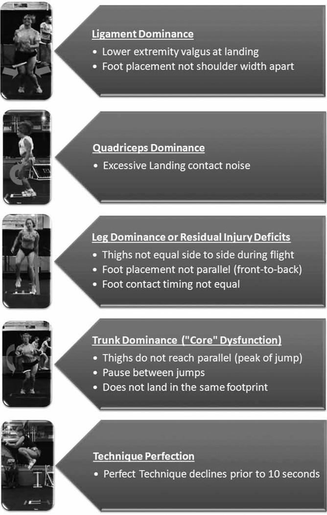
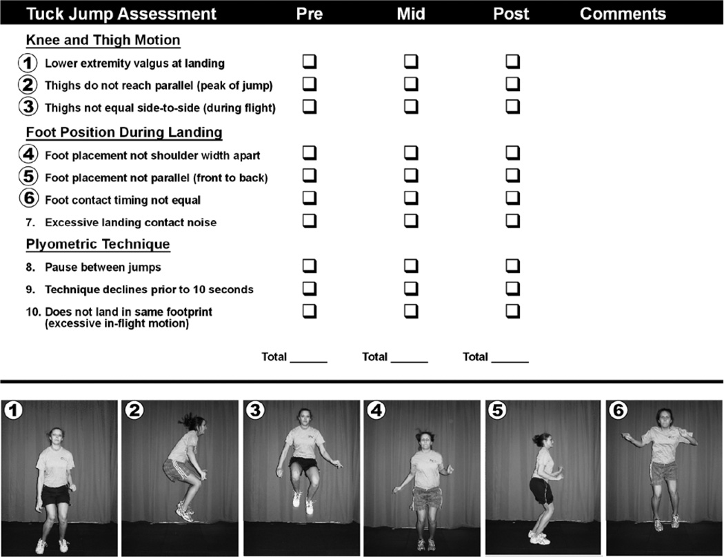
Biomechanical deficiencies can be identified in a simplified screening of a tuck-jump maneuver (Figure 1). The tuck-jump exercise provides the opportunity to assess flaws in athletes that relate to predictors for ACL injury (Figure 2).6 The deficiencies that can be observed are ligament dominance, quadriceps dominance, leg dominance or residual injury deficits, trunk dominance, and technique perfection. All these factors have neuromuscular-control components and may have some contribution to ACL injury. These neuromuscular-control imbalances can often be corrected through various forms of feedback during and immediately after training.12–14
Figure 1.
Tuck-jump criteria grouped by modifiable risk-factor categorizations.
Figure 2.
The tuck-jump assessment can be used to score deficits during a jumping and landing sequence. Figure reproduced from Myer GD, Ford KR, Hewett TE. Tuck jump assessment for reducing anterior cruciate ligament injury risk. Athl Ther Today. 2008;13(5):39–44.
McNair et al15 reported that impact forces were highly correlated with anterior tibial accelerations (r = .86), which has the possibility of being corrected. Two studies have reduced ground-force reaction by giving verbal feedback.12,14 The reduction of the ground force may be caused by increased activation of the hamstrings, causing the knee to be positioned at an increased degree of flexion. External knee-abduction moment is also a predictor for future injury and likely contributes to the stress on an ACL during jumping and landing tasks.9 Decreased hamstrings strength relative to the quadriceps is implicated as a potential mechanism for increased lower extremity injuries and potentially ACL injury risk in female athletes.16–20 The decreased strength and neuromuscular control of the hamstrings can cause increased ground forces.19 However, if the ground force can be reduced, then the anterior tibial acceleration may be reduced, as well.
The neuromuscular deficiencies discussed herein can be altered for the positive. Rucci and Tomporowski21 observed biomechanical changes in athletes who were provided with feedback during the power-clean movement. The feedback was in the form of video, verbal, and self-analysis. Self-analysis consisted of a biomechanical checklist of the most frequently observed technical flaws in performing a power-clean movement. Subjects consulted the checklist to properly diagnose and solve flaws in the movement. Both control and experimental groups showed improvement in their power-clean technique. The greatest improvement was noticed in the verbal- and video-feedback group. Herman et al22 looked at how a strength-training protocol and feedback would affect a stop-jump task. Across all participants, there was decreased peak vertical ground-reaction force, knee-valgus moment, and hip-abduction moment and increased knee-flexion angle, hip-flexion angle, and hip-abduction angle subsequent to the feedback protocol.22 These studies indicate that complex multijoint movements such as the tuck jump can be improved on through the use of augmented feedback.
The purpose of this study was to determine if visual and verbal feedback would improve neuromuscular control during 10 seconds of continuous tuck jumps. Our hypothesis was that visual and verbal feedback would improve neuromuscular control during performance of the tuck-jump exercise for 10 seconds. These changes may help reduce factors related to knee injury. There is strong evidence to support the efficacy of the chosen feedback modes in improvement of neuromuscular control related to these factors.9,12,14,16–19
Methods
Subjects
The study subjects consisted of 37 female high school soccer players randomly assigned to 1 of 2 intervention groups based on the type of feedback they received (augmented feedback [AF] n = 19, control n = 18). The (mean ± SD) age of the participants was 14.7 ± 1.7 years for the control group and 14.7 ± 1.4 years for the AF group. Height and mass means for the subjects were 160.8 ± 5.1 cm and 54.6 ± 7.8 kg for the control group and 160.9 ± 8.1 cm and 54.1 ± 6.8 kg for the AF group. Participants with history of previous ACL injury (n = 1) were allowed to participate with their team in the training program but were excluded from the final data analysis. This study was approved by the institutional review board. Parents or guardians signed informed consent before participation in the study.
Design
The study was a double-blind randomized controlled trial to determine the effects of augmented feedback on tuck-jump performance. The dependent variables of interest included the total tuck-jump deficiencies assessed (Figure 1).
Testing
The study was an 8-week (3 sessions/wk, 90 min/session) pretest and posttest analysis of the effects of augmented feedback on tuck-jump performance. This was accomplished by taking a baseline measure in both sagittal and frontal views before augmented feedback. Then, during each subsequent training session, subjects received the same training protocol and were videotaped performing both the tuck jump and treadmill running. The 2 groups (AF vs control) were then given augmented feedback according to their respective group during each training session. During the last training session additional sagittal and frontal views were taken for the posttest results.
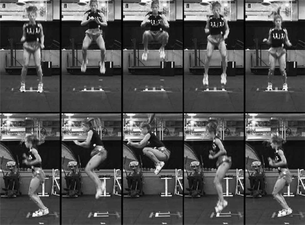
The subjects were first required to perform a baseline repeated tuck jump for 10 seconds. They were instructed by video and verbal commands regarding how to properly perform a tuck jump before performing the 10-second tuck jump. The video consisted of representative images from frontal view and sagittal view of a tuck jump (Figure 3). This was displayed for each subject. The subjects were instructed to place their feet on the 2 vertical strips (41 cm in length) of tape with the tip of their toes behind the intersecting horizontal line (35 cm in length) that connected the 2 vertical strips, which formed an H shape. They were instructed to initiate the jump with a slight crouch downward while they extended their arms behind. They then swung their arms forward as they simultaneously jumped straight up and pulled their knees up as high as possible. At the highest point of the jump the athletes are instructed to pull their thighs parallel to the ground. When landing, they should immediately begin the next tuck jump. Subjects were encouraged to land softly, using a toe to midfoot rocker landing and landing in the same footprint with each jump. The athletes were instructed to perform the tuck-jump exercise for 10 seconds and to not continue this jump if they demonstrated a sharp decline in technique during the allotted time frame. During the baseline process, 1 rater was used to present the video and verbal instructions for the tuck-jump test to the subjects. Another rater captured the video of the baseline tuck jump from frontal and sagittal views using a Panasonic Leica Dicomar 3.1-megapixel camera shooting at 60 frames/s. During the posttraining test session, subjects were also videotaped in the sagittal and frontal planes. They were not instructed how to properly perform a tuck jump during this period. The only information received by the subjects was for proper foot placement on the H mark and start and stop time.
Figure 3.
Desired technical performance of the tuck-jump exercise.
Standardized Training
Both groups of athletes were trained 3 times per week over 7 weeks using the same protocol designed to accommodate the off-season needs of high school soccer players. Therefore, the mode of feedback was the only difference between groups. The training sessions were 90 minutes in duration and contained 2 of the following modes of training, which rotated in a cyclical fashion: strength training, plyometrics, and energy-systems development.
The strength-training component focused on teaching proper biomechanics in several of the common weightlifting and power-lifting exercises including hang clean, hang snatch, box squat, back squat, and dead lift, as well as a variety of other accessory exercises. Sets and repetitions were organized in an undulating, linear fashion throughout the 7-week period. Additional emphasis was placed on improving hamstring and posterior-chain strength due to their relationship to both injury prevention and sports performance.10
The plyometric-training protocol focused on providing the athletes with feedback on their movement quality. Special time and attention were paid to teaching each athlete to jump and land with movement biomechanics that are consistent with avoiding the risk factors outlined previously in the article, including control of valgus knee motion and position, dissipation of landing forces through increased hip and knee flexion, and control of the center of mass relative to the base of support. The protocol progressed in difficulty by the addition of multiple jumps, multiple planes of movement, single-leg movements, and external perturbations. The final component of the training was repeated-sprint training performed on a high-speed treadmill. The sets, repetitions, and rest intervals were chosen to help improve anaerobic and aerobic fitness over the course of the summer to adequately prepare the athletes for the energy demands of soccer competition.
Augmented Feedback Training
For all sessions every subject was recorded during both tuck jump and treadmill-running warm-up. Subjects were randomly allocated to either the AF group or to the control feedback training group. Subjects assigned to the AF group were selected at random to review their tuck-jump video from their previous session, excluding the first session as no video feedback was given. The video feedback was given on a Dell Inspiron 1720 using QuickTime Player at half speed. QuickTime player is a multimedia framework for handling various formats of digital videos and was used as such. Videos were analyzed using a 10-point scale for the tuck jump (Figure 2).10 The subjects were observed for ligament dominance, quadriceps dominance, leg dominance or residual injury deficits, trunk dysfunction, and technique perfection. Ligament dominance is the inability to control frontal-plane motion during cutting or landing. These imbalances can be observed in lower extremity valgus at the knee and in foot placement not being shoulder width apart. Quadriceps dominance is an imbalance between knee-extensor and -flexor strength, recruitment, and coordination.6 Excessive landing noise predicts quadriceps dominance. Leg dominance is the dissymmetry in strength or coordination between both extremities, while leg-dominance dysfunction is most noticeable in thighs not being equal side to side during flight, foot placement not parallel (front to back), and foot-contact timing not equal.10 Thighs not equal is observed when both thighs do not follow the same flight pattern when not in contact with the ground. Foot placement not parallel is recognized when the feet are staggered or not directly in line with one another. Trunk or core dysfunction can be defined as an imbalance between the inertial demands of the trunk and core control and coordination to resist it.10 Trunk dysfunction can be noticed in subjects pausing between jumps, thighs not reaching parallel, and not landing in the same footprint. Thighs should be parallel with the ground during the peak of the tuck jump. The final assessment was technique perfection, which must last for 10 seconds. This demonstrates the athlete’s strength and ability to recreate the same pattern multiple times.
In order for a movement to be considered a dysfunction, the Specific flaw had to appear 3 or more times or have 1 extreme occurrence during the 10-second tuck jump. The subjects were informed of the 2 most apparent flaws they demonstrated and how to correct them. The 2 most apparent flaws were selected based on severity. This means the 2 flaws that appear the most flagrant or frequent were selected to provide feedback on. After the feedback, each subject performed a repeated tuck-jump exercise for 10 seconds. The tuck jump was recorded from the frontal view using a Cisco flip video camera mounted on a tripod. The frontal view was selected because it allows the grader to view all necessary deficits while obtaining practical application for clinical use. The camera and the stopwatch were controlled by the same person. This was performed by setting the camera to record and then informing the subject to begin the tuck jump.
Control Feedback Training
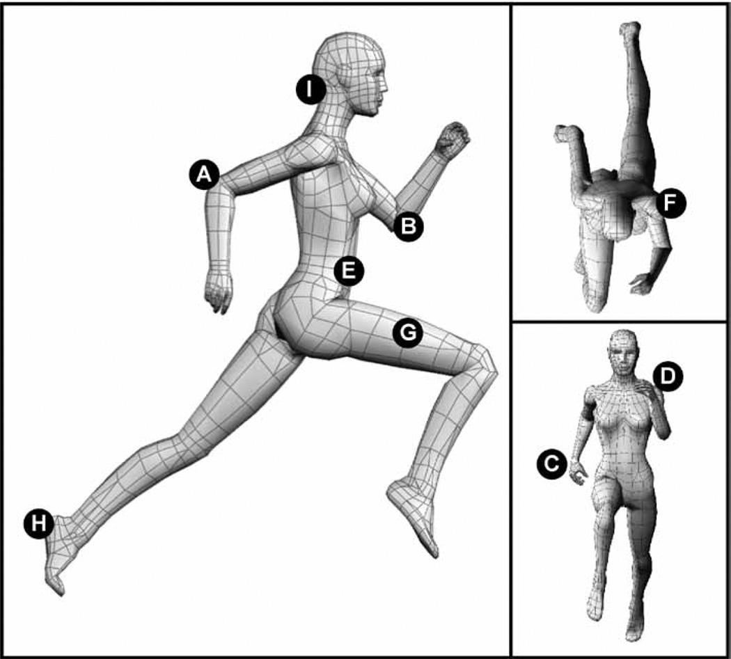
For all training sessions, all subjects were recorded during both tuck jump and treadmill-running warm-up. However, only subjects randomized into the control group received feedback based on running mechanics (Figure 4). The treadmill running was performed at the beginning of each training session at a standardized speed of 8 miles/h and recorded in the sagittal view with a Cisco flip video camera. Before using the treadmill, the subjects were instructed on how to properly get on and off it to avoid possible injury. The instructions were as follows: Grasp the bar firmly, then with your left foot push off several times to get accustomed to the speed of the treadmill, and, finally, once comfortable, step onto the treadmill and release the bar when ready. Subjects were then recorded for 10 seconds running on the treadmill. The start time was initiated when the subject released both hands from the bar located at the front of the treadmill. The stopwatch was controlled by a spotter located on the left side of the treadmill.
Figure 4.
An 8-point biomechanical representation of the control biofeedback provided to the study subjects.
During the 90-minute training session, the control subjects were randomly chosen to review their treadmill video from their previous session, excluding the first session as no video feedback was given. The video feedback was give on a Dell Inspiron 1720 using Quick Time Player at half speed. Videos were analyzed using an 8-point scale for treadmill (Figure 4).10 Athletes were instructed to flex their elbows to 90° and maintain this position (although slightly increased elbow extension was acceptable at the endpoint range of motion) during the back swing. They were instructed to maintain 90° elbow position (although slightly increased elbow flexion was acceptable at the endpoint range of motion) during the forward arm swing. Athletes were instructed to extend their shoulder to a point where their wrist would swing past their hip. In addition, during the back swing the athletes were encouraged to minimize shoulder horizontal abduction by keeping their “elbows in” and “brush your hip pocket with your wrist.” During the forward arm swing the athletes were told to continue to keep their elbows close to their body but not to horizontally adduct or flex their shoulder to a point that their “wrist should not cross the midline of your torso” or “take your wrist higher than your chin.” Athletes were instructed to “maintain upright position of your torso” during sprint training. Both the inclined treadmill training and resistive ground-based training can influence forward trunk flexion beyond optimal positions. This teaching cue for torso positions was used often for both sprint groups during training. Athletes were encouraged to “keep your shoulders square to the direction of travel” to limit torso rotation during training. They were encouraged to “drive your thigh through” and “attempt to get your thigh parallel to the ground” (90° hip flexion) during the forward leg swing. Athletes were instructed to initiate foot strike “on the balls of your feet” and “push off with full ankle extension.” They were encouraged to be relaxed and to avoid trying to “strain” through their sprint-training bouts. They were instructed to “relax your upper torso” and to “avoid clinching or straining your jaw and neck” during training bouts. The 2 most apparent flaws were selected for each subject by the same individual, on which they would receive feedback. The 2 most frequently observed flaws were selected to give feedback on. The subjects were informed of the 2 most apparent flaws they demonstrated and how to correct the flaws.
Statistics
Outcome measurements of tuck-jump deficits were scored by a rater blinded to both training group (control vs AF) and testing session (pretesting vs posttesting). The 10 deficits (Figure 2) were analyzed for each subject from frontal and sagittal video views at the same time. The subject then received 1 score on a scale from 1 to 10. A higher score indicated that more deficits were present. Descriptive data (eg, incidence of deficit occurrence) were totaled for pretest and posttest tuck-jump assessment and anthropometric measures. A 1-way ANOVA was used to evaluate potential group differences in height, mass, and age. A 2 × 2 repeated-measures ANOVA was employed to compare the within-subject effects of time (pretraining vs posttraining) and between-subjects factor of condition (control vs AF) on totaled task-Specific deficits measured during the tuck-jump assessment. Nonparametric confirmatory analysis was performed with the Wilcoxon matched-pairs signed-rank test. Statistical significance was established a priori at P < .05 to test the directional hypothesis that AF would be more effective than control feedback in improving deficits measured during the tuck-jump assessment. Statistical analyses were conducted in SPSS (SPSS, version 17.0, Chicago, IL).
Results
To test the hypothesis that the augmented feedback would reduce task-Specific deficits evaluated with the tuck-jump assessment, we employed a double-blind randomized trial with a control feedback group. The current study included 37 female high school soccer players randomly assigned to 1 of 2 intervention groups based on the type of feedback they received (AF vs control). The study groups were not different in age, height, or mass at either pretest or posttest assessment (P > .05). A significant interaction of time by group was noted with the task-Specific feedback training (P = .03). The AF group reduced deficits measured during the tuck-jump assessment by 23.6%, while the control training reduced deficits by 10.6%. A nonparametric confirmatory analysis was performed with the Wilcoxon matched-pairs signed-rank test, confirming the reduction of deficits at posttest in AF group (P = .001).
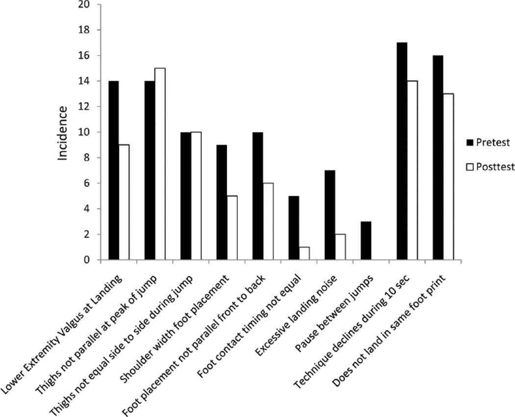
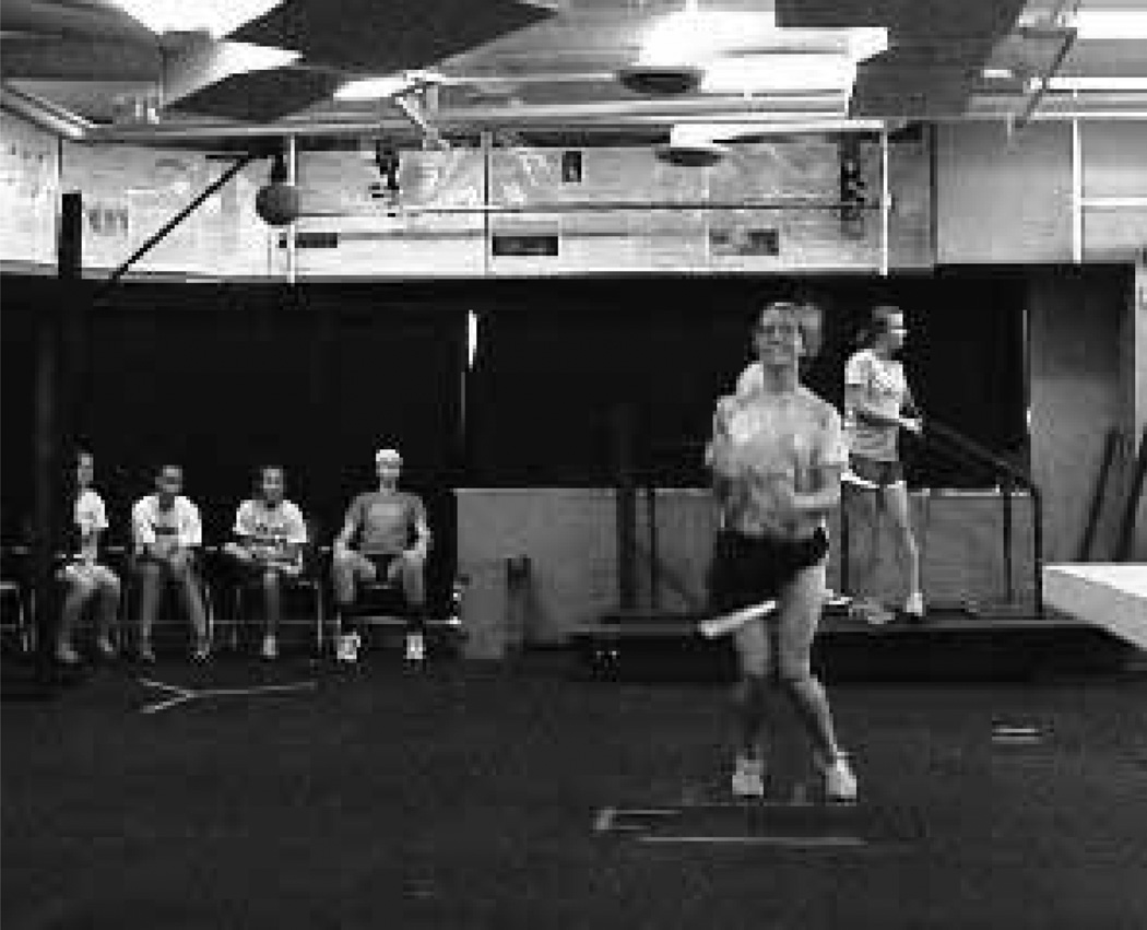
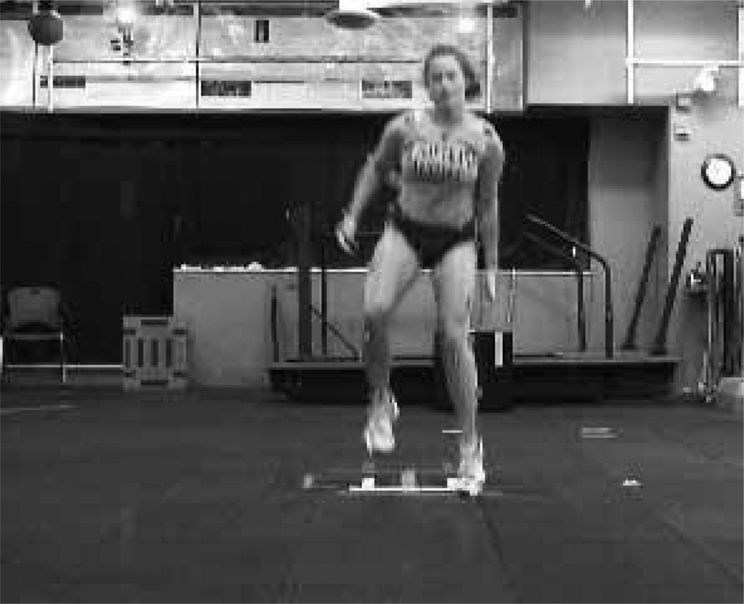
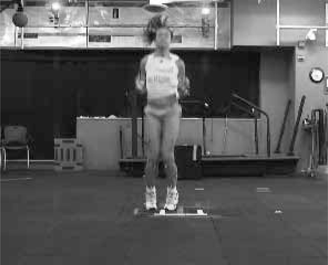
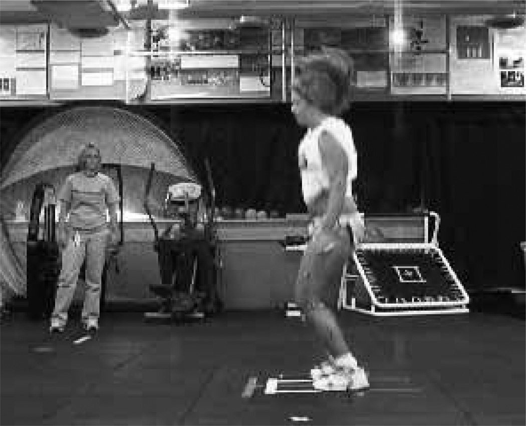
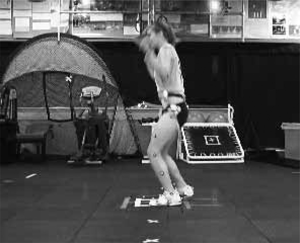
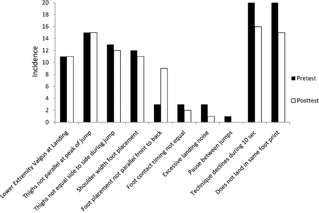
Figure 5 presents the pretest and posttest variables for the AF group. The variables that appear to have changed the most over the course of the training are represented by Figures 6–10. Each of these images represents an athlete who would receive a point for incorrectly performing 1 part of the test. Valgus knee position (Figure 6) experienced a 35.7% decrease in the number of athletes who exhibited this deficit. Feet not shoulder width apart (Figure 7) showed a 44.4% decrease. Excessive ground-contact noise (Figure 8) decreased in 71.4% of the participants. Feet not parallel front to back (Figure 9) was evident in 40% fewer cases after training. Foot timing not equal (Figure 10) was reduced by 80% in the feedback group. Conversely, the deficits that consisted of landing with excessive knee valgus, excessive landing-contact noise, and foot timing not equal at landing appeared to be the most amenable to change with the addition of augmented feedback training. Figure 11 shows a representation of the pretest and posttest assessments in the control group. This group, who did not receive the augmented feedback, did not appear to show a similar response to improve deficits in measures that may be related to increased ACL injury risk.
Figure 5.
Task-Specific changes associated with the augmented feedback intervention from pretest to posttest.
Figure 6.
Lower extremity valgus at landing. This is indicated by the athlete displaying a knock-kneed position while in contact with the ground.
Figure 10.
Foot-contact timing not equal. Similar to not placing the feel parallel, the athlete will occasionally change the timing of the foot contacts to protect an injured or weaker limb.
Figure 7.
Foot placement not shoulder width apart. This deficit can be manifested with feet either closer together or farther apart.
Figure 8.
Excessive landing contact noise. This is typically displayed by the athlete through landing with fat feet and is typically accompanied by a lack of knee and hip flexion during the stance phase.
Figure 9.
Foot placement not parallel (front to back). Often an athlete will “drop” one foot behind the other while on the ground to help minimize forces on an injured or weaker limb.
Figure 11.
Task-Specific changes associated with the control feedback group from pretest to posttest.
Discussion
The purpose of this study was to determine if visual and augmented feedback would improve neuromuscular control during a 10-second tuck jump. The tested hypothesis was supported by the finding of significantly greater improvements in the intervention group than in the control group. Proper technique for basic movements such as jumping are absent from many training protocols. Both groups received identical training with the exception of the type of augmented feedback (tuck jump or running). This highlights the importance of implementing feedback in a standardized training protocol.
Previous studies support the use of different forms of feedback.14 In one study, 35 female and 56 male athletes performed a jump from a box onto a force plate that measured the ground-reaction force (GRF).14 The subjects were divided into an augmented-feedback group and control group that did not receive augmented feedback. The augmented condition significantly reduced their GRF scores from prejump (4.53 ± 1.51) to postjump (3.57 ± 1.10), whereas those in the control group did not (mean pre GRF = 4.51 ± 1.77, mean post GRF = 4.33 ± 1.54).14 In another investigation, augmented feedback appeared to have significantly reduced GRF in both posttest conditions (2-minute posttest reduction, 0.85 ± 0.62; 1-week posttest reduction, 0.74 ± 0.58) as compared with the sensory, control I, and control II feedback groups.12 The feedback in the previous studies was well responded to, which gives the tuck-jump assessment the capability to be used as a training tool that is guided from the identified deficits.10
Modification of landing technique through feedback modalities will likely reduce risk factors associated with ACL injury. Withrow et al23 reported that increased hamstrings force during the flexion phase of simulated jump landings greatly decreased the tension placed on the ACL. The increased force could possibly have been attained by gains made with neuromuscular control initiated from feedback based on tuck-jump performance. This would be achieved by providing feedback for decreasing GRF. Decreasing the GRF has a direct correlation with decreasing the tibial acceleration that can cause knee injuries.14 Knee-abduction moment has been linked to deficits in hamstrings recruitment, which has also been linked to the increased risk of ACL injury in female athletes.7,9 Chappell et al24 came to a conclusion similar to that of Withrow et al23 and Myer et al,19 except that decreased knee flexion was cited to potentially cause anterior shearing forces on the knee. Dynamic stability of an athlete’s knee depends on accurate sensory input, not only from the surrounding musculature but also in trunk position during cutting, stopping, and landing movements.25,26 Decreased core neuromuscular control may contribute to increased valgus positioning in lower extremity collapse.27,28 If neuromuscular-control increases are produced through feedback on GRF during the tuck jump, important knee-injury predictors could be reduced, which may potentially reduce risk of knee-related injuries.
Use of a tuck jump as a plyometric exercise to provide augmented feedback in a training protocol has many benefits. One is the increased neuromuscular recruitment gained during the exercise. The greatest amount to be gained with biofeedback training may be in development of core and hamstrings neuromuscular control. This can be accomplished as the core attempts to stabilize the extremities during flight in a tuck jump. The hamstrings can be recruited during the landing phase as knee flexion is increased and landing noise is decreased. However, since a 10-second tuck jump is an advanced plyometric exercise, it must be progressed in a safe and challenging manner. This involves instructing the subject on the proper technique of a jump-landing pattern. Once the subject can successfully execute a single tuck jump with minimal difficulty, a double tuck jump can be initiated. Again, as this exercise is mastered with minimal flaws, the subject can begin tuck jumps for time, steadily increasing the time as improvements are noted. A key component to making neuromuscular advancements is altering the difficulty, not the intensity. The tuck jump is a maximum-effort exercise. If training capacity is overloaded, technical flaws will ensue. This defeats the purpose of the exercise if proper technique cannot be achieved due to an overload of intensity. Pairing tuck jumps with accessory exercises in a periodized strength program can be used to develop areas of weakness. Including a tuck-jump feedback program with a strength program will provide subjects the best circumstance to reduce deficits.
Biofeedback during a tuck jump can possibly prevent knee injury and also has implications for improving skill development from gained control of the core and trunk. The core activates before the extremities carry out a coordinated movement.29,30 This provides a stable base for the extremities to move about within the body’s center of gravity. If the core is activated late or does not have the ability to counteract the extremities’ force, 3 deficits will be observed in the tuck jump: thighs not reaching parallel, pauses between jumps, and not landing in same footprint.10 These cannot be directly corrected or improved by feedback. This is where accessory exercises can improve neuromuscular deficiencies and weaknesses within the core. When core advancements are made, this enables the extremities to move quicker and with more force. This is in response to the core activating in a more timely fashion and being able to absorb more force while still keeping the extremities in the body’s center of gravity. The sooner core stabilization can be initiated, the more quickly the extremities can move freely. This could result in a subject being faster and more efficient in movements.
One possible implication for improvements in neuromuscular control involves the conceptualization and activation of mirror neurons. Mirror neurons are the link between viewing and hearing something and then performing the action.31 The activation occurs in the motor regions of brain, which supports the idea that mirror neurons play a role in motor patterns. Kawato and Wolpert32 believe one possibility is that these neurons create an internal model for control. Current evidence indicates that the central nervous system uses internal models for movement planning, control, and learning.32 The exact role that mirror neurons play in motor movements is still being studied in a variety of animal models. The mirror neurons have been pinpointed to certain areas of the premotor cortex and parietal lobe when monkeys see or hear a complex task.33 However, studies have yet to map out the individual mirror neurons for certain stimuli in humans. Therefore, the human mirror-neuron system and how it functions are still under heavy debate. The proposed idea is that the mirror neurons fire when a complex task or movement is observed. The mirror neurons fire again just before the movement is completed by the individual once he or she observes the action. However, the actual mirror neuron itself does not fire to make the movement. Instead, the mirror neurons work by encoding the observed movement and then sending the impulse on to the motor neurons, which can then reproduce the complex motor task observed.33 The mirror-neuron function may further explain why complex movements can be improved through visual and verbal feedback. However, mirror neurons still need to be thoroughly researched to use their full potential in improving neuromuscular control.
Despite the many possible benefits of the tuck jump, there are still some limitations. The tuck jump is an advanced plyometric exercise that may be initially difficult for certain subjects to properly perform. To master a difficult movement, a progression from a single tuck jump to the more complex 10-second tuck jump should be initiated. This will allow for maximum improvement while keeping the athlete free from injury. Different subjects’ skill sets allow some to grasp a difficult movement more readily. Therefore, a few subjects may experience a quicker response time to the feedback. This does not imply that other subjects will not see improvements, only that the improvements may take longer. Greater advancement may result if additional time is allocated. However, when the Specific items in the test are separated and evaluated independently, it appears that the most modifiable changes are occurring in several of the variables related to bilateral asymmetries and valgus knee position. Cumulatively, these deficit assessments indicate that augmented feedback guided by the tuck-jump assessment tool can be valuable to reduce task-Specific deficits.
Conclusions
The current results demonstrate that providing athletes with task-Specific feedback can effectively reduce biomechanical risk factors associated with ACL injury. The data also indicate that Specific components of the tuck-jump assessment are potentially more modifiable than others. These would include valgus moment, feet shoulder width apart, feet parallel front to back, excessive landing noise, and pauses between jumps. These findings indicate that providing Specific feedback to athletes on jumping tasks may reduce their risk factors for injury through improving their biomechanics. Valgus moment, excessive landing noise, and pauses between jumps are potentially more malleable and may yield the greatest improvement for the time investment.
Acknowledgments
The authors would like to acknowledge funding support from National Institutes of Health/NIAMS Grants R01-AR049735, R01-AR05563, and R01-AR056259.
References
- 1.NCAA. NCAA Injury Surveillance System Summary. Indianapolis, IN: National Collegiate Athletic Association; 2002. [Google Scholar]
- 2.Arendt E, Dick R. Knee injury patterns among men and women in collegiate basketball and soccer. NCAA data and review of literature. Am J Sports Med. 1995;23(6):694–701. doi: 10.1177/036354659502300611. [DOI] [PubMed] [Google Scholar]
- 3.Malone TR, Hardaker WT, Garrett WE, Feagin JA, Bassett FH. Relationship of gender to anterior cruciate ligament injuries in intercollegiate basketball players. J South Orthop Assoc. 1993;2(1):36–39. [Google Scholar]
- 4.Myklebust G, Maehlum S, Holm I, Bahr R. A prospective cohort study of anterior cruciate ligament injuries in elite Norwegian team handball. Scand J Med Sci Sports. 1998;8(3):149–153. doi: 10.1111/j.1600-0838.1998.tb00185.x. [DOI] [PubMed] [Google Scholar]
- 5.Grindstaff TL, Hammill RR, Tuzson AE, Hertel J. Neuromuscular control training programs and noncontact anterior cruciate ligament injury rates in female athletes: a numbers-needed-to-treat analysis. J Athl Train. 2006;41(4):450–456. [PMC free article] [PubMed] [Google Scholar]
- 6.Myer GD, Ford KR, Hewett TE. Rationale and clinical techniques for anterior cruciate ligament injury prevention among female athletes. J Athl Train. 2004;39(4):352–364. [PMC free article] [PubMed] [Google Scholar]
- 7.Besier TF, Lloyd DG, Cochrane JL, Ackland TR. External loading of the knee joint during running and cutting maneuvers. Med Sci Sports Exerc. 2001;33(7):1168–1175. doi: 10.1097/00005768-200107000-00014. [DOI] [PubMed] [Google Scholar]
- 8.Li G, Rudy TW, Sakane M, Kanamori A, Ma CB, Woo SL. The importance of quadriceps and hamstring muscle loading on knee kinematics and in-situ forces in the ACL. J Biomech. 1999;32(4):395–400. doi: 10.1016/s0021-9290(98)00181-x. [DOI] [PubMed] [Google Scholar]
- 9.Hewett TE, Myer GD, Ford KR, et al. Biomechanical measures of neuromuscular control and valgus loading of the knee predict anterior cruciate ligament injury risk in female athletes: a prospective study. Am J Sports Med. 2005;33(4):492–501. doi: 10.1177/0363546504269591. [DOI] [PubMed] [Google Scholar]
- 10.Myer GD, Brent JL, Ford KR, Hewett TE. Real-time assessment and neuromuscular training feedback techniques to prevent ACL injury in female athletes. Strength Cond J. 2011;33(3):21–35. doi: 10.1519/SSC.0b013e318213afa8. [DOI] [PMC free article] [PubMed] [Google Scholar]
- 11.Myer GD, Ford KR, Khoury J, Succop P, Hewett TE. Biomechanics laboratory-based prediction algorithm to identify female athletes with high knee loads that increase risk of ACL injury. Br J Sports Med. 2011;45(4):245–252. doi: 10.1136/bjsm.2009.069351. [DOI] [PMC free article] [PubMed] [Google Scholar]
- 12.Onate JA, Guskiewicz KM, Sullivan RJ. Augmented feedback reduces jump landing forces. J Orthop Sports Phys Ther. 2001;31(9):511–517. doi: 10.2519/jospt.2001.31.9.511. [DOI] [PubMed] [Google Scholar]
- 13.Oñate JA, Guskiewicz KM, Marshall SW, Giuliani C, Yu B, Garrett WE. Instruction of jump-landing technique using videotape feedback: altering lower extremity motion patterns. Am J Sports Med. 2005;33(6):831–842. doi: 10.1177/0363546504271499. [DOI] [PubMed] [Google Scholar]
- 14.Prapavessis H, McNair PJ. Effects of instruction in jumping technique and experience jumping on ground reaction forces. J Orthop Sports Phys Ther. 1999;29(6):352–356. doi: 10.2519/jospt.1999.29.6.352. [DOI] [PubMed] [Google Scholar]
- 15.McNair PJ, Prapavessis H, Callender K. Decreasing landing forces: effect of instruction. Br J Sports Med. 2000;34(4):293–296. doi: 10.1136/bjsm.34.4.293. [DOI] [PMC free article] [PubMed] [Google Scholar]
- 16.Ford KR, Myer GD, Schmitt LC, Van den Bogert AJ, Hewett TE. Effect of drop height on lower extremity bio-mechanical measures in female athletes. Med Sci Sports Exerc. 2008;40(5):S80. [Google Scholar]
- 17.Ford KR, van den Bogert AJ, Myer GD, Shapiro R, Hewett TE. The effects of age and skill level on knee musculature co-contraction during functional activities: a systematic review. Br J Sports Med. 2008;42(7):561–566. doi: 10.1136/bjsm.2007.044883. [DOI] [PMC free article] [PubMed] [Google Scholar]
- 18.Knapik JJ, Bauman CL, Jones BH, Harris JM, Vaughan L. Preseason strength and flexibility imbalances associated with athletic injuries in female collegiate athletes. Am J Sports Med. 1991;19(1):76–81. doi: 10.1177/036354659101900113. [DOI] [PubMed] [Google Scholar]
- 19.Myer GD, Ford KR, Barber Foss KD, Liu C, Nick TG, Hewett TE. The relationship of hamstrings and quadriceps strength to anterior cruciate ligament injury in female athletes. Clin J Sport Med. 2009;19(1):3–8. doi: 10.1097/JSM.0b013e318190bddb. [DOI] [PMC free article] [PubMed] [Google Scholar]
- 20.Hewett TE, Stroupe AL, Nance TA, Noyes FR. Plyometric training in female athletes: decreased impact forces and increased hamstring torques. Am J Sports Med. 1996;24(6):765–773. doi: 10.1177/036354659602400611. [DOI] [PubMed] [Google Scholar]
- 21.Rucci JA, Tomporowski PD. Three types of kinematic feedback and the execution of the hang power clean. J Strength Cond Res. 2010;24(3):771–778. doi: 10.1519/JSC.0b013e3181cbab96. [DOI] [PubMed] [Google Scholar]
- 22.Herman DC, Onate JA, Weinhold PS, et al. The effects of feedback with and without strength training on lower extremity biomechanics. Am J Sports Med. 2009;37(7):1301–1308. doi: 10.1177/0363546509332253. [DOI] [PubMed] [Google Scholar]
- 23.Withrow TJ, Huston LJ, Wojtys EM, Ashton-Miller JA. Effect of varying hamstring tension on anterior cruciate ligament strain during in vitro impulsive knee flexion and compression loading. J Bone Joint Surg Am. 2008;90(4):815–823. doi: 10.2106/JBJS.F.01352. [DOI] [PMC free article] [PubMed] [Google Scholar]
- 24.Chappell JD, Yu B, Kirkendall DT, Garrett WE. A comparison of knee kinetics between male and female recreational athletes in stop-jump tasks. Am J Sports Med. 2002;30(2):261–267. doi: 10.1177/03635465020300021901. [DOI] [PubMed] [Google Scholar]
- 25.Hewett TE, Paterno MV, Myer GD. Strategies for enhancing proprioception and neuromuscular control of the knee. Clin Orthop Relat Res. 2002;(402):76–94. doi: 10.1097/00003086-200209000-00008. [DOI] [PubMed] [Google Scholar]
- 26.Hewett TE, Zazulak BT, Myer GD, Ford KR. A review of electromyographic activation levels, timing differences, and increased anterior cruciate ligament injury incidence in female athletes. Br J Sports Med. 2005;39(6):347–350. doi: 10.1136/bjsm.2005.018572. [DOI] [PMC free article] [PubMed] [Google Scholar]
- 27.McConnell J. The physical therapist’s approach to patello-femoral disorders. Clin Sports Med. 2002;21(3):363–387. doi: 10.1016/s0278-5919(02)00027-3. [DOI] [PubMed] [Google Scholar]
- 28.Zazulak BT, Ponce PL, Straub SJ, Medvecky MJ, Avedisian L, Hewett TE. Gender comparison of hip muscle activity during single-leg landing. J Orthop Sports Phys Ther. 2005;35(5):292–299. doi: 10.2519/jospt.2005.35.5.292. [DOI] [PubMed] [Google Scholar]
- 29.Hodges PW, Richardson CA. Contraction of the abdominal muscles associated with movement of the lower limb. Phys Ther. 1997;77(2):132–142. doi: 10.1093/ptj/77.2.132. disc 142–134. [DOI] [PubMed] [Google Scholar]
- 30.Hodges PW, Richardson CA. Feedforward contraction of transversus abdominis is not influenced by the direction of arm movement. Exp Brain Res. 1997;114(2):362–370. doi: 10.1007/pl00005644. [DOI] [PubMed] [Google Scholar]
- 31.Kohler E, Keysers C, Umilta MA, Fogassi L, Gallese V, Rizzolatti G. Hearing sounds, understanding actions: action representation in mirror neurons. Science. 2002;297(5582):846–848. doi: 10.1126/science.1070311. [DOI] [PubMed] [Google Scholar]
- 32.Kawato M, Wolpert D. Internal models for motor control. Novartis Found Symp. 1998;218:291–304. doi: 10.1002/9780470515563.ch16. disc 304–307. [DOI] [PubMed] [Google Scholar]
- 33.Oztop E, Kawato M, Arbib M. Mirror neurons and imitation: a computationally guided review. Neural Netw. 2006;19(3):254–271. doi: 10.1016/j.neunet.2006.02.002. [DOI] [PubMed] [Google Scholar]


