Abstract
Background
The prevalence of obesity is rising. Obesity can lead to cardiovascular and ventilatory complications through multiple mechanisms. Cardiac and pulmonary function in asymptomatic subjects and the effect of structured dietary programs on cardiac and pulmonary function is unclear.
Objective
To determine lung and cardiac function in asymptomatic obese adults and to evaluate whether weight loss positively affects functional parameters.
Methods
We prospectively evaluated bodyplethysmographic and echocardiographic data in asymptomatic subjects undergoing a structured one-year weight reduction program.
Results
74 subjects (32 male, 42 female; mean age 42±12 years) with an average BMI 42.5±7.9, body weight 123.7±24.9 kg were enrolled. Body weight correlated negatively with vital capacity (R = −0.42, p<0.001), FEV1 (R = −0.497, p<0.001) and positively with P 0.1 (R = 0.32, p = 0.02) and myocardial mass (R = 0.419, p = 0.002). After 4 months the study subjects had significantly reduced their body weight (−26.0±11.8 kg) and BMI (−8.9±3.8) associated with a significant improvement of lung function (absolute changes: vital capacity +5.5±7.5% pred., p<0.001; FEV1+9.8±8.3% pred., p<0.001, ITGV+16.4±16.0% pred., p<0.001, SR tot −17.4±41.5% pred., p<0.01). Moreover, P0.1/Pimax decreased to 47.7% (p<0.01) indicating a decreased respiratory load. The change of FEV1 correlated significantly with the change of body weight (R = −0.31, p = 0.03). Echocardiography demonstrated reduced myocardial wall thickness (−0.08±0.2 cm, p = 0.02) and improved left ventricular myocardial performance index (−0.16±0.35, p = 0.02). Mitral annular plane systolic excursion (+0.14, p = 0.03) and pulmonary outflow acceleration time (AT +26.65±41.3 ms, p = 0.001) increased.
Conclusion
Even in asymptomatic individuals obesity is associated with abnormalities in pulmonary and cardiac function and increased myocardial mass. All the abnormalities can be reversed by a weight reduction program.
Introduction
The worldwide prevalence of obesity is rising [1]. Obesity leads to increasing morbidity [1], [2], [3] and mortality [4], a loss of potential and quality adjusted years of life and is a major economic challenge for health care systems [5]. It contributes to an increasing risk for a large variety of diseases including arterial hypertension, diabetes mellitus, dyslipidemia, coronary artery disease, stroke, cancer and obstructive sleep apnea [1]. A negative influence of obesity on diastolic and systolic ventricular function was reported as well as “obesity cardiomyopathy” [6], [7]. Obesity is also an independent predictor for the development of pulmonary hypertension in patients with diastolic left ventricular dysfunction [8]. In patients with obstructive sleep apnea, body weight correlates with pulmonary artery pressure [9]. Further, obesity can cause obesity hypoventilation syndrome (OHS) [10], [11] and may contribute to pulmonary hypertension in these patients [10], [11]. In subjects with OHS and severe PH, pulmonary artery pressure is correlated to body mass index, pCO2 and reduced power of breathing [11]. However, not all obese subjects develop obesity hypoventilation syndrome [12], and neither all patients with OHS are affected by pulmonary hypertension [13]. The cardiac and pulmonary function in asymptomatic obese subjects is not well studied.
Obesity can affect pulmonary function in multiple ways. Static lung volumes and maximal power of breathing is reduced. Airway resistance and work of breathing is increased [14], [15]. A contributory role of these abnormalities to the pathophysiology of asthma and pulmonary arterial hypertension in obese patients is likely [16]–[24]. Humoral interactions, e.g. leptin resistance contributing to hypoventilation [29], and a major role of adiponectine in the pathophysiology of PAH and asthma have been discussed [25]–[28]. However, it remains unclear whether body fat contributes to functional abnormalities in the cardiopulmonary system via an underlying systemic inflammatory process or if ventilation and pulmonary circulation are only influenced by the mechanical influences of increased body fat [30]. Because interactions and the sequence of events remain unclear, it is of great interest to determine possible early events on the path to clinically evident disease. It is unclear whether obesity has an impact on lung and cardiac function in asymptomatic subjects.
Interventional programs are promising tools to reduce obesity associated morbidity and mortality. The benefit of surgically induced weight loss on lung and cardiac function has been convincingly demonstrated [13], [31]. In contrast, there are few studies that have assessed the effect of structured weight reduction programs on pulmonary function and echocardiographically measured cardiac function, blood pressure and pulmonary hemodynamics assessed by echocardiography or right heart catheterization and they have shown conflicting results [32]–[34]. Especially the effect of dietary induced weight loss on lung and cardiac function of asymptomatic subjects has not been studied.
We studied and report the effect of a weight reduction program which is based on the use of a formula diet on lung and cardiac function. It includes a supervision of all participants by a multidisciplinary team of physicians, dietary specialists, physiotherapists and psychologists specialized in the long-term treatment of obese patients.
Objective
We aimed to determine the influence of body weight on lung and cardiac function in asymptomatic obese adults and to evaluate the effect of a structured weight reduction program on lung and cardiac function.
We show that even in asymptomatic individuals obesity is associated with cardiopulmonary functional abnormalities and these abnormalities can be reversed by weight reduction.
Methods
In a prospective observational study lung function tests and echocardiography were performed in 74 subjects who underwent an interdisciplinary 52-week weight reduction program. The structure of this weight loss program addressing people with a BMI>30 kg/m2 was described before [35]. It consists of a one-week run-in-period, a twelve week fasting period followed by an eight week changeover and a 31-week stabilization period. The study was approved by our local Ethics Committee. Baseline data were collected during the one-week run-in-period. A follow-up examination 16 weeks after start of the dietary program was offered to all patients. The time point four weeks after completion of the fasting period was chosen to analyze the effect of potential maximum weight loss on cardiopulmonary function.
Study subjects
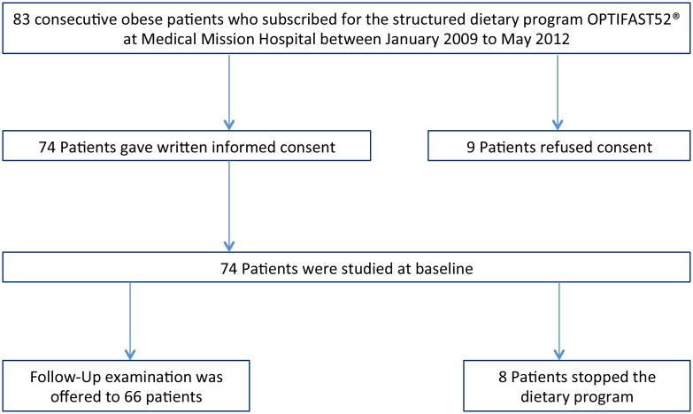
Study subjects were recruited from 83 consecutive subjects who registered for the structured dietary program at Medical Mission Hospital between January 2009 and May 2012. 74 patients gave written informed consent and were studied at baseline and at follow-up 12 weeks later. Nine patients declined to participate. Eight patients who had stopped the dietary program during the first twelve weeks were not examined at the 16-week follow-up. Some patients did not complete all measurements of the follow-up examination. Only patients with data available from both baseline and three month follow-up period were included for analysis of changes in each parameter.
Procedures
Bodyplethysmography including measurement of transfer factor (Masterscreen Body/Diff CareFusion, Germany) was performed according to the European Respiratory Society Statement [36]. Inspiratory mouth pressures were measured as described [37]–[41]. Echocardiography (Vivid7, GE Medical Systems, Solingen, Germany) was performed to measure left and right ventricular function, myocardial mass, systolic right ventricular pressure, right ventricular outflow tract acceleration time and to rule out cardiac shunts and significant valve pathology as recommended [42], [43]. We further analyzed the available data of Bioelectrical Impedance analysis, which was introduced into the weight reduction program during the study. Bioelectrical impedance analysis (BIA 2000-S, Data Input GmbH Darmstadt, Germany) was performed as recommended and according to the manufacturers guideline [44], [45]. The same diagnostic tests used at baseline were performed at the follow-up.
Statistical analysis
Statistical analysis was done using the program SPSS, Version 19.0 (IBM SPSS Statistics). Data are presented as mean and standard deviation. Baseline and follow-up values were compared and significance was evaluated by the paired t-test and assumed if p-value was <0.05. Pearson correlation of the parameters was calculated. Statistical significance was assumed if p-value was <0.05.
Results
Anthropometric data and bioelectrical impedance analysis
The subjects were selected as shown in figure 1. Table 1 summarizes the anthropometric data of the 74 individuals at baseline. The results of the bioelectric impedance analysis at baseline are shown in Table S1. The cohort comprised 32 males and 42 females. Mean age was 43±12 years. The subjects presented with severe obesity with a mean body-mass-index (BMI) of 42.5±7.9 kg/m2 and a mean body weight of 123.7±24.9 kg. The waist-to-hip ratio was 0.94±0.1. Body fat was 55.8±16.6 kg and 44.4±8.4% respectively. Resting metabolic rate was 1756.4±293.9 kcal. Body fluid was calculated with 50.7±11.5 l. The majority of subjects presented without dyspnea: NYHA I = 68/74, NYHA II = 6/74, NYHA III = 0/74, NYHA IV = 0/74. Seven of the 74 subjects were smokers. Comorbidities are shown in table S2.
Figure 1. Subject selection for the analysis.
Table 1. Anthropometric data.
| Parameter | N = 74, Value Mean ± SD | |
| Anthropometric data | ||
| female/male | 74 | 32/42 |
| Age (years) | 74 | 43±12 |
| BMI (kg/m2) | 74 | 42.5±7.9 |
| Body weight (kg) | 74 | 123.7±24.9 |
| Waist/hip ratio | 74 | 0.94±0.1 |
| Systolic blood pressure | 74 | 133±14 |
| Diastolic blood pressure | 74 | 89±9 |
| Heart rate | 74 | 73±9 |
| NYHA Class I/II/III/IV | 74 | 68/6/0/0 |
Data are shown as mean ± standard deviation.
Baseline data
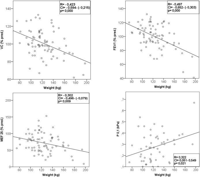
The results of the pulmonary function tests are summarized in table 2. Mean expiratory flow 25, intrathroracic gas volume (ITGV), total lung capacity and transfer factor of the lung measured by the single breath method (TLCO-SB) were mildly decreased compared to the, not weight specific normal values, published by Quanjer for a cohort of subjects aged 18–70 years with a height of 1.45–1.95 m [36]. P0.1 and P0.1/Pimax were increased compared to a cohort with normal weight reported by Koch et al [38] and non weight-specific reference values proposed by the German Airway League [39] based on data published by Hautmann and Windisch [40], [41] indicating an increased respiratory load. There was a significant negative correlation between body weight and vital capacity (R = −0.42, p<0.001), FEV1 (R = −0.49, p<0.001), MEF25 (R−0.30, p = 0.009) (Fig. 2) and FEV1/VC (R = 0.37, p = 0.001). Body weight (R = 0.32, p = 0.02) (Figure 2) and body fat (R = 0.316, p = 0.03) was positively correlated to P0.1. FEV1 and MEF25 were negatively correlated to body fat. We found a significant negative correlation of waist-to-hip-ratio with vital capacity (R = −0.43, p<0.001) and with total lung capacity (R = −0.34, p = 0.003).
Table 2. Baseline data: Bodyplethysmography including mouth occlusion pressure and echocardiography.
| N | Baseline (Mean ± SD) | |
| Bodyplethysmography | ||
| VC (% pred.) | 74 | 100.4±15.6 |
| FEV1 (% pred.) | 74 | 99.8±17.7 |
| FEV1/VC (%) | 74 | 79.2±7.7 |
| MEF 25 (% pred.) | 74 | 71.9±28.6 |
| ITGV (% pred.) | 74 | 91.2±16.5 |
| RV (% pred.) | 74 | 102.1±32.7 |
| TLC (% pred.) | 74 | 102.0±12.2 |
| TLC-He (% pred) | 30 | 90.0±11.0 |
| SR tot (% pred.) | 74 | 109.1±63.5 |
| TLCO SB (% pred.) | 47 | 87.3±14.0 |
| P0.1 (kPa) | 51 | 0.28±0.13 |
| PI max (kPa) | 36 | 6.8±2.9 |
| P0.1/PI max | 29 | 0.05±0.05 |
| BF (1/min) | 35 | 18.0±5.5 |
| Echocardiography | ||
| IVSd (mm) | 54 | 10.9±2.5 |
| LVPWd (mm) | 54 | 10.6±2.3 |
| EF biplan Simpson (%) | 53 | 63.1±7.2 |
| LIMP | 46 | 0.54±0.28 |
| MAPSE (cm) | 51 | 1.8±0.4 |
| E/E’ | 54 | 6.74±1.67 |
| TAPSE (mm) | 54 | 28.8±5.1 |
| RIMP | 51 | 0.40±0.22 |
| RA area (cm2) | 54 | 13.9±5.5 |
| RV (cm) | 54 | 3.2 0.7 |
| e/e’ | 51 | 4.82±1.67 |
| TDI-TVA (cm/s) | 51 | 14.5±3.0 |
| RVSP (mmHg) | 7 | 20.4±8.6 |
| PV AccT (ms) | 50 | 117.8±29.6 |
Data are shown as mean ± standard deviation.
Figure 2. Correlations of body weight with pulmonary function.
VC = vital capacity. FEV1 = forced expired volume at one second. MEF 25 = Mean expiratory flow 25. P 0.1 = mouth occlusion pressure at 0.1 second.
Echocardiographic data at baseline are shown in table 2. We found an increased left and right myocardial performance index compared to the normal values proposed by the American (ASE) and European Society of Echocardiography (ESE) [43] that are not weight specific. e/e’ was increased compared to these normal values indicating diastolic dysfunction of the right ventricle. Mean diameter of the interventricular septum and posterior wall of the left ventricle were mildly enlarged in comparison to the normal values recommended by ASE and ESE published by Lang et al [42]. However, diameter of all cardiac chambers, ejection fraction, mitral flow pattern and tissue Doppler parameter of the left ventricle were normal.
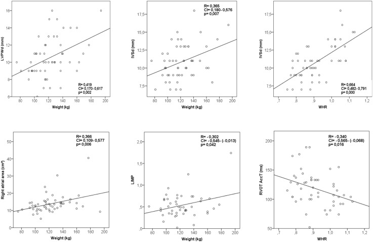
We found a positive correlation between body weight and myocardial wall thickness (IVSd, R = 0.37, p = 0.007; LPWd (R = 0.42, p = 0.002), body weight and left myocardial performance index (R = 0.30, p = 0.04) and body weight and right atrial area (R = 0.37, p = 0.006), (Figure 3). Waist-to-hip ratio was positively correlated with myocardial wall thickness (IVSd, R = 0.66, p<0.001) and acceleration time in the right ventricular outflow tract (R = −0.34, p = 0.02; Figure 3).
Figure 3. Correlations of body weight with cardiac function and myocardial wall thickness.
LVPWd = diastolic left ventricular posterior wall diameter; IVSd = diastolic interventricular septal wall thickness; LIMP = left myocardial performance index; RVOT AccT = acceleration time of flow in the right ventricular outflow tract.
Follow-Up data
Follow-up data and absolute changes from baseline are shown in table 3. After 4 months of the dietary program, body weight decreased dramatically (−26.0±11.8 kg, p<0.001), as well as BMI (−8.9±3.8, p<0.001) and body fat (−18.5±7.7, p<0.001). Table 3 shows the significant increase of lung volumes measured by bodyplethysmography and spirometric flow parameters. P0.1/Pimax decreased indicating improving breathing capacity.
Table 3. Baseline and follow-up data, absolute changes and p values.
| N | Baseline(Mean ± SD) | Follow-Up(Mean ± SD) | abs. change(Mean ± SD) | p-value | |
| Weight, Height and Waist to hip ratio | |||||
| Weight (kg) | 55 | 125.6±24.5 | 99.6±17.9 | −26.0±11.8 | <0,001 |
| BMI (kg/m2) | 55 | 43.4±8.1 | 34.5±6.3 | −8.9±3.8 | <0,001 |
| WHR | 62 | 0.93±0.11 | 0.90±0.09 | −0.03±0.1 | <0,001 |
| Bodyplethysmography | |||||
| VC (% pred.) | 51 | 98.5±15.7 | 104±13.7 | +5.5±7,5 | <0,001 |
| FEV1 (% pred.) | 51 | 97.7±18.8 | 107.5±17.4 | +9.8±8.3 | <0,001 |
| FEV1/VC (%) | 50 | 78,9±8.4 | 80.9±7.9 | +2.0±4.9 | 0,006 |
| MEF 25 (% pred.) | 51 | 68.7±29,008 | 82.3±30.5 | +13.6±20.6 | <0,001 |
| ITGV (% pred.) | 51 | 90.6±17.2 | 107.0±21.0 | +16.4±16.0 | <0,001 |
| RV (% pred.) | 50 | 100.6±32.9 | 87.2±38.0 | −13.4±35.4 | 0,01 |
| TLC (% pred.) | 51 | 100.78±12.1 | 101.1±12.1 | +0,25±11.0 | 0,86 |
| SR tot (% pred.) | 51 | 110.7±63.0 | 93.3±36.6 | −17.4±41.5 | <0,01 |
| TLCO SB (% pred.) | 32 | 89.7±13.1 | 91.84±13.2 | +2,2±10.5 | 0,26 |
| TLCO/VA (% pred.) | 32 | 106.2±13.8 | 101.9±13.1 | −4.3±9.7 | 0,018 |
| TLC-He (% pred.) | 30 | 89.9±10.6 | 95.2±10.0 | +5.3±6.0 | <0,001 |
| P0.1 (kPa) | 35 | 0.31±0.12 | 0.23±0.11 | −0.07±1.0 | <0,001 |
| PI max (kPa) | 36 | 6.9±3.0 | 7.2±2.4 | +0.3±2,3 | 0.59 |
| P0.1/PI max | 29 | 0.06±0.05 | 0.04±0.03 | −0.02±0.05 | <0.01 |
| BF (1/min) | 35 | 19.1±5.8 | 18.2±4.6 | −0.9±5.5 | 0.35 |
| Echocardiography | |||||
| IVSd (cm) | 35 | 1.05±0.23 | 0.97±0.18 | −0.08±0.20 | 0.02 |
| LVPWd (cm) | 35 | 1.05±0.24 | 0.97±0.19 | −0.08±0.20 | 0.03 |
| EF biplan Simpson (%) | 35 | 64±6.8 | 62.9±7.2 | −1.06±9.2 | 0.5 |
| LIMP | 27 | 0.61±0.32 | 0.45±0.31 | −0.16±0.35 | 0.02 |
| MAPSE (cm) | 34 | 1.81±0.35 | 1.95±0.40 | +0.14±0.36 | 0.03 |
| LVEI | 34 | 0.96±0,09 | 0,93±0.10 | −0.03±0.10 | 0,09 |
| e/e’ | 35 | 5.07±1.81 | 4.76±1.19 | −0.31±1.48 | 0,22 |
| TDI-TVA (cm/s) | 35 | 14.3±2.9 | 13.1±2.62 | −1.2±3.1 | 0,03 |
| RVSP (mmHg) | 2 | 28.3±11.7 | 20.5±4.5 | −7.8±6.7 | 0,35 |
| PV AccT (ms) | 31 | 123.0±29.4 | 149.6±37.9 | +26.6±41.3 | 0,001 |
| BIA measurements | |||||
| Resting metabolic rate (Kcal) | 49 | 1750±295 | 1554±171 | −195±197 | <0,001 |
| Phase angle (°) | 49 | 5.99±0.75 | 5.36±0.86 | −0.63±0.99 | <0,001 |
| Body fluid (l) | 49 | 50.6±11.4 | 45.1±8.5 | −5,5±4,2 | <0,001 |
| Lean body mass (kg) | 49 | 69.1±15.6 | 61.7±11.6 | −7.4±5.8 | <0,001 |
| ECM (kg) | 49 | 33.2±7.1 | 32.0±7.3 | −1.2±2.5 | 0,002 |
| BCM (kg) | 49 | 35.9±9.3 | 29.7±5.4 | −6.2±6.2 | <0,001 |
| ECM/BCM-Index | 49 | 0.94±0.13 | 1.08±0.18 | +0.14±0.19 | <0,001 |
| Cell amount (%) | 49 | 51.7±3.6 | 48.4±4.3 | −3.3±4.7 | <0,001 |
| Body fat (kg) | 49 | 57.4±16.6 | 38.9±15.3 | −18.5±7.7 | <0,001 |
| Body fat (%) | 49 | 45.1±8.0 | 37.8±9.6 | −7.3±4.9 | <0,001 |
| Body fat corr. (kg) | 49 | 59.1±16.6 | 37.8±14.6 | −21.3±7 | <0,001 |
Data are shown as mean ± standard deviation. This table shows data of subjects with corresponding data at both timepoints, baseline and follow-up examination.
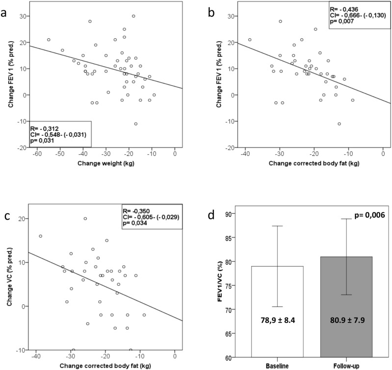
The change of vital capacity (R = −0.35, p 0.03) and FEV1 (R = −0.44, <p 0.01) negatively correlated with the change of body fat. (Figure 4). The change of FEV1 (R = –0.31, p = 0.03) (Figure 4), but not of vital capacity was correlated to the change of body weight.
Figure 4. Correlations of changes of pulmonary function with changes of body weight and changes of body fat and change of FEV1/VC from baseline to follow-up.
a–c: Correlations of changes of pulmonary function with changes of body weight and changes of body fat (a–c) d: Change of FEV1/VC (%) ratio from baseline to follow-up (d), (data as mean ± SD), p = 0.006. VC = vital capacity. FEV1 = forced expired volume at one second.
Additionally we noticed a decrease of myocardial wall thickness (−0.08±0.20 cm, p = 0.02), an improvement of left ventricular myocardial performance index (−0.16±0.35, p = 0.02), and mitral annular plane systolic excursion MAPSE (+0.14 cm±0.36, p = 0.03). Pulmonary outflow acceleration time increased (AT +26.6 ms±41.3, p = 0.001) indicating a decrease of pulmonary vascular resistance.
The decrease of myocardial wall thickness and the improvement of left myocardial performance index were positively correlated with the decrease of body weight (Figure 5).
Figure 5. Correlations of changes of left myocardial performance index and myocardial wall thickness with changes of body weight.
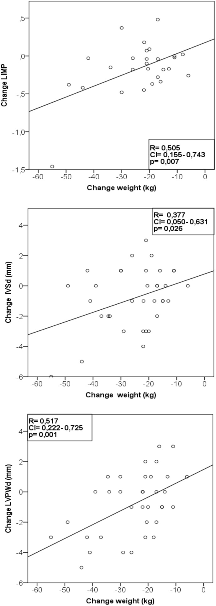
LIMP = left myocardial performance index. LVPWd = diastolic left ventricular posterior wall diameter; IVSd = diastolic interventricular septal wall thickness.
Furthermore, the subjects with a history of asthma (n = 5) showed an absolute increase of FEV1 (+13.8%, p<0.001). An absolute decrease of residual volume (RV) of −15.8% led to an absolute increase of VC (+10.6%, p = 0.005). Due to the increase of FEV1 and VC the absolute increase of FEV1/FV (+1.1%, p = 0.6) was not significant (Table S3).
The patients with OSA showed an absolute increase of FEV1 (+12.3%, p = 0.02) and VC (+10.3%, p = 0.007). P 0.1 decreased absolutely from 0.34 kPa to 0.26 kPa and P 0.1/Pi max from 0.06 to 0.04. Due to the small sample size (n = 2) the difference did not reach significance (Table S3).
The subgroup analysis of the pooled subjects with cardiocirculatory conditions (arterial hypertension n = 23, chronic heart failure n = 1, coronary artery disease n = 1) showed an absolute increase of FEV1 (+11.6%, p<0.001), VC (+7.5, p<0.001), FEV1/VC (+3.0, p = 0.046) and an absolute increase of P 0.1 (+0.07 kPa, p = 0 006) (Table S3).
Discussion
There is no doubt that obesity is associated with a large variety of diseases including cardiopulmonary dysfunction [46], [47], and is a major contributor to morbidity [2], [3] and mortality [3], [4], [46]. However, surprisingly little is known about the influence of obesity on cardiac and pulmonary function in obese individuals who have none or very mild symptoms. Importantly, it is unclear whether dietary weight loss programs lead to improved cardiopulmonary function similar to what has been reported for surgical weight loss programs [34].
The study cohort consisted of severely obese individuals the majority presenting asymptomatic in NYHA Class I without dyspnea. Although some subjects had comorbidities such as arterial hypertension, diabetes and dyslipidemia, which may have contributed to the increase in LV wall thickness, they had no obvious end organ damage. In our cohort we detected functional pulmonary and cardiac abnormalities at baseline which significantly improved after weight loss.
Obesity can lead to OHS and contribute to elevated pulmonary artery pressure in OHS. However, not all obese subjects develop OHS [12] and not all patients with OHS show PH [13]. Although in our cohort maximal inspiratory mouth pressures was normal [38], [39] and there was only a mild decrease of airflow the subjects showed an impairment of P 0.1 and P0.1/Pi max [38], [39]. The increased P0.1 and P0.1/Pi max in asymptomatic subjects highlights the increased respiratory load early on in obese subjects. The results are in line with data reporting abnormal breathing patterns in obese subjects with no history of pulmonary disease [15]. Despite the mild changes of ventilatory parameters at baseline the correlation of improving VC and FEV1 with loss of body fat further strengthens the assumption that obesity has a major impact on ventilatory function in these individuals.
Although an association of asthma and overweight has been proposed [17], [18], there is still an ongoing debate on the degree of the interaction between obesity and asthma [21], [30]. Our subjects showed a significant correlation of weight and body fat with pulmonary functional impairment. The importance of body fat distribution for ventilatory function has been described previously [17]. Our subjects showed a correlation of waist-to-hip ratio with vital capacity and TLC indicating a predominant role of central obesity for ventilatory abnormalities.
Obesity has a high impact on cardiac morbidity and mortality [1], [4]. An association between obesity and pulmonary mortality has also been proposed [46]. Cardiovascular disease [47], obstructive sleep apnea [9] and hypoventilation syndrome [10] are associated with obesity. Recent data suggest a contribution of obesity to the development of hypoventilation associated severe pulmonary hypertension [11], an independent influence of body weight on pulmonary hypertension in diastolic left ventricular function [8] and also obesity cardiomyopathy [6], [7].
The evaluation of myocardial function in obese subjects needs to differentiate hypercontractility [6], [7] in early stages of obesity, impaired left ventricular function in patients affected by manifest cardiomyopathy and additional parameters of myocardial function such as left and right myocardial performance index [48], [49], annular tissue velocity and annular valve flow parameters reflecting diastolic function. Despite left ventricular ejection fraction and TAPSE being normal, we found elevated left and right myocardial performance index as an early sign of cardiac (diastolic and systolic) functional distress [43], [48], [49]. Myocardial wall thickness of the interventricular septum and posterior wall was mildly enlarged [42]. Echocardiography in severely obese individuals is difficult to perform, but all echocardiographic procedures were performed by a highly experienced physician. Due to absence of detectable tricuspid valve insufficiency, right ventricular systolic pressure could not be estimated in the majority of tests. However, right ventricular outflow acceleration time (RVOT AT) could be measured in nearly all subjects. Therefore RVOT AT is a helpful non-invasive parameter for a qualitative analysis of pulmonary blood flow and its reduction is associated with decreasing pulmonary artery pressure and vascular resistance [50]. The acceleration time of the right ventricular outflow tract was mildly decreased in our cohort, suggesting the presence of increased pulmonary artery pressure and pulmonary vascular resistance [50].
The positive correlation of myocardial wall thickness and the negative correlation of pulmonary outflow tract acceleration time with the waist- to-hip ratio suggest an influence of central obesity on cardiac function and pulmonary perfusion. The morphological cardiac and functional cardiopulmonary changes may have been mild, but the improvement of these parameters as well as the correlation of weight and body fat loss with myocardial wall thickness and improvement of myocardial performance index highlight the clinical significance of the results. The correlation between decreased body fat and improved vital capacity and FEV1 following weight loss further suggests that the abnormalities are part of an early pathophysiological process leading ultimately to disease in obese individuals.
Weight loss is strongly recommended for patients with obesity related diseases such as obstructive sleep apnea [51], [52], but the effect of early intervention on lung and cardiac function in asymptomatic obese subjects is less clear. It has previously been shown that surgical induced weight loss has positive effects on cardiac function [31], [53], [54], [55], [56]. Syed reported a decrease of myocardial mass, but no significant improvement of functional parameters following a structured program combining diet, exercise and surgical procedures. However, in this study left myocardial performance index and acceleration time in the right ventricular outflow tract was not investigated [34]. There are only few reports on dietary induced weight loss and its effect on cardiac and pulmonary function. Reduction of body fat improved lung function after a Mediterranean diet [32]. A modest improvement of lung volumes at rest after a moderately successful dietary weight loss was shown and a relationship with the amount of chest fat was discussed [33].
In our study, the participants achieved a remarkable weight loss, that was substantially higher than in previous reports [32], [33] leading to a highly significant increase in lung volumes, ventilatory flow parameters and a decrease of respiratory load. We found significant improvement of early indicators of disturbed myocardial performance such as LIMP and MAPSE. Additionally our subjects had an improvement of myocardial wall thickness following successful weight loss. We noticed an improvement of the RVOT AT following weight loss in our study subjects. This may indicate reduced pulmonary vascular resistance and improved blood flow following a dietary induced weight loss.
Our cohort consisted of asymptomatic or almost asymptomatic obese patients. There were only few patients with relevant comorbidities. Subjects with a history of asthma and sleep apnea showed an improvement of pulmonary function comparable to the improvement of the whole cohort. The lack of significance of FEV1/FVC in the asthmatics is the result of an increase of FEV1 and vital capacity and of the small sample size of subjects with comorbidities. Although there is a clear signal towards an improvement of lung function in the few subjects with a history of asthma and OSA and cardiocirculatory conditions, this should be investigated in further studies especially addressing subjects with comorbidities.
Our study has several limitations. Not all subjects seen at baseline were examined after the weight loss program, but only patients with data available at both baseline and three month follow-up period were included into the analysis of changes in each parameter. Bioelectrical impedance analysis was not performed in all patients. However, the robustness of our data still suggest that asymptomatic individuals without end organ damage entering a structured weight loss program show mild cardiac and pulmonary functional impairment associated with weight and body fat. This can be reversed solely by a structured dietary induced weight loss.
Conclusion
In a cohort of severely obese asymptomatic adults we found evidence for early functional pulmonary and cardiac distress. Lung volumes and left ventricular myocardial performance index were negatively correlated to body weight. Airway flow is negatively and respiratory load is positively correlated to body fat. Myocardial wall thickness is positively correlated to body weight. Right ventricular outflow tract acceleration time and myocardial wall thickness is correlated to waist-to-hip ratio. A structured dietary program resulted in significant improvement of ventilatory and myocardial parameters in these obese individuals strongly suggesting that aggressive weight loss programs as early as possible may be able to prevent cardiopulmonary morbidity.
Supporting Information
Results of Bioelectric Impedance analysis at baseline.
(DOCX)
History of comorbidities.
(DOCX)
Weight, BMI and pulmonary function test data of subjects with a history of asthma, obstructive sleep apnea or cardiocirculatory conditions. (arterial hypertension n = 23, chronic heart failure n = 1, coronary artery disease n = 1).
(DOCX)
Acknowledgments
We thank Mrs. Monika Nagel and Mrs. Kathrin Wolf for the support with the data acquisition.
The data were presented as a poster at the ATS Annual Meeting 2013.
Data Availability
The authors confirm that all data underlying the findings are fully available without restriction. All relevant data are within the paper and its Supporting Information files.
Funding Statement
The authors have no support or funding to report.
References
- 1.World Health Organisation (2011) Global status report on noncommunicable diseases 2010 Chapter 1 burden, mortality, morbidity and risk factors. Geneva, World Health Organisation. Publication details: Editors: World Health OrganizationNumber of pages: 176 Publication date: April 2011, Languages: English. ISBN: 978 92 4 156422 9.
- 2. Seidell JC, de Groot LC, van Sonsbeek JL, Deurenberg P, Hautvast JG (1986) Associations of moderate and severe overweight with self-reported illness and medical care in Dutch adults. Am J Public Health 76: 264–269. [DOI] [PMC free article] [PubMed] [Google Scholar]
- 3. Negri E, Pagano R, Decarli A, La Vecchia C (1988) Body weight and the prevalence of chronic diseases. J Epidemiol Community Health 42: 24–29. [DOI] [PMC free article] [PubMed] [Google Scholar]
- 4. Flegal KM, Kit BK, Orpana H, Graubard BI (2013) Association of all-cause mortality with overweight and obesity using standard body mass index categories: a systematic review and meta-analysis. JAMA 309(1): 71–82. [DOI] [PMC free article] [PubMed] [Google Scholar]
- 5. Konnopka A, Bödemann M, König HH (2011) Health burden and costs of obesity and overweight in Germany. Eur J Health Econ 12(4): 345–52. [DOI] [PubMed] [Google Scholar]
- 6. Pascual M, Pascual DA, Soria F, Vicente T, Hernández AM, et al. (2003) Effects of isolated obesity on systolic and diastolic left ventricular function. Heart 89(10): 1152–1156. [DOI] [PMC free article] [PubMed] [Google Scholar]
- 7. Alpert MA (2001) Obesity cardiomyopathy: pathophysiology and evolution of the clinical syndrome. Am J Med Sci 321(4): 225–36. [DOI] [PubMed] [Google Scholar]
- 8. Leung CC, Moondra V, Catherwood E, Andrus BW (2010) Prevalence and risk factors of pulmonary hypertension in patients with elevated pulmonary venous pressure and preserved ejection fraction. Am J Cardiol 106(2): 284–6. [DOI] [PubMed] [Google Scholar]
- 9. Hawrylkiewicz I, Sliwinski P, Gorecka D, Plywaczewski R, Zielinski J (2004) Pulmonary haemodynamics in patients with OSAS or an overlap syndrome. Monaldi Arch Chest Dis 61: 148–152. [DOI] [PubMed] [Google Scholar]
- 10. Kessler R, Chaouat A, Schinkewitch P, Faller M, Casel S, et al. (2001) The Obesity Hypoventilation syndrome revisited: a prospective study of 34 consecutive cases. Chest 120: 369–376. [DOI] [PubMed] [Google Scholar]
- 11. Held M, Walthelm J, Baron S, Roth C. Jany BH (2014) Functional impact of pulmonary hypertension due to hypoventilation and changes under NIPPV. Eur Respir J 43: 156–165. [DOI] [PubMed] [Google Scholar]
- 12. Sugerman HJ, Fairman RP, Baron PL, Kwentus JA (1986) Gastric surgery for respiratory insufficiency of obesity. Chest 90: 81–96. [DOI] [PubMed] [Google Scholar]
- 13. Sugerman HJ, Baron PL, Fairman RP, Evans CR, Vetrovec GW (1988) Hemodynamic Dysfunction in Obesity Hypoventilation Syndrome and the effect of treatment with surgically induced weight loss. Ann Surg 207: 604–613. [DOI] [PMC free article] [PubMed] [Google Scholar]
- 14. Koenig SM (2001) Pulmonary complications of obesity. Am J Med Sci 321(4): 249–279. [DOI] [PubMed] [Google Scholar]
- 15. Chlif M, Keochkerian D, Choquet D, Vaidie A, Ahmaidi S (2009) Effects of obesity on breathing pattern, ventilatory neural drive and mechanics. Respir Physiol Neurobiol 2009 168(3): 198–202. [DOI] [PubMed] [Google Scholar]
- 16. Dixon AE, Hoguin F, Sood A, Salome CM, Pratley RE, et al. (2010) on behalf of the American Thoracic Society Ad Hoc Subcommittee on Obesity and Lung Disease (2010) American Thoracic Society Documents. An Official American Thoracic Society Workshop report: Obesity and Asthma. Proc Am Thor Soc 7: 325–335. [DOI] [PubMed] [Google Scholar]
- 17. Brumpton B, Langhammer A, Romundstad P, Chen Y, Mai XM (2013) General and abdominal obesity and incident asthma in adults: the HUNT study. Eur Respir J 41(2): 323–9. [DOI] [PubMed] [Google Scholar]
- 18. Shore SA (2013) Obesity and asthma: location, location, location. Eur Respir J 41: 253–254. [DOI] [PMC free article] [PubMed] [Google Scholar]
- 19. Camargo CA Jr, Weiss ST, Zhang S, Willett WC, Speizer FE (1999) Prospective study of body mass index, weight change, and risk of adult-onset asthma in women. Arch Intern Med 159(21): 2582–8. [DOI] [PubMed] [Google Scholar]
- 20. Beuther DA, Sutherland ER (2007) Overweight, obesity, and incident asthma: a meta-analysis of prospective epidemiologic studies. Am J Respir Crit Care Med 175(7): 661–6. [DOI] [PMC free article] [PubMed] [Google Scholar]
- 21. Aaron SD, Vandemheen KL, Boulet LP, Mc Ivor RA, Fitzgerald JM, et al. (2008) Canadian Respiratory Clinical Research Consortium. Overdiagnosis of asthma in obese and nonobese adults. CMAJ 179(11): 1121–31. [DOI] [PMC free article] [PubMed] [Google Scholar]
- 22. Boulet LP (2013) Asthma and obesity. Clin Exp Allergy. 2013 Jan 43(1): 8–21. [DOI] [PubMed] [Google Scholar]
- 23. Farah CS, Salome CM (2012) Asthma and obesity: a known association but unknown mechanisms. Respirology 17: 12–21. [DOI] [PubMed] [Google Scholar]
- 24. Burger CD, Foreman AJ, Miller DP, Safford RE, McGoon MD, et al. (2011) Comparison of body habitus in patients with pulmonary arterial hypertension enrolled in the Registry to Evaluate Early and Long-term PAH Disease Management with normative values from the National Health and Nutrition Examination Survey. Mayo Clin Proc 86(2): 105–12. [DOI] [PMC free article] [PubMed] [Google Scholar]
- 25. Medoff BD, Okamoto Y, Leyton P, Weng M, Sandall BP, et al. (2009) Adiponectin deficiency increases allergic airway inflammation and pulmonary vascular remodeling. Am J Respir Cell Mol Biol 41(4): 397–406. [DOI] [PMC free article] [PubMed] [Google Scholar]
- 26. Summer R, Fiack CA, Ikeda Y, Sato K, Dwyer D, et al. (2009) Adiponectin deficiency: a model of pulmonary hypertension associated with pulmonary vascular disease. Am J Physiol Lung Cell Mol Physiol 297(3): L432–8. [DOI] [PMC free article] [PubMed] [Google Scholar]
- 27. Weng M, Raher MJ, Leyton P, Combs TP, Scherer PE, et al. (2011) Adiponectin decreases pulmonary arterial remodeling in murine models of pulmonary hypertension. Am J Respir Cell Mol Biol 45(2): 340–7. [DOI] [PMC free article] [PubMed] [Google Scholar]
- 28. Summer R, Walsh K, Medoff BD (2011) Obesity and pulmonary arterial hypertension: Is adiponectin the molecular link between these conditions? Pulm Circ 1(4): 440–7. [DOI] [PMC free article] [PubMed] [Google Scholar]
- 29. Rabec C, de Lucas Ramos P, Veale D (2011) Respiratory complications of obesity. Arch Bronconeumol 47(5): 252–61. [DOI] [PubMed] [Google Scholar]
- 30. Schachter LM, Salome CM, Peat JK, Woolcock AJ (2001) Obesity is a risk for asthma and wheeze but not airway hyperresponsivness. Thorax 56: 4–8. [DOI] [PMC free article] [PubMed] [Google Scholar]
- 31. Cavarretta E, Casella G, Calì B, Dammaro C, Biondi-Zocai G, et al. (2013) Cardiac Remodeling in Obese Patients After Laparoscopic Sleeve Gastrectomy. World J Surg 37: 565–72. [DOI] [PubMed] [Google Scholar]
- 32. De Lorenzo A, Petrone-De Luca P, Sasso GF, Carbonelli MG, Rossi P, et al. (1999) Effects of weight loss on body composition and pulmonary function. Respiration 66: 407–412. [DOI] [PubMed] [Google Scholar]
- 33. Babb TG, Wyrick BL, Chase PJ, De Lorey DS, Rodder SG, et al. (2011) Weight loss via dieat and exercise improves exercise breathing mechanics in obese men. Chest 140(2): 454–460. [DOI] [PubMed] [Google Scholar]
- 34. Syed M, Rosati C, Torosoff MT, El-Hajjar M, Feustel P, et al. (2009) The impact of weight loss on cadiac structureand function in obese patients. Obes Surg 19: 36–40. [DOI] [PubMed] [Google Scholar]
- 35. Bischoff SC, Damms-Machado A, Betz C, Herpertz S, Legenbauer T, et al. (2012) Multicenter evaluation of an interdisciplinary 52-week weight loss program for obesity with regard to body weight, comorbidities and quality of life – a prospective study. Internationla Journal of Obesity 36: 614–624. [DOI] [PMC free article] [PubMed] [Google Scholar]
- 36.Quanjer PH, Tammeling GJ, Gotes JE, Pedersen OF, Peslin R, et al. (1993) Lung volumes and forced ventilatory flows. Report Working Party Standardization of Lung Functional Tests, European Community for Steel and Coal. Official Statement of the European Respiratory Society. Eur Respir J Suppl. 16: 5–40. [PubMed]
- 37. ATS/ERS Statement on Respiratory Muscle Testing (2002) AM J Respir Crit Care Med. 166: 518–624. [DOI] [PubMed] [Google Scholar]
- 38. Koch B, Schäper C, Ittermann T, Bollmann T, Völzke H, et al. (2010) Reference values for respiratory pressures in a general adult population – results of the Study of Health in Pomerania (SHIP). Clin Physiol Funct Imaging 30: 460–465. [DOI] [PubMed] [Google Scholar]
- 39. Criée CP (2003) Recommendations of the German Airway League. Pneumologie 57: 98–100. [DOI] [PubMed] [Google Scholar]
- 40. Hautmann H, Hefele S, Schotten K, Huber RM (2000) Maximal inspiratory mouth pressures (Pimax) in healthy subjects. Respir Med 94: 689–693. [DOI] [PubMed] [Google Scholar]
- 41.Windisch W, Hennings E, Sorichter S, Hamm H, Criée CP (2002) Peak and plateau inspiratory mouth pressures in healthy subjectsat different lung volumes. Eur Respir J 20 Suppl. 38: 498 s.
- 42. Lang RM, Bierig M, Devereux RB, Flachskampf FA, Foster E, et al. (2006) Recommendations for chamber quantification. Eur J Echocardiography 7: 79–108. [DOI] [PubMed] [Google Scholar]
- 43. Rudski LG, Lai WW, Afilalo J, Hua L, Handschumacher MD, et al. (2010) Guidelines for the echocardiographic assessment of the right heart in adults: a report from the American Society of Echocardiography. Endorsed by the European Association of Echocardiography, a registered branch of the European Society of Cardiology, and the Canadian Society of Echocardiography. J Am Soc Echocardiography 23: 685–713. [DOI] [PubMed] [Google Scholar]
- 44. Kyle UG, Bosaeus I, De Lorenzo AD, Deurenberg P, Elia M, et al. (2004) Composition of the ESPEN Working Group. Bioelectrical impedance analysis – part I: review of principles and methods. Clinical Nutrition 23: 1430–1453. [DOI] [PubMed] [Google Scholar]
- 45. Kyle UG, Bosaeus I, De Lorenzo AD, Deurenberg P, Elia M, et al. (2004) Bioelectrical impedance analysis – part II: utilization in clinical practice. Clinical Nutrition 23: 1430–1453. [DOI] [PubMed] [Google Scholar]
- 46. Haque AK, Gadre S, Taylor J, Haque SA, Freeman D, et al. (2008) Pulmonary and cardiovascular complications of obesity – an autopsy study of 76 obese subjects. Arch Pathol Lab Med 132: 1397–1404. [DOI] [PubMed] [Google Scholar]
- 47. Hubert HB, Feinlieb M, Mc Namara PM, Castelli WP (1983) Obesity as an independent risk factor for cardiovascular diasease: a 26-year follow-up of participants in the Framingham heart Study: Circulation. 67: 968–977. [DOI] [PubMed] [Google Scholar]
- 48. Tei C, Ling LH, Hodge DO, Oh JK, Rodeheffer RJ, et al. (1995) New index of combined systolic and diastolic myocardial performance: a simple and reproducible measure of cardiac function–a study in normals and dilated cardiomyopathy. J Cardiol 26(6): 357–66. [PubMed] [Google Scholar]
- 49. Tei C, Dujardin KS, Hodge DO, Bailey KR, McGoon MD, et al. (1996) Doppler echocardiographic index for assessment of global right ventricular function. J Am Soc Echocardiogr. 9(6): 838–47. [DOI] [PubMed] [Google Scholar]
- 50. Chan K-L, Currie PJ, Seward JB, Hagler DJ, Mair DD, et al. (1987) Comparison of three Doppler ultrasound methods in the prediction of pulmonary artery pressure. JACC 9(3): 549–554. [DOI] [PubMed] [Google Scholar]
- 51. Tuomilehto HP, Seppä JM, Partinen MM, Peltonen M, Gylling H, et al. (2009) Lifestyle intervention with weight reduction: first-line treatment in mild obstructive sleep apnea. Am J Respir Crit Care Med 179: 320–7. [DOI] [PubMed] [Google Scholar]
- 52. Qaseem A, Holty J-EC, Owens DK, bDallas P, Starkey M, et al. (2013) Management of Obstructive Sleep Apnea in Adults: A Clinical Practice Guideline From the American College of Physicians and Clinical Guidelines Committee of the American College of Physicians Ann Intern Med. 159(7): 471–483 doi:10.7326/0003-4819-159-7-201310010-00704 [DOI] [PubMed] [Google Scholar]
- 53. Kardassis D, Bech-Hanssen O, Schöander M, Sjöström L, Petzold M, et al. (2012) Impact of body composition, fat distribution and sustained weight loss on cardiac function in obesity. Int J Cardiol 150: 128–133. [DOI] [PubMed] [Google Scholar]
- 54. Thomas PS, Cowen ERT, Hulands G, Milledge JS (1989) Respiratory function in the morbidly obese before and after weight loss. Thorax 44: 382–386. [DOI] [PMC free article] [PubMed] [Google Scholar]
- 55. Santana ANC, Souza R, Martins AP, Macedo F, Rascovski A, et al. (2006) The effect of massive weight loss on pulmonary function of morbid obese patients. Respiratory Medicine 100: 1100–1104. [DOI] [PubMed] [Google Scholar]
- 56. Valencia-Flores M, Orea A, Herrera M, Santiago V, Rebollar V (2004) Effect of Bariatric Surgery on obstructive Sleep apnea and hypopnea syndrome, electrocardiogram, and pulmonary arterial hypertension. Obesity Surgery 14: 755–762. [DOI] [PubMed] [Google Scholar]
Associated Data
This section collects any data citations, data availability statements, or supplementary materials included in this article.
Supplementary Materials
Results of Bioelectric Impedance analysis at baseline.
(DOCX)
History of comorbidities.
(DOCX)
Weight, BMI and pulmonary function test data of subjects with a history of asthma, obstructive sleep apnea or cardiocirculatory conditions. (arterial hypertension n = 23, chronic heart failure n = 1, coronary artery disease n = 1).
(DOCX)
Data Availability Statement
The authors confirm that all data underlying the findings are fully available without restriction. All relevant data are within the paper and its Supporting Information files.


