Abstract
A case of abdominal pregnancy in a 39 year old female gravida 4, para 0+3 is presented. Ultrasonography revealed a viable abdominal pregnancy at 15 weeks gestational age. She was initially managed conservatively. Surgical intervention became necessary at 20 weeks gestational age following Ultrasound detection of foetal demise. The maternal outcome was favourable. This case is presented to highlight the dilemma associated with diagnosis and management of abdominal pregnancy with a review of literature.
Introduction
Ectopic pregnancy is very rarely sited in the abdominal cavity. It is normal for implantation of the fertilized ovum to occur within the uterine cavity. When implantation takes place anywhere outside the uterine cavity it is referred to as an ectopic pregnancy. Abdominal pregnancy is a rare type of ectopic pregnancy where the developing embryo implants and grows within the peritoneal cavity. Abdominal pregnancy can further be classified as being primary or secondary. Primary abdominal pregnancy which is extremely rare occurs when a fertilized ovum implants itself initially on some abdominal organ. Most cases of abdominal pregnancy are secondary in that the ovum first implants in the fallopian tube, ovary or uterus and subsequently escapes through a rupture into the peritoneal cavity.
There are reported cases of abdominal pregnancy developing to term with delivery of a live foetus through an abdominal incision1. There is a significant risk of maternal intra-peritoneal haemorrhage with fatal consequences. The overall foetal survival rate remains low2
Reports
Case Report
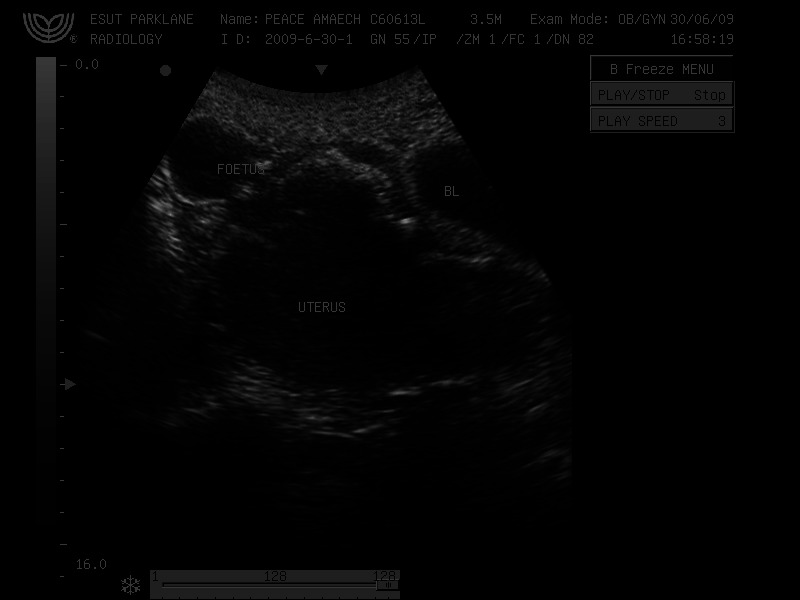
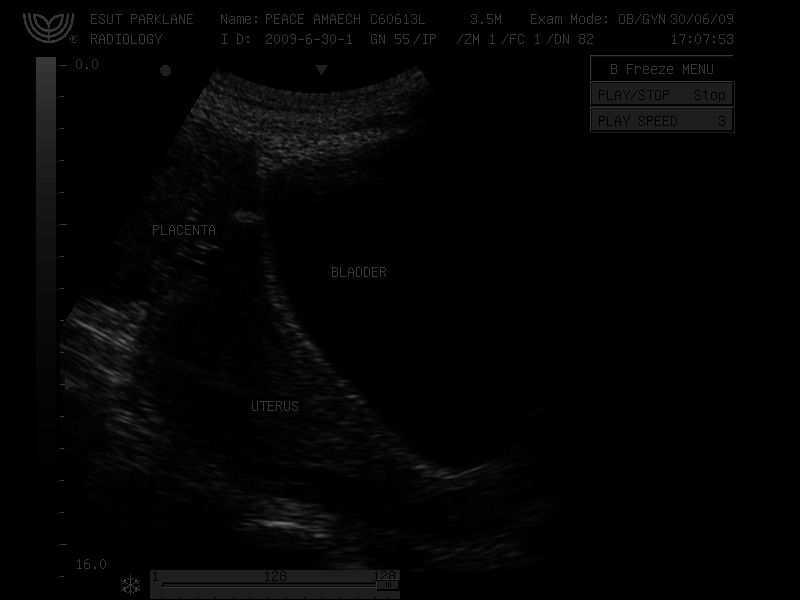
Mrs. A., a 39-year old gravida 4, para 0+3 presented to the accident and emergency department with a 2-month history of abdominal discomfort, persistent vomiting, diarrhea and jaundice. She could not remember her last menstrual date but claimed she was three months pregnant. She had taken some herbal concoctions prior to presentation. On examination she was found to be febrile (37.7oc), very pale (PCV-19%), hypotensive (BP-80/50mmHg) and clinically jaundiced. Investigation confirmed a haemolytic jaundice with a total bilirubin of 63.2mmol/L and normal liver enzymes. Her serum Urea and Creatinine levels were normal. Her urine contained blood elements and protein. Her blood group was A+ and she tested negative for HIV. Urgent abdominopelvic Ultrasonography was requested as part of her initial gynaecological assessment. Trans-abdominal Ultrasonography revealed a live and active intra-abdominal foetus floating freely in the peritoneal cavity close to the maternal anterior abdominal wall [FIG 1]. The biometric parameters estimated foetal gestational age at 15 weeks. Other Ultrasound findings include a significant amount of clear peritoneal fluid, a left-sided suprapubic mass representing ectopic placenta, a bulky empty uterus separate from the placenta and foetus [FIG 2], normal liver size with unaltered echo-pattern. Subsequent Trans-abdominal Ultrasound using Doppler confirmed placental and umbilical cord colour-flow pattern and active movements of the extremities were recorded on real time Cine images.
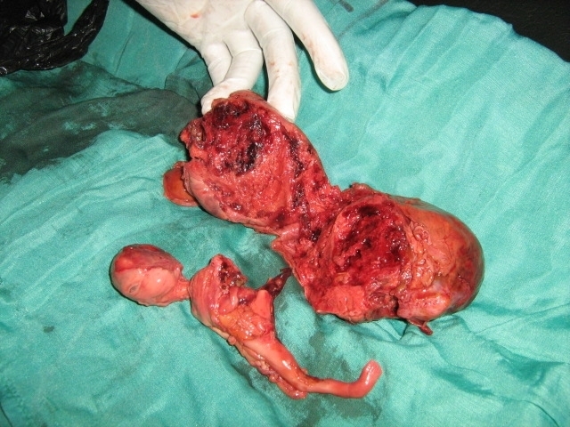
Mrs. A. was counseled, transfused with screened whole blood and further stabilized with conservative management over the next four weeks. She was regularly monitored using Ultrasonography. Laparotomy was performed at 20 weeks gestation following Ultrasound diagnosis of foetal demise. A sub-umbilical midline incision was made after making sure that there was no clinical vascular bruit. The placenta was attached outside the uterus at the fundus and received a mass of blood vessels from unidentified branches of the left internal iliac artery. The umbilical cord was seen lying free in the peritoneal cavity and was traced to the foetus in the right iliac fossa. The placenta was completely removed from its ectopic location at the peritoneal surface of the uterine fundus and its size correlated with pre-operative ultrasound dimensions. The left fallopian tube and left ovary were matted with the placental vascular supply in an inflammatory mass which was also excised. This finding led us to believe that this was a case of secondary abdominal pregnancy. Rupture of the amniotic sac was suspected as the leading cause of foetal demise. The lifeless foetus was delivered from the right iliac fossa. The foetus appeared malformed with a missing left lower limb [FIG 3]. This was not detected by ultrasound done before 20 weeks. The Uterus and right fallopian tube were found to be intact and normal in appearance. Intra-operative blood transfusion was not required even though 2 pints of cross-matched blood were on standby. Blood loss was estimated to be 400 mls. The abdomen was closed in layers and the recovery was uneventfully. She was discharged ten days later and was reviewed after six weeks with no fresh complaints.
Conclusions
Early accurate diagnosis of abdominal pregnancy remains vital for reducing morbidity and mortality in this potentially life-threatening entity. Ultrasonography in experienced hands has very high sensitivity and specificity as a diagnostic tool and continues to play a significant monitoring role in the pre-operative and post-operative periods. The management method is influenced by adequate patient counseling and the overall clinical picture. Prompt surgical intervention with careful evaluation of the placenta before its removal offers the best prognosis.
Discussion
A high index of suspicion is needed to make a first time diagnosis of abdominal pregnancy3. Diagnosis is missed in one-fourth of reported cases4.
The reported incidence of abdominal pregnancy varies widely with geographical location ranging between 1: 10,000 deliveries in the USA5,6 and 1:654 deliveries in Ibadan-Nigeria2. Multiparity and poor socio-economic status are implicated as epidemiological factors7.
Clinical presentation can be variable with abdominal pain occurring at 16-17 weeks gestation8,9 as was observed in our patient. The finding of clinical jaundice and severe anaemia as part of presentation in our patient was unusual. Jaundice associated with pregnancy has been described by Holzbach10 in three disease entities which include: 1) recurrent cholestasis of pregnancy (RCP); 2) viral hepatitis coincident with pregnancy; 3) acute fatty liver of pregnancy (AFLP). Although our patient was not screened for Hepatitis B surface antigen (HBs Ag), the finding of blood elements in her urine against a background of normal liver enzymes suggested that her jaundice was due to a haemolytic crises probably caused by ingestion of herbal concoctions.
The diagnosis of early abdominal pregnancy is by β-hCG estimation and Ultrasonography. In the case of our patient, Ultrasonography was the single stand-alone test used to diagnose abdominal pregnancy.
Allibone GW et al 11 described major criteria for sonographic diagnosis of intra-abdominal pregnancy. These include:
1) demonstration of foetus in a gestational sac outside the uterus, or the depiction of an abdominal or pelvic mass identifiable as the uterus separate from the foetus; 2) failure to see a uterine wall between the foetus and the urinary bladder; 3) recognition of a close approximation of the foetus to the maternal abdominal wall; and 4) localization of the placenta outside the confines of the uterine cavity. All of these features were recognized in our patient. More recent literature listed other additional criteria such as oligohydramnios, abnormal foetal lie, placenta previa appearance and maternal bowel gas impeding foetal visualization4. Magnetic resonance imaging (MRI T2-WI), or colour Doppler Ultrasound could be used to localize the placenta8,12. Where resources abound placental localization by Magnetic resonance imaging offers the best method of diagnosis. In our case, colour Doppler Ultrasound was used with accuracy. Evaluation of gross foetal morphology can be further assisted by use of 3-D Ultrasonography where this is available. The outlook for the foetus in abdominal pregnancy is poor13. The perinatal mortality varies from 85 to 95%14, and the rate of foetal deformation is reported to range from 20 to 90%15,16. The most common deformations and malformations were observed in the exposed areas of the foetus such as the head and the extremities16. The intraoperative finding of a missing left lower limb from the foetus in this case was not suggested by earlier ultrasound images where cine-recordings depicted active movement of both lower extremities. The final Ultrasound scan at 20 weeks gestational age when foetal demise was detected revealed only one lower limb in extended fixed position. Cathy A. Stevens15 proposed two etiological mechanisms for the foetal limb defects in abdominal pregnancy. These mechanisms which are extrinsic compression and vascular disruption may have resulted in foetal auto-amputation in this case.
Current concepts on management of abdominal pregnancy support immediate active surgical intervention with termination of the pregnancy if diagnosed before 24 weeks gestation5,17,18. In patients who present after 24 weeks, the appropriateness of conservative management5 is debatable. There is need to assess each individual case and adopt the most appropriate method with a view to limiting materno-foetal morbidity and mortality. A conservative approach requires close surveillance of the patient and regular monitoring using Ultrasonography. The patient should be admitted into hospital where blood bank facilities and resources needed for rapid surgical intervention are obtainable. Intra-operative management of the placenta poses another dilemma for the clinician. Although removal of the placenta offers a better prognosis2, this should not be attempted if there is any risk of massive haemorrhage with a fatal outcome. Placentas left in-situ usually regress gradually and are monitored with serial serum β-hCG estimation and Ultrasonography. The prophylactic use of methotrexate in placenta management is no longer advocated by some clinicians19. In their view, the necrosed placental tissue is a potent culture medium with increased risk of serious intraperitoneal infection.
Fig 1.

Trans-abdominal Ultrasound showing foetus separate from uterus and close to maternal anterior abdominal wall. BL – Maternal Bladder
Fig 2.

Trans- abdominal Ultrasound showing empty uterus and placenta separate from uterus. There is no myometrium between the placenta and the bladder.
Fig 3.

The malformed foetus and the placenta (cut open)
Contributor Information
II Okafor, Enugu State University Teaching Hospital, Department of Obstetrics & Gynaecology.
AC Ude, Enugu State University Teaching Hospital, Department of Clinical Radiology.
ASO Aderibigbe, Enugu State University Teaching Hospital, Department of Clinical Radiology.
OC Amu, Enugu State University Teaching Hospital, Department of Surgery.
PE Udeh, Enugu State University Teaching Hospital, Department of Surgery.
NEN Obianyo, Enugu State University Teaching Hospital, Department of Surgery.
COC Ani, Enugu State University Teaching Hospital, Department of Clinical Radiology.
References
- 1.Zeck W, Kelters I, Winter R, Lang U, Petru E. Lessons learned from four advanced abdominal pregnancies at an East African Health Center . J. Perinat Med. 2007;35(4):278–281. doi: 10.1515/JPM.2007.075. [DOI] [PubMed] [Google Scholar]
- 2.Stanley JH, Horger EO,, Fagan CJ, Andriole JG, Fleischer AC. Sonographic findings in abdominal pregnancy. AJR Am J Roentgenol . 1986;147(5):1043–1046. doi: 10.2214/ajr.147.5.1043. [DOI] [PubMed] [Google Scholar]
- 3.Ayinde OA, Aimakhu CO, Adeyanju OA, Omigbodun AO. Abdominal pregnancy at the University College Hospital, Ibadan : a ten-year review. Afr J. Reprod Health. 2005 ;9(1):123–127. [PubMed] [Google Scholar]
- 4.Lamina MA, Akinyemi BO, Fakoya TA, Shorunmu TO, Oladapo OT. Abdominal pregnancy: a cause of failed induction of labour . Niger J Med. 2005;14(2):213–217. doi: 10.4314/njm.v14i2.37183. [DOI] [PubMed] [Google Scholar]
- 5.Zhang J,, Sheng Q. Full-term abdominal pregnancy: a case report and review of literature . Gynecol Obstetrics Invest. 2008;65(2):139–141. doi: 10.1159/000110015. [DOI] [PubMed] [Google Scholar]
- 6.Sherer DM, Dalloul M, Gorelick C, Kheyman M, Abdelmalek E, Zinn H, Abulafia O. Unusual maternal vasculature in the placenta periphery leading to diagnosis of abdominal pregnancy at 25 weeks’ gestation . JOURNAL OF Clinical Ultrasound. 2007;35(5):268–273. doi: 10.1002/jcu.20375. [DOI] [PubMed] [Google Scholar]
- 7.Sfar E, Kaabar H, Marrakechi O, Zouari F, Chelli M, Kharouf M. Abdominal pregnancy, a rare anatomoclinical entity. 4 case reports . Rev Fr Gynecol Obstet. 1993;88(4):261–265. [PubMed] [Google Scholar]
- 8.Rahman MS, Al Suleiman SA, Rahman J, Al-Sibai MH. Advanced abdominal pregnancy- observation in 10 cases. Obstetrics Gynecol . 1982;59:366–372. [PubMed] [Google Scholar]
- 9.Paternoster DM, Santarossa C. Primary Abdominal Pregnancy- a case report. Minerva Ginecol. 1999;51(6):251–253. [PubMed] [Google Scholar]
- 10.Kun KY, Wong PY, Ho MW, Tai CM, Ng TK. Abdominal pregnancy presenting as a missed abortion at 16 weeks gestation. Hong Kong Medicine JOURNAL. 2000;6(4):425–427 . [PubMed] [Google Scholar]
- 11.Jazayeri A, Davis TA, Contreras DN. Diagnosis and management of abdominal pregnancy: A case report. J. Reprod Med. 2002;47(12):1047–1049. [PubMed] [Google Scholar]
- 12.Hsieh CH, Hsu TY, Changchien CC. Abdominal pregnancy- report of two cases and review of literature. Changgeng Yi Xue Za Zhi. 1994 ;17(3):268–275. [PubMed] [Google Scholar]
- 13.Holzbach RT. Jaundice in pregnancy. Am JOURNAL OF Medicine. 1976. pp. 367–376. [DOI] [PubMed]
- 14.Cathy A, Malformations and deformations in pregnancy. American Journal of Medicineical Genetics. 1993;47(8):1189–1195. doi: 10.1002/ajmg.1320470812. [DOI] [PubMed] [Google Scholar]
- 15.Bright AS, Maser AH. Advanced abdominal pregnancy- review of the recent literature and report of a case. Obstetrics Gynecol . 1961;17:316–324. [Google Scholar]
- 16.Brandt AL, Tolson D. JOURNAL OF Emerg Medicine. Vol. 2. 30; 2006. Missed abdominal ectopic pregnancy; pp. 171–174. [DOI] [PubMed] [Google Scholar]
- 17.Bertrand G, Le Ray C, Simand-Emond L, Dubois J, L Leduc. Imaging in the management of abdominal pregnancy: a case report and review of the literature. JOURNAL OF Obstetrics Gynaecology Can. 2009;31(1):57–62. doi: 10.1016/s1701-2163(16)34055-5. [DOI] [PubMed] [Google Scholar]
- 18.Allibone GW, Fagan CJ, Porter SC. The Sonographic features of intra-abdominal pregnancy . JOURNAL OF Clinical Ultrasound . 1981;9(7):383–387. doi: 10.1002/jcu.1870090706. [DOI] [PubMed] [Google Scholar]
- 19.Kun KY, Wong PY, Ho MW, Tai CM, Ng TK. Abdominal pregnancy presenting as a missed abortion at 16 weeks gestation . Hong Kong Med J. 2000;6(4):425–427. [PubMed] [Google Scholar]


