

Abstract
Complications that may occur while performing myomectomy in pregnancy can be prevented in a well-optimised surgery. Counselling and comprehensive peri-operative preparations are mandatory to minimise litigations and untoward events. Myomectomy in pregnancy remains a contentious issue. Degeneration of fibroid during pregnancy is common. However, conservative management suffices in majority of cases. In non-responsive conservative treatment, myomectomy may be an option. This article discusses our experience in treating a 38-year-old woman in her fourth pregnancy at 15 weeks gestation with symptomatic uterine fibroid. She had persistent abdominal pain since nine weeks gestation. She developed fever and acute abdomenat at 15 weeks and the uterus was larger than dates.Ultrasound scan confirmed single pregnancy with a large intramural fibroid showing degenerative changes. A myomectomy was performed as a preventive measure to prevent massive haemorrhage. Although performing myomectomy during pregnancy is considered controversial, complications can be minimised with properly-planned surgery.
Keywords: Fibroid, Myomectomy, Obstetrics, haemorrhage, Pregnancy
Introduction
Performing antenatal myomectomy remains controversial. Primary concerns include massive haemorrhage requiring blood transfusion, hysterectomy and foetal loss.
Uterine fibroid is usually asymptomatic. The surge in placental hormone resulting in an increase in fibroid size may complicate the pregnancy. There are possible risks of miscarriage, foetal growth restriction, preterm delivery and abnormal foetal presentation requiring operative delivery.1 Foetal anomalies, namely limb deformities and contractures, may occur as a result of uterine fibroid.2
Fibroids-related complications happen in 5-8% of all pregnancies and conservative management suffices in majority of cases.1 Hyalinisation and red degeneration contribute to 50% of the complications, leading to intolerable pain.3 With adequate analgesia, most pregnancies have a favourable outcome. Myomectomy in selected cases is reported to be safe to mother and her foetus. Lelis et al reported only one case (out of 13) where a woman had a miscarriage without any complication of hysterectomy or massive bleeding.3 Justifying myomectomy remains contentious; doctors should take preventive steps to minimise complications that can lead to deleterious outcomes. Although uterine fibroid in pregnancy is not uncommon, there were no cases of antenatal myomectomy reported in our centre in the past five years. Detailed assessment at the primary care level is important to risk stratify and identify cases that will be benefit from surgery.
Case Summary
A 38-year old woman, in her fourth pregnancy, presented at 15 weeks gestation, with an acute abdomen to a maternity unit in a district hospital in Makassar, Indonesia. She was diagnosed as having an anterior uterine fibroid measuring 10cm x 10cm at 9 weeks gestation. Although she experienced occasional abdominal pain, she had never sought medical attention. The pain became more persistent and severe severe at 10th week of gestation 10th week gestation. It lasted until 15th week when she experienced intolerable and acute abdominal pain and developed fever.
Upon assessment, she was febrile but haemodynamically stable. The size of the uterus corresponded to 22 weeks gestation. Minimal abdominal examination was done due to tenderness. An ultrasound scan showed an intramural fibroid of 15x10 cm size and a viable foetus. Both ovaries appeared normal and there was no evidence of placental abruption. Her haemoglobin level was 13 g/dL, and the total were white cell count and C-reactive protein was 15 × 109/L and 2.1 mg/dL, respectively; this indicated possible inflammatory reaction. She was admitted for observation and treated with regular parenteral opiods and antibiotics for possible infection. As the pain was severe, the patient responded poorly to the medical treatment. Although myomectomy is invasive, it is an option to curb debilitating pain in degenerative fibroid. The couple were informed of the risk of bleeding requiring transfusion as well as possible hysterectomy and miscarriage.
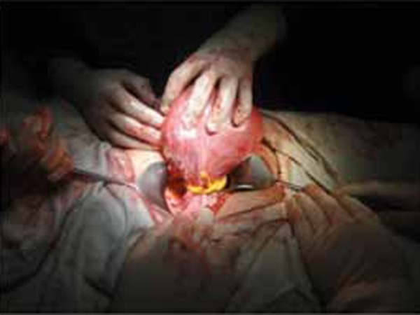
Maylard incision was performed and a huge uterine fibroid at the anterior wall was confirmed. A Foleys catheter was used to occlude both uterine arteries aſter the broad ligaments were penetrated at its base using a clamp, avoiding the vascular area (Figure 1). The tie was released every eight minutes to allow perfusion to the viable foetus. The fibroid was enucleated using sharp dissection and the peripheral feeding vessels were occluded by “double stitches” suturing technique (Figure 2).
Figure 1.
Application of tourniquet at the base of broad ligament to occlude bilateral uterine arteries
Figure 2.

An intramural fibroid is being enucleated
Following removal of the single 10x10 cm intramural fibroid, the cavity was obliterated by interrupted sutures and the serosal layer was closed by “continuous baseball” sutures. Haemostasis was secured and the tie was released with estimated blood loss of 100ml. The red regeneration of leiomyomata was confirmed histologically. Tocolysis and administing progesterone were considered but due to limited resources, none was administered. Patient had an elective caesarean section at 38 weeks due to presence of previous myomectomy.
Discussion
Management of uterine fibroid in pregnancy, especially those showing degenerative changes, remains controversial as there are concerns of detrimental effects on maternal health jeopardising foetal viability and growth. Generally, conservative management is recommended in uncomplicated cases. However, in symptomatic cases showing poor response to medical treatment, invasive procedure is an option.4,5 Myomectomy can be considered afrer comprehensive assessment and counselling to avoid untoward sequelae and risk of litigation.
Myomectomy is justified when there are degenerative changes of the fibroid in pregnancy causing intolerable pain. Heavy bleeding needing transfusion leading to risks of hysterectomy is a major concern in myomectomy procedure though this was not demonstrated in certain studies.2,5 This patient experienced persistent pain since nine weeks of pregnancy and it was associated with fever at 15 weeks which did not resolve with regular analgesia.
Injection of vasoconstrictor and Boney clamps application to the feeding vessels were found to be effective in minimising haemorrhage. The same principle was practiced in the current case by using a Foleys catheter to compress both uterine arteries at the base of the broad ligaments. Some medical practitioners recommended inclusion of the infundibulopelvic ligaments for fundal fibroid.6,7 However, another major concern was the reduction of foetal oxygenation if occlusion was prolonged. Even though, theoretically, there are collateral circulations following occlusion of the uterine supply, we were sceptical as the perfusion was less, leading to reduced bleeding during myomectomy in other cases.7,8 At present, there is no established consensus on this issue.
Traditionally, laparoscopic myomectomy in pregnancy is believed to be associated with foetal acidosis, foetal loss and bleeding. This could be due to carbon dioxide retention following pneumoperitoneum at surgery.8 However, this modality has become a trend with successful outcomes.9 A drastic improvement in learning curve may have contributed to its popularity. This treatment method was not applied in this case due to limited resources in this particular centre.
A meticulous surgery was performed to minimise haemorrhage, with double stitches suturing at the peripheral feeding vessels. Haemostasis was secured before closure and anti-adhesive gel was used to reduce adhesion. Although the endometrial lining was not encroached, an elective Caesarean section was is performed at 38 weeks to prevent uterine rupture in labour.
Prostaglandin inhibitor namely, indomethacin, had been prophylactically used to prevent preterm labour as a conservative medical treatment in uterine fibroid; it works by relaxing myometrial tissues. However, a prolonged use especially in advanced pregnancy can lead to complications with premature closure of ductus arteriosus, and hence, not recommended.10 The use of this agent in post myomectomy has not produced conclusive results. As progesterone quiescents the myometrial activity and prevents premature labour, its use is strongly recommended. A large meta-analysis significantly revealed a reduction of premature birth before 34 weeks in high-risk cases.11 However, it was not prescribed in this case due to limited resources.
Conclusion
Complications of myomectomy during pregnancy can be avoided in optimised surgery. Risk-benefit assessment must be entirely evaluated to justify the surgical option, especially when conservative management has failed. Proper counselling on possible risks, comprehensive peri-operative preparations are mandatory to minimise possible litigation and untoward events.
References
- 1.Phelan JP. Myomas pregnancy. Obstet. Gynecol. Clin. North Am. 1995;22:801–805. [PubMed] [Google Scholar]
- 2.Hasbargen U, Strauss A, Summerer-Moustaki M. et al. Myomectomy as a pregnancy-preserving option in the carefully selected patient. Fetal Diagn. Ther. 2002;17:101–103. doi: 10.1159/000048017. [DOI] [PubMed] [Google Scholar]
- 3.DE Lolis, SN Kalantaridou, G Makrydimas. et al. Successful myomectomy during pregnancy. Hum Reprod. 2003;18((8)):1699–1702. doi: 10.1093/humrep/deg318. [DOI] [PubMed] [Google Scholar]
- 4.Wittich AC, Salminen ER, Yancey MK. et al. Myomectomy during early pregnancy. Mil. Med. 2000;165:162–164. [PubMed] [Google Scholar]
- 5.Hasan F, Arumugam K, Sivanesaratnam V. Uterine leiomyomata in pregnancy. Int. J. Gynecol. Obstet. 1990;34:45–49. doi: 10.1016/0020-7292(91)90537-f. [DOI] [PubMed] [Google Scholar]
- 6.Omigbodun AO, Fawole AO. Myomectomy during Pregnancy and Delivery. Is it safe? Trop J Obstet Gynaecol. 2005;22((1)):1–3. [Google Scholar]
- 7.Sapmaz E, Celik H, Altungul A.. Bilateral ascending uterine artery ligation vs. tourniquet use for hemostasis in cesarean myomectomy. A comparison. J Reprod Med. 2003;48:950–4. [PubMed] [Google Scholar]
- 8.Sinha R, Sundaram M, Mahajan C. et al. Laparoscopic Myomectomy with Uterine Artery Ligation: Review Article and Comparative Analysis. J Gynecol Endosc Surg. 2011;2((1)):3–10. doi: 10.4103/0974-1216.85272. [DOI] [PMC free article] [PubMed] [Google Scholar]
- 9.Mario A, Italo A, Maria A. et al. Laparosopic myomectomy of a subserous pedunculated fibroid at 14 weeks of pregnancy: a case report. Journal of Medical Case Reports. 2011;5 doi: 10.1186/1752-1947-5-545. [DOI] [PMC free article] [PubMed] [Google Scholar]
- 10.Vermillion ST, Scardo JA, Lashus AG. et al. The effect of indomethacin tocolysis on fetal ductus arteriosus constriction with advancing gestational age. Am J Obstet Gynecol. 1997;177 doi: 10.1016/s0002-9378(97)70184-4. [DOI] [PubMed] [Google Scholar]
- 11.Dodd JM, Vicki JR, Caroline A. et al. Progesterone for the Prevention of Preterm Birth: A Systematic Review. Obstetrics & Gynecology. 2008;112((1)):127–134. doi: 10.1097/AOG.0b013e31817d0262. [DOI] [PubMed] [Google Scholar]