Abstract
Objectives:
The aim of this CBCT investigation on the detection of caries was to assess the influence of artefacts produced by the presence of amalgam fillings located in the vicinity.
Methods:
102 non-cavitated pre-molar and molar teeth were placed in blocks of silicone with approximal contacts consisting of 3 sound or carious teeth and 1 mesial–occlusal–distal amalgam-filled tooth in-between. Radiographs of all the teeth were recorded using the CBCT system (NewTom™ 3G; QR Srl, Verona, Italy; field of view, 9 inches). Data from the CBCT unit were reconstructed and sectioned in the mesiodistal tooth plane. Images were evaluated twice by two observers, using a five-step confidence scale. After the CBCT examination, the teeth were individually sectioned in the mesiodistal direction with a diamond saw. Using a light microscope at ×40 magnification, the true morphological status of all approximal surfaces was established.
Results:
Sensitivity of the CBCT for the detection of caries on surfaces located proximally and distally to an amalgam filing ranged from 0.27 to 0.30 for enamel and from 0.47 to 0.56 for dentin. Specificity values for enamel proximal and distal lesions were 0.48 and 0.53, respectively, for enamel and 0.33 to 0.38, respectively, for proximal and distal dentin cases. Intra-observer reliability was 0.84, and interobserver reliability was 0.49.
Conclusions:
Owing to its low specificity, scans from a CBCT examination should not be used to determine the presence of demineralization of the tooth surface when amalgam fillings are present in the region of interest.
Keywords: CBCT, caries, amalgam, artefacts
Introduction
CBCT has become a common method of visualization in the maxillofacial region.1 It is used to evaluate the upper and lower jaws for surgical, endodontic, orthodontic and prosthodontic reasons. Some studies have shown that the accuracy of caries detection with CBCT scanning may be similar to that obtained by radiographs, phosphor plates and CCD sensor methods.2–5 In the Haiter-Neto et al6 study, performed on 160 approximal and occlusal surfaces, the average sensitivity in the detection of approximal caries was 0.14 for NewTom™ 3G (QR Srl, Verona, Italy) field of view (FOV) 9 inches and 0.21 with the use of 3D Accuitomo® (3DX) FOV 4-cm unit (J. Morita Mfg. Corp., Kyoto, Japan). Young et al7 examined the performance of a 3DX Accuitomo CBCT unit in the detection of non-cavitated lesions. The average sensitivities for proximal enamel and dentin lesions in their study were 0.24 and 0.61, respectively, while the specificities were 0.94 and 0.95, respectively. Unfortunately, when CBCT examination is performed in the presence of metal objects in the oral cavity, such as metal fillings, posts and crowns, artefacts can often be superimposed on surrounding tissues, including the teeth. These artefacts can severely compromise the result of CBCT examination.8 In their study, Costa et al9 noted that the presence of a metallic post in a canal significantly reduced the specificity and sensitivity of root fracture diagnosis. They stated that the artefacts produced by metallic posts had reduced the observer's confidence at the time of diagnosis, leading to levels of interobserver agreement ranging from no agreement to weak agreement. Pauwels et al10 in their study concluded that regions in the vicinity of metal rods were moderately or gravely affected by artefacts, with values between 6.1% and 27.4% for titanium and between 10.0% and 43.7% for lead. Esmaeili et al11 noted that artefacts produced by metal objects are the result of a beam-hardening phenomenon that occurs in all CT and CBCT imaging systems. These authors state that evaluation of these artefacts is required to assess their potential influence on the diagnostic process. Our goal was to analyse the ability of CBCT to detect non-cavitated carious lesions located on the approximal mesial and distal surfaces of teeth located next to a tooth with an amalgam filling, under in vitro conditions.
Methods and materials
102 non-cavitated human pre-molar and molar teeth, extracted for orthodontic reasons, were chosen for this study. Only teeth with visually intact proximal surfaces qualified for investigation. Teeth with loss of tissue and teeth with fillings were excluded from the investigation.
As a source of possible artefacts in CBCT examination, a single tooth with a mesial–occlusal–distal cavity was prepared and filled with an amalgam material.
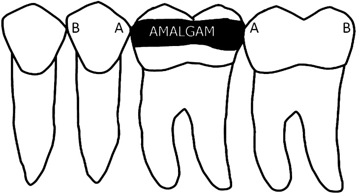
Pre-molar and molar teeth were placed in blocks of silicone with approximal contacts consisting three visually non-cavitated teeth and with the one mesial–occlusal–distal amalgam-filled tooth in between (Figure 1). This single amalgam-filled tooth was used throughout the investigation, it was removed after each set of teeth had been subjected to CBCT examination and used in every subsequent set of teeth. A total of 34 of these 3 + 1 sets of teeth were prepared for CBCT examination. Each set of teeth was immersed in a water container and examined by means of a CBCT unit (NewTom 3G; FOV, 9 inches; pixel size, 0.25 mm) at a fixed setting of 110 kV, automatically adjusted milliamperes and a scanning time of 36 s. Volumetric CBCT data were reconstructed and sectioned at 0.25 mm in the mesiodistal tooth plane, by means of proprietary NewTom software. All the images thus prepared were independently examined by two experienced dental radiologists each with a minimum 5 years' experience in the radiology field.
Figure 1.
Arrangement of four-tooth sets. A, surfaces next to filling; B, surfaces distal to filling.
The approximal mesial and distal surfaces of the two teeth located medially and distally to the amalgam-filled tooth were evaluated. For each proximal surface, the calibrated observers were asked to state whether they could identify caries, using a five-step confidence scale namely, 1, caries definitely not present; 2, caries probably not present; 3, unsure if caries is present or absent; 4, caries probably present; and 5, caries definitely present. The observations and scoring were performed for enamel and dentine during separate sessions. All the examinations were performed independently twice, with a 2-week interval. Both observers assessed the images, in random order, in a dimmed room on the same computer with a 19-inch monitor with the distance from viewer to monitor ranging between 50 and 60 cm. Proprietary NewTom 3G software was used to display images for evaluation. The observers were able to modify the brightness, contrast and gamma parameters of each radiograph, according to their personal needs, but without access to the raw data from the CBCT unit. For evaluation purposes, each observer could choose any or all of the images provided and focus on those preferred for visualizing any possible caries on proximal surfaces. After the CBCT examination, the teeth previously examined by the radiologist were individually mounted in acrylic resin and sectioned into 700-μm thick sections, in the mesiodistal plane, using a diamond saw. The sections were attached to a glass slide by means of transparent varnish. Using a light microscope at ×40 magnification, the tooth sections were examined to establish the true histological status of the approximal surfaces, in terms of the presence and extent of carious demineralization. A five-step scale was used to determine the extent of carious demineralization on proximal surfaces, namely: sound surface, carious demineralization to half the thickness of enamel, carious demineralization completely through the enamel, demineralization through the enamel and into one-third of the dentin and, finally, deeper dentin demineralization. The presence of carious demineralization on microscopic images was recorded when a white, radio-opaque or brown discolouration was observed in the region of interest.
Based on the location of the surface being investigated in relation to the amalgam filling, two categories of data were created, namely Group A, surfaces located next to an amalgam filling (n = 68) and Group B, surfaces distal to an amalgam filling (n = 68) (Figure 1).
Data analysis
Parameters expressing various aspects of accuracy (sensitivity, specificity, accuracy and positive and negative predictive values) were computed for each observer by validating the results obtained from the radiographic examination with those obtained from the histological examination of Groups A and B.
For this, the data were aggregated in the following way: the radiologist's scores for the presence of caries were collapsed into two groups as follows: Score 4 (caries probably present) and 5 (caries definitely present) creating a “caries group”, while Scores 1 (caries definitely not present), 2 (caries probably not present) and 3 (unsure if caries is present or absent) formed the “caries-free group”. The histological scores were aggregated into three categories: “sound surface”, “carious demineralization in enamel” and “carious demineralization through the enamel and present in the dentin”. The κ coefficiency values of intra- and interobserver reliability were calculated, to test the ability of the radiologists to give a correct and repeatable diagnosis.
Results
The true status of the 136 tested approximal surfaces, according to the histological examination, was as follows: 75 sound surfaces (36 in Group A and 39 in Group B), 36 with carious lesions in the enamel (20 in Group A and 16 in Group B) and 25 with carious demineralization through the enamel and present in the dentin (12 in Group A and 13 in Group B).
The sensitivities, specificities, positive and negative predictive values and accuracy were calculated for each of the proximal surfaces and are presented in Tables 1 and 2.
Table 1.
Sensitivity, specificity, positive predictive values (PPV+), negative predictive values (PPV−) and accuracy calculated for each of the proximal surfaces, for enamel lesions
| Surface | Sensitivity |
Specificity |
PPV+ |
PPV− |
Accuracy |
|||||
|---|---|---|---|---|---|---|---|---|---|---|
| First observer | Second observer | First observer | Second observer | First observer | Second observer | First observer | Second observer | First observer | Second observer | |
| A | 0.27 | 0.28 | 0.64 | 0.48 | 0.47 | 0.46 | 0.56 | 0.59 | 0.44 | 0.43 |
| B | 0.30 | 0.34 | 0.60 | 0.53 | 0.52 | 0.58 | 0.61 | 0.58 | 0.43 | 0.49 |
Table 2.
Sensitivity, specificity, positive predictive values (PPV+), negative predictive values (PPV−) and accuracy calculated for each of the proximal surfaces, for dentin lesions
| Surface | Sensitivity |
Specificity |
PPV+ |
PPV− |
Accuracy |
|||||
|---|---|---|---|---|---|---|---|---|---|---|
| First observer | Second observer | First observer | Second observer | First observer | Second observer | First observer | Second observer | First observer | Second observer | |
| A | 0.47 | 0.56 | 0.58 | 0.33 | 0.84 | 0.80 | 0.80 | 0.85 | 0.49 | 0.52 |
| B | 0.56 | 0.74 | 0.61 | 0.38 | 0.85 | 0.83 | 0.74 | 0.73 | 0.57 | 0.67 |
The calculated κ coefficient value of intra-observer reliability was 0.84, while that for interobserver reliability was 0.49.
Discussion
The objective of this study was to determine the potential of CBCT in the detection of approximal caries on the surfaces of teeth located next to a tooth with an amalgam restoration. In general, CBCT examination is not the system of choice for diagnosing caries and is mainly used for surgical and orthodontic reasons. Our experience, however, shows that when clinicians have had this form of examination carried out for other reasons, most have used it additionally to check for the presence of other pathological findings, including the presence of caries. In our study, we were trying to simulate a very common situation in the oral cavity, namely the checking of teeth abutting those filled with amalgam restorations.
In our study, the sensitivity values obtained for enamel lesions in both the A and B groups were low, from which it can be concluded that enamel demineralization, when present on an approximal surface, is not likely to be detected if an amalgam filling is in the region of interest. The specificity in both Groups A and B is low, meaning that one cannot be sure that there is truly a carious lesion when the CBCT indicates one. The distribution of sound vs diseased surfaces, based on histological examination, was quite similar in both groups. In Group A, there were 36 sound and 32 diseased teeth, and in Group B, there were 39 sound and 29 diseased surfaces. The pattern of answers given by the two observers was also somewhat similar. The observers gave more correct answers about the presence of caries for both Groups A and B in cases when caries were histologically present on the proximal surface but gave more incorrect answers about the presence of caries when it was histologically absent.
The sensitivity for dentin lesions was higher than it was for enamel lesions in both A and B groups. The values found do not differ from data reported previously.6,7 However, these authors had investigated the use of CBCT units for the detection of caries when amalgam or other sources of potential noise and artefacts were not present. Furthermore, unlike other authors, we noticed a problem with low specificity values in relation to dentin lesions similar to those detected in enamel lesions. The dentinal specificity values of Group A reported by our first and second observers were 0.47 and 0.56, respectively. In Group B, they were 0.56 and 0.74, respectively. It has to be stressed that the number of histologically confirmed carious dentin lesions was small—only 12 out of 68 and 13 out of 68 in Groups A and B, respectively.
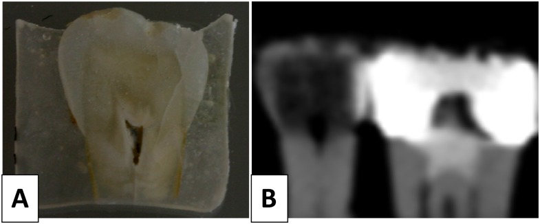
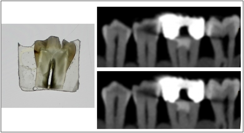
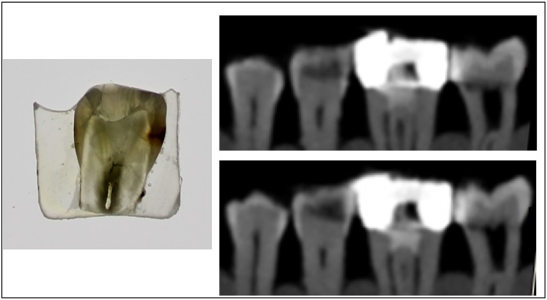
Although the findings of the observers were generally repeatable in respect of their own observations, the correlation between them was only moderate. An observer's experience and calibration in the diagnosis of carious lesions on radiographs was not very helpful. The first and second observers interpreted what they saw on a computer screen in different ways. Sometimes what they saw they believed to be an artefact and sometimes they believed it to be demineralization of the tooth surface. Both observers complained about their low confidence at the time of diagnosis. The problem of the interpretation of a CBCT scan is visualized in Figures 2–4.
Figure 2.
A sound pre-molar tooth used in the study: (a) the microscopic section reveals a sound enamel and dentine on both the mesial and distal sides; (b) corresponding cropped image of CBCT examination that was interpreted by one observer as caries on both enamel and dentin on the mesial side. The second observer stated that there was only enamel demineralization but on both mesial and distal sides.
Figure 4.
Microscopic image of a section of a pre-molar tooth and the corresponding CBCT scans. Microscopic examination reveals the presence of enamel caries on the mesial side. CBCT examination is severely compromised by the presence of diagonally shaped artefact that makes diagnosis of enamel demineralization very uncertain.
Figure 3.
Microscopic image of a section of a pre-molar tooth and the corresponding CBCT scans. Microscopic examination reveals the presence of enamel and dentine caries on distal side. CBCT examination suggests enamel and dentine caries on distal side but the extent of demineralization is more severe in comparison with histological view.
Factors that we found to be particularly important in our study were scatter and beam hardening caused by the presence of high-density metallic structures, such as amalgam fillings. We believe that no studies have been published that can be directly compared with the one reported here. It follows that analysis of our data can only be compared with that from articles in which caries detection by CBCT with no beam-hardening effects were evaluated, such as the articles of Haiter-Neto et al6 and Young et al7 mentioned in the introduction of this article. Of importance might also be those papers focusing on the CBCT examination of teeth with other metallic inclusions such as metallic root posts.
In the study by Costa et al,12 the possible impact of the presence of an intracanal metallic post on the diagnosis of a root fracture in CBCT was examined. In this study, where a small FOV (5 × 5 cm; PaX Uni3D/Vatech unit; Vatech Co., Hwasung, Korea) with 0.2 voxel size was used, they noted that the presence of a metallic post in a canal significantly reduced the specificity and sensitivity of root fracture diagnosis, in comparison with the situation where a metallic post was not present in the canal. Moreover, they noticed that intra- and interobserver agreement in the interpretation of roots with a metallic post was weak (0.36–0.39 and 0.157–0.453, respectively), while for roots without metallic posts, it was moderate to high. In another study by Costa et al9 a large FOV CBCT unit (20 × 15 cm Picasso Master 3D®/Vatech unit; Vatech Co.) with 0.2 voxel size was used. These authors observed a high level of noise on the images of both those roots with, and without, metallic restorations. This, they claimed, was owing to increased scattered radiation produced by the FOV of the unit itself. They also noted that the artefacts produced by metallic posts had reduced the observer's confidence at the time of diagnosis, leading to the “no agreement level” of interobserver agreement.
The voxel size and FOV used in our study were high, which might be a weakness and suggest that a smaller voxel of a smaller FOV machine might produce better results. It is known that image voxel size plays an important role in the quality of image, because it is correlated with spatial resolution. A smaller voxel size will increase spatial resolution which, in normal anatomical conditions, will be helpful in depicting fine details. With increased spatial resolution, however, other components of image quality, such as image noise and the contrast-to-noise ratio, are also changed.
Bechara et al13 noted that reducing the voxel size (from 0.2000 to 0.0076) increased noise and decreased the signal-to-noise ratio in a small FOV CBCT unit. It can be expected that a similar situation will occur when a CBCT scan is performed with a metallic object in the region of interest.
A number of attempts have been made to overcome the problem of beam-hardening artefacts. Some authors suggest that increasing the tube potential setting of the CBCT machine can reduce the number of artefacts present.14–17 Hunter and McDavid18 examined the possibility of reducing the cupping artefacts seen when an aluminium cylinder is examined by means of CBCT. They proved that the degree of this effect can be reduced by pre-filtration of the X-ray beam by means of a copper filter, which, they claimed, was responsible for narrowing the X-ray spectrum by the elimination of low-energy photons. They also emphasized the fact that the cupping effect, which is a result of both beam hardening and scatter radiation, was responsible for underestimating the true density of the object. Other authors have concentrated on the pre- and post-processing aspects of data acquisition.16,19 However, clinical validation of these methods remains unclear.
It has to be mentioned that our results were obtained by means of a CBCT unit that utilizes image intensifier technology. This method of data acquisition has been recently replaced by flat panel technology. It is known that, in general, flat panel detectors have higher resolution, lower noise and better image characteristics than the image intensifier technology-based devices.20 One can therefore expect that a similar study performed with a flat panel CBCT unit could produce different results.
Moreover, our research is an in vitro study, which like most such studies attempts to simulate the clinical, in vivo situation. We chose human teeth for our examination, but we did not imitate human behaviour during a clinical CBCT examination. Our sets of teeth were static during the entire examination, although patients may not be able to remain still for the several seconds required for CBCT examination. As explained by Schulze et al15 even a patient's minimal movement during a CBCT examination will negatively influence the quality of the images obtained and limit its final spatial resolution. These limitations may lead to the conclusion that in vivo results may actually be even worse than those achieved in vitro.
With the limitations of the type of unit used here, we can conclude that scans from a CBCT examination should not be used to determine the presence or absence of caries when amalgam fillings are present in direct contact with an area being diagnosed.
Acknowledgments
Acknowledgments
We would like to express our special thanks to Professor Geoffrey Shaw for his linguistic correction and valuable professional guidance.
References
- 1.Mozzo P, Procacci C, Tacconi A, Martini PT, Andreis IA. A new volumetric CT machine for dental imaging based on the cone-beam technique: preliminary results. Eur Radiol 1998; 8: 1558–64. [DOI] [PubMed] [Google Scholar]
- 2.Tsuchida R, Araki K, Okano T. Evaluation of a limited cone-beam volumetric imaging system: comparison with film radiography in detecting incipient proximal caries. Oral Surg Oral Med Oral Pathol Oral Radiol Endod 2007; 104: 412–16. doi: 10.1016/j.tripleo.2007.02.028 [DOI] [PubMed] [Google Scholar]
- 3.Senel B, Kamburoglu K, Uçok O, Yüksel SP, Ozen T, Avsever H. Diagnostic accuracy of different imaging modalities in detection of proximal caries. Dentomaxillofac Radiol 2010; 39: 501–11. doi: 10.1259/dmfr/28628723 [DOI] [PMC free article] [PubMed] [Google Scholar]
- 4.Qu X, Li G, Zhang Z, Ma X. Detection accuracy of in vitro approximal caries by cone beam computed tomography images. Eur J Radiol 2011; 79: e24–7. doi: 10.1016/j.ejrad.2009.05.063 [DOI] [PubMed] [Google Scholar]
- 5.Kayipmaz S, Sezgin ÖS, Saricaoğlu ST, Çan G. An in vitro comparison of diagnostic abilities of conventional radiography, storage phosphor, and cone beam computed tomography to determine occlusal and approximal caries. Eur J Radiol 2011; 80: 478–82. [DOI] [PubMed] [Google Scholar]
- 6.Haiter-Neto F, Wenzel A, Gotfredsen E. Diagnostic accuracy of cone beam computed tomography scans compared with intraoral image modalities for detection of caries lesions. Dentomaxillofac Radiol 2008; 37: 18–22. doi: 10.1259/dmfr/87103878 [DOI] [PubMed] [Google Scholar]
- 7.Young SM, Lee JT, Hodges RJ, Chang TL, Elashoff DA, White SC. A comparative study of high-resolution cone beam computed tomography and charge-coupled device sensors for detecting caries. Dentomaxillofac Radiol 2009; 38: 445–51. doi: 10.1259/dmfr/88765582 [DOI] [PubMed] [Google Scholar]
- 8.MacDonald D. Oral and maxillofacial radiology: a diagnostic approach. Chichester, UK: John Wiley & Sons; 2011. p. 367. [Google Scholar]
- 9.Costa FF, Gaia BF, Umetsubo OS, Pinheiro LR, Tortamano IP, Cavalcanti MG. Use of large-volume cone-beam computed tomography in identification and localization of horizontal root fracture in the presence and absence of intracanal metallic post. J Endod 2012; 38: 856–9. doi: 10.1016/j.joen.2012.03.011 [DOI] [PubMed] [Google Scholar]
- 10.Pauwels R, Stamatakis H, Bosmans H, Bogaerts R, Jacobs R, Horner K, et al. ; SEDENTEXCT Project Consortium. Quantification of metal artifacts on cone beam computed tomography images. Clin Oral Implants Res 2013; 24(Suppl. A100): 94–9. doi: 10.1111/j.1600-0501.2011.02382.x [DOI] [PubMed] [Google Scholar]
- 11.Esmaeili F, Johari M, Haddadi P. Beam hardening artifacts by dental implants: comparison of cone-beam and 64-slice computed tomography scanners. Dent Res J (Isfahan) 2013; 10: 376–81. [PMC free article] [PubMed] [Google Scholar]
- 12.Costa FF, Gaia BF, Umetsubo OS, Cavalcanti MG. Detection of horizontal root fracture with small-volume cone-beam computed tomography in the presence and absence of intracanal metallic post. J Endod 2011; 37: 1456–59. [DOI] [PubMed] [Google Scholar]
- 13.Bechara B, McMahan CA, Moore WS, Noujeim M, Geha H, Teixeira FB. Contrast-to-noise ratio difference in small field of view cone beam computed tomography machines. J Oral Sci 2012; 54: 227–32. [DOI] [PubMed] [Google Scholar]
- 14.Chindasombatjaroen J, Kakimoto N, Murakami S, Maeda Y, Furukawa S. Quantitative analysis of metallic artifacts caused by dental metals: comparison of cone-beam and multi-detector row CT scanners. Oral Radiol 2011; 27: 114–20. [Google Scholar]
- 15.Schulze R, Heil U, Gross D, Bruellmann DD, Dranischnikow E, Schwanecke U, et al. Artefacts in CBCT: a review. Dentomaxillofac Radiol 2011; 40: 265–73. doi: 10.1259/dmfr/30642039 [DOI] [PMC free article] [PubMed] [Google Scholar]
- 16.Bechara B, McMahan CA, Geha H, Noujeim M. Evaluation of a cone beam CT artefact reduction algorithm. Dentomaxillofac Radiol 2012; 41: 422–8. doi: 10.1259/dmfr/43691321 [DOI] [PMC free article] [PubMed] [Google Scholar]
- 17.Schulze RK, Berndt D, d' Hoedt B. On cone-beam computed tomography artifacts induced by titanium implants. Clin Oral Implants Res 2010; 21: 100–7. doi: 10.1111/j.1600-0501.2009.01817.x [DOI] [PubMed] [Google Scholar]
- 18.Hunter AK, McDavid WD. Characterization and correction of cupping effect artefacts in cone beam CT. Dentomaxillofac Radiol 2012; 41: 217–23. doi: 10.1259/dmfr/19015946 [DOI] [PMC free article] [PubMed] [Google Scholar]
- 19.Zhang Y, Zhang L, Zhu XR, Lee AK, Chambers M, Dong L. Reducing metal artifacts in cone-beam CT images by preprocessing projection data. Int J Radiat Oncol Biol Phys 2007; 67: 924–32. doi: 10.1016/j.ijrobp.2006.09.045 [DOI] [PubMed] [Google Scholar]
- 20.Kalender WA, Kyriakou Y. Flat-detector computed tomography (FD-CT). Eur Radiol 2007; 17: 2767–79. doi: 10.1007/s00330-007-0651-9 [DOI] [PubMed] [Google Scholar]


