Abstract
Cancer cells with constitutive phosphatidylinositol 3-kinase (PI3K)/Akt pathway activation have been associated with overexpression of the lipogenic enzyme fatty acid synthase (FAS) as a means to provide lipids necessary for cell growth. In contrast, K-Ras-driven cancer cells suppress utilization of de novo synthesized fatty acids and rely on exogenously supplied fatty acids for cell growth and membrane phospholipid biosynthesis. Consistent with a differential need for de novo fatty acid synthesis, cancer cells with activated PI3K signaling were sensitive to suppression of FAS; whereas mutant K-RAS-driven cancer cells continued to proliferate with suppressed FAS. Surprisingly, in response to FAS suppression, we observed robust increases in both Akt and ERK phosphorylation. Akt phosphorylation was dependent on the insulin-like growth factor-1 receptor (IGF-1R)/PI3K pathway and mTOR complex 2. Intriguingly, K-Ras-mediated ERK activation was dependent on N-Ras. Pharmacological inhibition of PI3K and MEK in K-Ras-driven cancer cells resulted in increased sensitivity to FAS inhibition. These data reveal a surprising sensitivity of K-Ras-driven cancer cells to FAS suppression when stimulation of Akt and Erk was prevented. As K-Ras-driven cancers are notoriously difficult to treat, these findings have therapeutic implications.
Keywords: Fatty acid synthase, K-Ras, Akt, ERK
Introduction
Several studies have shown that endogenous lipid production is necessary for growth and survival in cancer cells of various tissue types and mutation signature [1–6]. A subset of research has focused on cancer cells with dysregulation of the phosphatidylinositol-3-kinase (PI3K)2/Akt pathway [7–9]. Among the many targets of PI3K signals, Akt induces lipogenic enzymes, including fatty acid synthase (FAS) [10], which catalyzes the terminal step in de novo lipogenesis, the anabolic conversion of glucose into fatty acids. Increased glucose uptake by cancer cells with constitutive PI3K/Akt signaling has been associated with high levels of FAS expression and increased fatty acid synthesis [11–13], thereby satisfying the demand for new membrane composition in rapidly proliferating cells. Constitutive Akt activation can be the result of a gain-of-function mutation in the PI3K gene (PIK3CA) [14] or more commonly, loss of PI3K antagonist, PTEN (phosphatase and tensin homologue deleted on chromosome 10). Loss of PTEN sensitizes cells to FAS inhibition [15,16] while induction of PTEN abrogates the effect [7,17]. The inference is that cancer cells with intact PTEN and corresponding low Akt activation and FAS expression are unaffected by FAS inhibition.
Despite intact PTEN, K-Ras-driven cancer cells can activate the PI3K/Akt pathway – making it difficult to target cancer cells harboring K-Ras mutations [18,19]. In addition to being able to activate the PI3K/Akt pathway [20,21], oncogenic K-Ras also activates the Raf/MEK/ERK pathway [22]. PI3K/Akt activation is also regulated by growth factors through a canonical insulin-like growth factor-1 receptor (IGF-1R)/PI3K/Akt pathway [23,24]. Whether cancer cells with oncogenic K-RAS are linked to the PI3K/Akt pathway directly (predictive of growth-factor independence) or indirectly (growth-factor dependent), the RAS/Raf/MEK/ERK and PI3K/Akt pathways are compensatory [25,26]. Thus, single agents targeting either pathway are not efficacious. Instead, combined inhibition of components in both pathways is necessary to compromise cancer cells with mutant K-RAS [27].
In this study, we investigated the effect of FAS inhibition on proliferation and viability of K-RAS mutant cancer cells. We used pharmacological and genetic means to inhibit FAS in human cancer cell lines harboring K-RAS mutations. We found a surprising tumorigenic advantage in that Fas inhibition led to Akt and ERK activation. Because tumors adapt to a nutrient-depleted microenvironment during tumorigenesis, these findings identify survival signals that may need to be compromised for therapeutic intervention.
Materials and methods
Cells, cell culture conditions and cell viability
The human cancer cell lines PANC-1, Mia PaCa-2, HCT116, MDA-MB-468 and PC3 cells were obtained from the American Tissue Type Culture Collection and cultured in Dulbecco's Modified Eagle Medium (Sigma) supplemented with 10% Fetal Bovine Serum (Sigma). Cell viability was determined as ratio of non-adherent cells to adherent cells after treatment using a Coulter counter.
Antibodies and reagents
The following antibodies against: PARP, PTEN, Akt, P-AktS473, P-ERK, ERK, P-P70 S6 kinase, mTOR, Raptor, Rictor, and IGF-1R were obtained from Cell Signaling; α-actin was from Sigma. The antibody for FAS was obtained from BD BioSciences. Negative control scrambled siRNA and siRNA targeted against mTOR, Raptor and Rictor were obtained from Dharmacon. siRNAs targeted against FAS were obtained from Santa Cruz Biotechnology. Cerulenin, LY294002 and PD0325901 were purchased from Sigma. Lipofectamine RNAiMax (Invitrogen) was used for transient transfections.
Cell proliferation
Cells were plated at 50% confluence and treated the next day. Cells were trypsinized at 24 and 48 hours and counted using a Coulter counter.
Western blot analysis
Cells were plated at 90% confluence. Extraction of proteins from cultured cells and Western blot analysis of extracted proteins was performed using the ECL system (Amersham) as described previously [28].
Transient transfections
Cells were plated in six-well plates in medium containing 10% FBS. The next day (50% confluence), transfections with siRNAs (100 nM) in Lipofectamine RNAiMAX were performed. After 6 hours, reagents were replaced with fresh media (0% or 10% FBS) and cells were allowed to incubate for an additional 48 hours.
Results
K-Ras mutant cells do not require de novo lipogenesis for cell growth
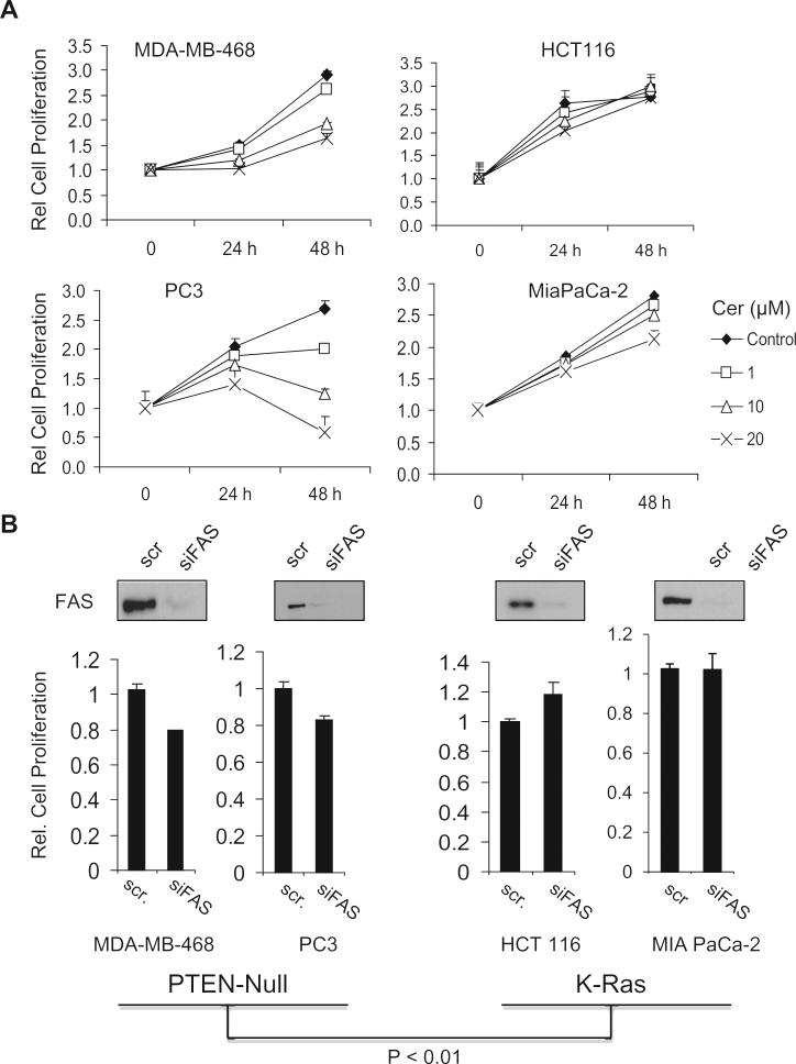
It was recently reported that K-Ras-driven cancer cells depend largely on exogenously supplied fatty acids rather than endogenously synthesized fatty acids for cell growth and proliferation [29,30]. In contrast, cancer cells with mutations in the PI3K/Akt pathway have elevated expression of lipogenic enzymes and de novo fatty acid synthesis [10]. We therefore compared the effect of suppressing FAS on the proliferation of PTEN-null and K-Ras-driven cancer cells. As shown in Fig. 1A, the proliferation of MDA-MB-468 breast and PC3 prostate cancer cells (PTEN-null) was inhibited by the irreversible FAS inhibitor cerulenin [31] – indicating a dependence on endogenous lipid production. This observation also suggested that the PTEN-null cancer cells were unable to utilize exogenously supplied serum lipids. In contrast, proliferation of the K-Ras-driven HCT116 colon and MiaPaCa-2 pancreatic cancer cells was not affected by cerulenin (Fig. 1A). Qualitatively similar results were obtained by knockdown of FAS with siRNA, where it can be seen that there is a significant difference in the response to FAS knockdown between the PTEN-null and the K-Ras-driven cancer cells lines (Fig. 1B). These data indicate that K-Ras mutant cells do not require endogenous lipid production for cell growth.
Fig. 1.
K-Ras mutant cancer cells proliferate independently of fatty acid synthase inhibition. (A) PTEN null cells lines, MDA-MB-468 and PC3 and K-RAS mutant cell lines, HCT116 and MIA PaCa-2 cells were plated at 50% confluence and treated with cerulenin (Cer) at indicated doses. After 24 and 48 hour time points, cells were trypsinized and counted by Coulter counter. Data represent mean ± SEM from three independent experiments with each sample counted two times. (B) Cells were transiently transfected with 100 nM siRNA against FAS (siFAS) or with negative control scrambled siRNA (scr). After 48 hours, cells were harvested and lysates were immunoblotted for FAS protein and counted by Coulter counter. Data represent mean ± SEM from three independent experiments with each sample counted two times. P-value represents combined data sets for the PTEN-null cell lines (MDA-MB-468 and PC3) and K-Ras mutant cell lines (HCT116 and MIA PaCa-2). The values for the ratios of cell number in cells treated with siFAS divided by cell number of controls in the PTEN-null and mutant K-Ras cell lines.
FAS inhibition induces pro-survival, Akt and ERK activation, in serum-deprived KRAS mutant cancer cells
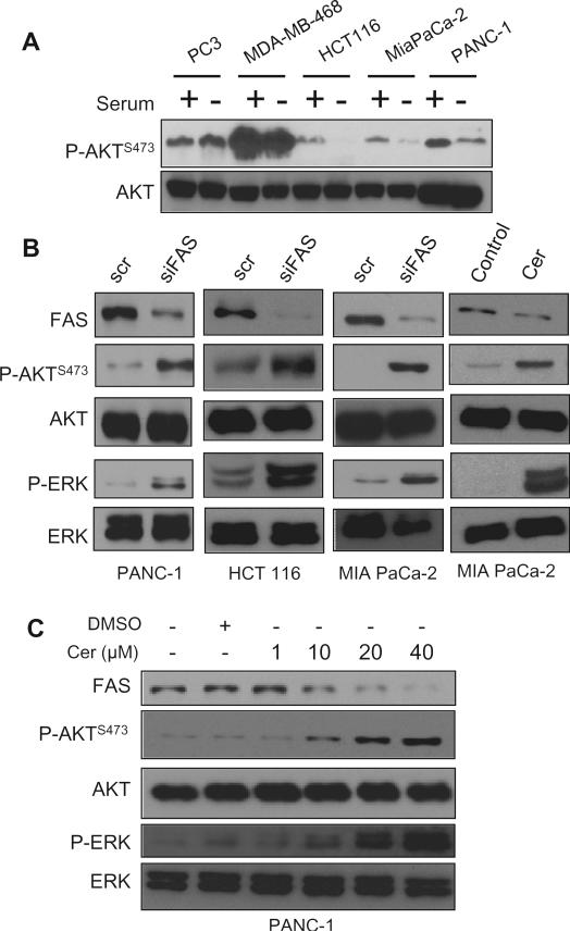
Kalaany and Sabatini previously reported a differential response to nutrient deprivation of human cancer cells with dysregulated PI3K signals relative to cancer cells with K-Ras mutations [24]. This was reflected by a differential sensitivity for Akt phosphorylation at Ser473 for PTEN-null and K-Ras-driven cancer cells upon serum withdrawal. Cancer cells with activating K-Ras mutations were more sensitive to nutrient restriction than those with dysregulated PI3K signaling. Similar to what was observed by Kalaany and Sabatini, there were higher basal levels of Akt-S473 phosphorylation in cells that were PTEN-null than K-Ras-driven cancer cells (Fig. 2A). In addition, phosphorylation of Akt was inhibited by serum deprivation in the mutant K-Ras-driven cancer cell lines, but not PTEN null cell lines (Fig. 2A) – indicating that Akt phosphorylation in the K-Ras driven cancer cells is dependent on serum. It was recently reported that K-Ras-driven cancer cells are dependent on exogenously supplied lipids [29,30]. Thus, we speculated that in the absence of serum lipids, there is a compensatory increase in fatty acid synthesis to compensate for the absence of serum lipids. We therefore examined the effect of suppressing FAS – either by siRNA knockdown or cerulenin on the phosphorylation of Akt and another Ras target Erk. Surprisingly, we found that FAS inhibition caused a robust increase in both Akt and ERK phosphorylation in three K-Ras-driven cancer cell lines – PANC1, HCT116, and MIA PaCa-2 (Fig. 2B). The effect was dose dependent and correlated well with reduced levels of FAS observed with increasing cerulenin dosage in PANC1 cells (Fig. 2C). These data reveal a novel feedback activation of Akt and ERK activation in K-Ras-driven cancer cells in response to FAS inhibition.
Fig. 2.
FAS inhibition induces Akt and ERK activation upon serum deprivation. (A) PTEN-null PC3, MDA-MB-468 cells, and K-Ras-driven HCT116, MiaPaCa-2, and PANC1 cells were plated and grown in 10% serum or 0% serum for 24 hours. Lysates were immunoblotted for Akt phosphorylated at Ser473 (P-AKTS473) and total AKT. (B) PANC-1, HCT116, and MIA PaCa-2 cells were transfected as in Fig. 1. After 48 hours, cells were grown in 0% serum for an additional 24 hours. Lysates were then immunoblotted for AKT phosphorylated at Ser473 and total Akt, and phosphorylated ERK (P-ERK) and total ERK. (C) PANC-1 cells were plated and grown in 0% serum medium at 90% confluence. After 24 hours, cells were treated with indicated concentrations of cerulenin for an additional 24 hours in 0% serum. Lysates were immunoblotted for P-AKT, total Akt, and P-ERK and total ERK as in (B). Experiments shown are representative of three independent experiments.
Insulin-like growth factor-1 receptor (IGF-1R) and mTORC2 mediate Akt activation and N-RAS compensates for K-RAS-mediated ERK activation upon FAS inhibition
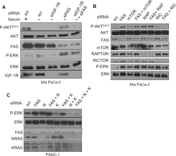
To address mechanism for activation of survival signals in response to FAS inhibition, we tested if Akt activation was via the canonical IGF-1R/PI3K/Akt pathway. Akt activation upon knockdown of FAS was suppressed by simultaneous knockdown of IGF-1R (Fig. 3A). Knockdown of the IGF-1R did not impact ERK phosphorylation. Mammalian/mechanistic target of rapamycin complex 2 (mTORC2) phosphorylates Akt at Ser473 [32]. We therefore inhibited expression of Rictor, signature component of mTORC2 along with mTOR and Raptor (signature protein of mTORC1), both of which served as controls (Fig. 3B). Upon FAS inhibition, neither inhibition of mTOR nor raptor compromised Akt activation. As expected, knockdown of Rictor prevented Akt activation. These data suggest that Akt activation induced by FAS inhibition is mediated by the IGF-1R/PI3K/Akt pathway. Thus, Akt activation is induced by a new means, but not mechanism.
Fig. 3.
IGF-1R and mTORC2 mediate Akt activation, while N-RAS mediates ERK activation. (A) MIA PaCa-2 cells were transiently transfected with siRNAs against FAS and/or IGF-1R or with scrambled control siRNA (scr) as in Fig. 2B. Lysates were then immunoblotted for AKT phosphorylated at Ser473 (P-AktS473) and total Akt, and phosphorylated ERK (P-ERK) and total ERK, and IGF-1R. (B) MIA PaCa-2 cells were transfected as in (A) with siRNAs against FAS and/or mTOR, Raptor (RAP), Rictor (RIC) or with negative control siRNA (scr). After 48 hours, cells were grown in 0% serum for an additional 24 hours. Lysates were then immunoblotted for the indicated proteins. (C) PANC-1 cells were transfected as in (A) with siRNAs against FAS, N-RAS (N), K-RAS (K) or in indicated combinations or negative control siRNA. After 48 hours, cells were grown in 0% serum for an additional 24 hours. Lysates were then immunoblotted for the indicated proteins. Experiments shown are representative of three independent experiments.
Since we observed an increase in ERK activation upon FAS inhibition, we investigated possible mechanisms for ERK activation. Since the MEK/ERK pathway is responsive to Ras GTPase and N-Ras has been implicated in K-Ras-driven cancers [33], we investigated the impact of both N-Ras and K-Ras on ERK activation stimulated by FAS inhibition. We suppressed FAS, N-RAS, or K-RAS expression with siRNAs alone or in combination (Fig. 3C). Inhibition of K-Ras expression reduced basal ERK phosphorylation; however, if FAS expression was suppressed along with K-Ras, there was still an increase in ERK phosphorylation. While suppression of N-Ras expression by itself did not impact on ERK phosphorylation induced FAS suppression, the combination of K-Ras and N-Ras suppression did block the induction of ERK phosphorylation induced by suppression of FAS. This suggests that N-Ras is required for the induction of ERK caused by FAS suppression, but in a K-Ras-dependent manner. In this case, both a new means and mechanism induces ERK activation.
Combined MEK and PI3K inhibition sensitizes K-Ras cells to FAS inhibition
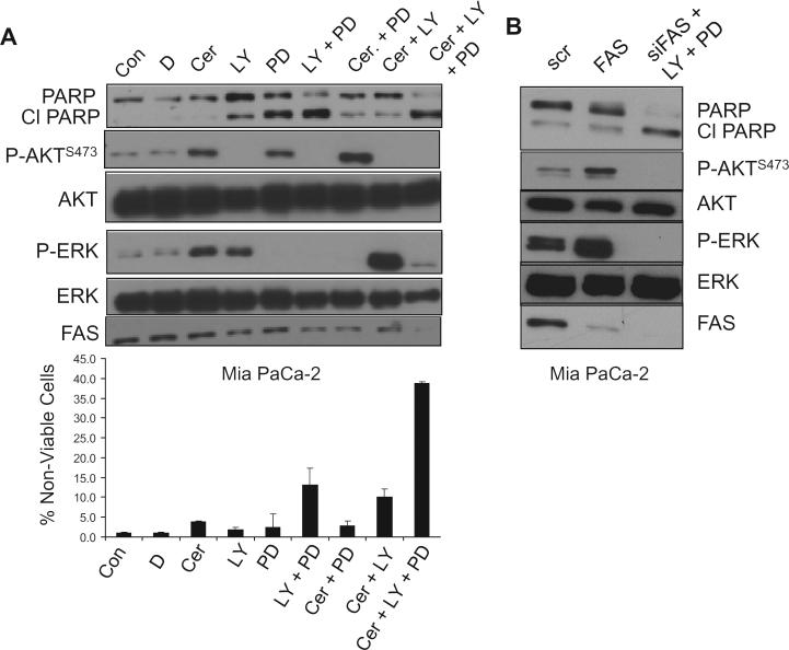
Recent studies have demonstrated that effective targeting of mutant K-Ras driven tumors in mice requires combined inhibition of both PI3K/Akt and RAS/MEK/ERK pathways [27,34]. We inhibited MEK and PI3K in Mia PaCa-2 cells to determine if the pro-survival signals stimulated by FAS inhibition are dominantly mediated through the activation of either pathway. As shown in Fig. 4A, the MEK inhibitor PD0325901 and the PI3K inhibitor LY294002 effectively inhibited cerulenin-induced Akt and ERK activation, respectively. There was limited PARP cleavage and cell death seen with either compound by itself – indicating limited apoptosis (Fig. 4A). Furthermore, phosphorylated ERK increased upon cerulenin and LY294002 treatment; and similarly, phosphorylated Akt increased upon cerulenin and PD0329051 treatment. In addition, cleaved PARP caused by either drug alone was suppressed by cerulenin (compare lanes 4 and 8 and lanes 5 and 7). However, FAS inhibition by cerulenin failed to promote survival when both MEK and PI3K were suppressed (Fig. 4A). This effect was also observed by the combination of FAS knockdown along with LY294002 and PD0325901 treatment (Fig. 4B). These data indicate that activation of the pro-survival kinases Akt and ERK induced by suppression of FAS provides a survival advantage and that suppression of the survival kinases creates a heightened sensitivity to FAS inhibition. These data are also consistent with a model where there is compensatory utilization of different survival pathways depending on what stresses are encountered.
Fig. 4.
FAS inhibition and serum deprivation heighten sensitivity to dual PI3K and MEK inhibition. (A) MIA PaCa-2 cells were plated and grown in 0% serum at 90% confluence. Twenty-four hours later, cells were either untreated (Con), treated with DMSO (D), 40 μM cerulenin (Cer), 20 μM LY29402 (LY), MEK inhibitor PD0325901 (PD) (100 nM), or with indicated combinations. After 24 hours, lysates were probed for indicated proteins or phospho-proteins (upper panel) and cell viability was determined as ratio of non-adherent cells to total number of cells (lower panel). (B) MIA PACA-2 cells were transfected with siRNA against FAS or with negative control siRNA (scr). Forty-eight hours later, cells were grown in 0% serum only or with 20 μM LY and 100 nM PD for an additional 24 hours, at which time lysates were probed for indicated proteins or phospho-proteins. Data for the cell viability assays represent the mean ± SEM from three independent experiments with each sample counted two times. Western blot experiments in (A) and (B) are representative of three independent experiments.
Discussion
In summary, this study implicates an unanticipated response of K-Ras-driven cancer cells to FAS suppression. Pro-survival effectors were induced upon FAS inhibition in cancer cells with mutant K-RAS. This response included Akt and ERK activation – both of which suppress apoptotic programs. Suppression of both Akt and Erk activation in response to FAS inhibition resulted in apoptosis – indicating that the stimulation of Akt and ERK promoted survival under the stress of lipid withdrawal. Stimulation of Akt was dependent on IGF-R1 and mTORC2, and intriguingly ERK activation was dependent on both K-Ras and N-Ras. Although feedback activation of Akt has been observed upon pharmacological inhibition of mTORC1 activation [35,36], we determined that cerulenin also stimulates phosphorylation of the mTORC1 substrate S6 kinase in MIA PaCa-2 cells (data not shown) – indicating that neither Akt nor ERK activation by FAS inhibition was due to inhibition of mTORC1. This novel feedback activation of survival kinases suggests both complications and opportunities for treating K-Ras-driven cancers.
While the lack of fatty acid production did correlate with growth inhibition in PTEN null cells, it had no effect on K-RAS mutant cells. Nonetheless, lipids are required to perpetuate cancer cell growth and proliferation [37], regardless of mutation status. Thus, the data suggest that cells harboring oncogenic K-Ras utilize exogenous lipids for cell growth. Supportively, a clinical study showed an association between dietary fatty acids and K-Ras driven colon tumors [38]. Two recent studies demonstrated that K-Ras-driven cancers depend on exogenously supplied lipids [29,30]. Here, the authors show that cells with RAS pathway activation uptake exogenous lipids, which supports their proliferation. In contrast, PTEN null cells were not capable of using available serum lipids to sustain proliferation rates. It seems that dependence on endogenous lipid supply mediates independence of exogenous nutrient supply in constitutively activated PI3K/Akt pathway cells. This effect is diametrically opposed in K-Ras-driven cancer cells in which independence of endogenous lipid supply indicates dependence on exogenous nutrient supply. Given that K-Ras-driven cancer cells depend largely on exogenously supplied and not on de novo lipid biosynthesis, it was very surprising that that K-Ras-driven cancer cells would respond so robustly to cerulenin, which inhibits de novo fatty acid synthesis. However, it has also been reported that FAS is expressed at high levels in pancreatic cancers [39], where more than 90% of the cancers are K-Ras-driven [40]. These findings suggest a more complex role for FAS in K-Ras-driven cancer cells that may involve more than fatty acid synthesis. In this regard, it may be of significance that cerulenin treatment leads to reduced levels of FAS (see Fig. 2B and C). Clearly there is more to be learned about the role of FAS in K-Ras-driven cancers.
Mutated K-Ras is found in approximately 30% of human cancers and is prevalent in pancreatic (90%) and colon cancer (50%) [40]. As such, there is great interest in developing effective targeting strategies. Basic and clinical research studies have demonstrated greater efficacy by simultaneously inhibiting nodes in both the RAS/Raf/MEK/ERK and PI3K/ Akt pathways as opposed to targeting these pathways singly [25,26,41]. Significantly, we observed a heightened sensitivity to MEK and PI3K inhibitors upon FAS inhibition. The effect of cerulenin was observed in the absence of serum, which is not so relevant in an animal; however, K-Ras-driven cancers have increased macropinocytosis [42] to help scavenge lipids [29]. We have been able to mimic the loss of serum lipids with EIPA [5-(N-ethyl-N-isopropyl) amiloride] [30]. Thus, it is possible to mimic serum free conditions in K-Ras-driven cancer cells where there is elevated macropinocytosis.
In contrast to tumors, lipogenesis is low in nearly all non-malignant tissues [43]. The majority of fatty acid content in normal cells is acquired from the diet. Thus, with the exception of liver and adipose tissue [44], there is little need to store energy and FAS expression is minimal. This differential expression of FAS between normal and cancer cells has incited a strong interest in developing FAS inhibitors with the expectation that normal cells will not be affected. The data provided here suggest that suppressing survival signals generated by suppression of FAS may be a critical for target in FAS in K-Ras mutant cancers.
Acknowledgments
We thank Darin Salloum for comments on the manuscript. This work was supported by a grant from the National Cancer Institute (CA46677). Research Centers in Minority Institutions (RCMI) award RR-03037 from the National Center for Research Resources of the National Institutes of Health, which supports infrastructure and instrumentation, is also acknowledged. Paige Yellen was supported by grant TL1RR024998 of the Clinical and Translational Science Center at Weill Cornell Medical College.
Abbreviations
- FAS
fatty acid synthase
- IGF-1R
insulin-like growth factor-1 receptor
- mTORC2
mammalian/mechanistic target of rapamycin complex 2
- PI3K
phosphatidylinositol 3-kinase
Footnotes
Conflict of interest
The authors declare no conflicts of interest.
References
- 1.Brusselmans K, De Schrijver E, Verhoeven G, Swinnen JV. RNA interference-mediated silencing of the acetyl-CoA-carboxylase-alpha gene induces growth inhibition and apoptosis of prostate cancer cells. Cancer Res. 2005;65:6719–6725. doi: 10.1158/0008-5472.CAN-05-0571. [DOI] [PubMed] [Google Scholar]
- 2.Calvisi DF, Wang C, Ho C, Ladu S, Lee SA, Mattu S, et al. Increased lipogenesis, induced by AKT-mTORC1-RPS6 signaling, promotes development of human hepatocellular carcinoma. Gastroenterology. 2011;140:1071–1083. doi: 10.1053/j.gastro.2010.12.006. [DOI] [PMC free article] [PubMed] [Google Scholar]
- 3.Hatzivassiliou G, Zhao F, Bauer DE, Andreadis C, Shaw AN, Dhanak D, et al. ATP citrate lyase inhibition can suppress tumor cell growth. Cancer Cell. 2005;8:311–321. doi: 10.1016/j.ccr.2005.09.008. [DOI] [PubMed] [Google Scholar]
- 4.Kuhajda FP, Jenner K, Wood FD, Hennigar RA, Jacobs LB, Dick JD, et al. Fatty acid synthesis: a potential selective target for antineoplastic therapy. Proc. Natl Acad. Sci. U.S.A. 1994;91:6379–6383. doi: 10.1073/pnas.91.14.6379. [DOI] [PMC free article] [PubMed] [Google Scholar]
- 5.Pizer ES, Thupari J, Han WF, Pinn ML, Chrest FJ, Frehywot GL, et al. Malonyl-coenzyme-A is a potential mediator of cytotoxicity induced by fatty-acid synthase inhibition in human breast cancer cells and xenografts. Cancer Res. 2000;60:213–218. [PubMed] [Google Scholar]
- 6.Swinnen JV, Vanderhoydonc F, Elgamal AA, Eelen M, Vercaeren I, Joniau S, et al. Selective activation of the fatty acid synthesis pathway in human prostate cancer. Int. J. Cancer. 2000;88:176–179. doi: 10.1002/1097-0215(20001015)88:2<176::aid-ijc5>3.0.co;2-3. [DOI] [PubMed] [Google Scholar]
- 7.Guo D, Prins RM, Dang J, Kuga D, Iwanami A, Soto H, et al. EGFR signaling through an Akt-SREBP-1-dependent, rapamycin-resistant pathway sensitizes glioblastomas to antilipogenic therapy. Sci. Signal. 2009;2:ra82. doi: 10.1126/scisignal.2000446. [DOI] [PMC free article] [PubMed] [Google Scholar]
- 8.Van de Sande T, Roskams T, Lerut E, Joniau S, Van Poppel H, Verhoeven G, et al. High-level expression of fatty acid synthase in human prostate cancer tissues is linked to activation and nuclear localization of Akt/PKB. J. Pathol. 2005;206:214–219. doi: 10.1002/path.1760. [DOI] [PubMed] [Google Scholar]
- 9.Wang HQ, Altomare DA, Skele KL, Poulikakos PI, Kuhajda FP, Di Cristofano A, et al. Positive feedback regulation between AKT activation and fatty acid synthase expression in ovarian carcinoma cells. Oncogene. 2005;24:3574–3582. doi: 10.1038/sj.onc.1208463. [DOI] [PubMed] [Google Scholar]
- 10.Porstmann T, Griffiths B, Chung YL, Delpuech O, Griffiths JR, Downward J, et al. PKB/Akt induces transcription of enzymes involved in cholesterol and fatty acid biosynthesis via activation of SREBP. Oncogene. 2005;24:6465–6481. doi: 10.1038/sj.onc.1208802. [DOI] [PubMed] [Google Scholar]
- 11.DeBerardinis RJ, Lum JJ, Hatzivassiliou G, Thompson CB. The biology of cancer: metabolic reprogramming fuels cell growth and proliferation. Cell Metab. 2008;7:11–20. doi: 10.1016/j.cmet.2007.10.002. [DOI] [PubMed] [Google Scholar]
- 12.Plas DR, Thompson CB. Akt-dependent transformation: there is more to growth than just surviving. Oncogene. 2005;24:7435–7442. doi: 10.1038/sj.onc.1209097. [DOI] [PubMed] [Google Scholar]
- 13.Swinnen JV, Brusselmans K, Verhoeven G. Increased lipogenesis in cancer cells: new players, novel targets. Curr. Opin. Clin. Nutr. Metab. Care. 2006;9:358–365. doi: 10.1097/01.mco.0000232894.28674.30. [DOI] [PubMed] [Google Scholar]
- 14.Vogt PK, Kang S, Elsliger MA, Gymnopoulos M. Cancer-specific mutations in phosphatidylinositol 3-kinase. Trends Biochem. Sci. 2007;32:342–349. doi: 10.1016/j.tibs.2007.05.005. [DOI] [PubMed] [Google Scholar]
- 15.Bandyopadhyay S, Pai SK, Watabe M, Gross SC, Hirota S, Hosobe S, et al. FAS expression inversely correlates with PTEN level in prostate cancer and a PI 3-kinase inhibitor synergizes with FAS siRNA to induce apoptosis. Oncogene. 2005;24:5389–5395. doi: 10.1038/sj.onc.1208555. [DOI] [PubMed] [Google Scholar]
- 16.Zhu X, Qin X, Fei M, Hou W, Greshock J, Bachman KE, et al. Combined phosphatase and tensin homolog (PTEN) loss and fatty acid synthase (FAS) overexpression worsens the prognosis of Chinese patients with hepatocellular carcinoma. Int. J. Mol. Sci. 2012;13:9980–9991. doi: 10.3390/ijms13089980. [DOI] [PMC free article] [PubMed] [Google Scholar]
- 17.Hao J, Zhu L, Zhao S, Liu S, Liu Q, Duan H. PTEN ameliorates high glucose-induced lipid deposits through regulating SREBP-1/FASN/ACC pathway in renal proximal tubular cells. Exp. Cell Res. 2011;317:1629–1639. doi: 10.1016/j.yexcr.2011.02.003. [DOI] [PubMed] [Google Scholar]
- 18.Castellano E, Downward J. RAS interaction with PI3K: more than just another effector pathway. Genes Cancer. 2011;2:261–274. doi: 10.1177/1947601911408079. [DOI] [PMC free article] [PubMed] [Google Scholar]
- 19.Shaw RJ, Cantley LC. Ras, PI(3)K and mTOR signalling controls tumour cell growth. Nature. 2006;441:424–430. doi: 10.1038/nature04869. [DOI] [PubMed] [Google Scholar]
- 20.Gupta S, Ramjaun AR, Haiko P, Wang Y, Warne PH, Nicke B, et al. Binding of ras to phosphoinositide 3-kinase p110α is required for ras-driven tumorigenesis in mice. Cell. 2007;129:957–968. doi: 10.1016/j.cell.2007.03.051. [DOI] [PubMed] [Google Scholar]
- 21.Rodriguez-Viciana P, Warne PH, Dhand R, Vanhaesebroeck B, Gout I, Fry MJ, et al. Phosphatidylinositol-3-OH kinase as a direct target of Ras. Nature. 1994;370:527–532. doi: 10.1038/370527a0. [DOI] [PubMed] [Google Scholar]
- 22.Baines AT, Xu D, Der CJ. Inhibition of Ras for cancer treatment: the search continues. Future Med. Chem. 2011;3:1787–1808. doi: 10.4155/fmc.11.121. [DOI] [PMC free article] [PubMed] [Google Scholar]
- 23.Ebi H, Corcoran RB, Singh A, Chen Z, Song Y, Lifshits E, et al. Receptor tyrosine kinases exert dominant control over PI3K signaling in human KRAS mutant colorectal cancers. J. Clin. Invest. 2011;121:4311–4321. doi: 10.1172/JCI57909. [DOI] [PMC free article] [PubMed] [Google Scholar]
- 24.Kalaany NY, Sabatini DM. Tumours with PI3K activation are resistant to dietary restriction. Nature. 2009;458:725–731. doi: 10.1038/nature07782. [DOI] [PMC free article] [PubMed] [Google Scholar]
- 25.Halilovic E, She QB, Ye Q, Pagliarini R, Sellers WR, Solit DB, et al. PIK3CA mutation uncouples tumor growth and cyclin D1 regulation from MEK/ERK and mutant KRAS signaling. Cancer Res. 2010;70:6804–6814. doi: 10.1158/0008-5472.CAN-10-0409. [DOI] [PMC free article] [PubMed] [Google Scholar]
- 26.Wee S, Jagani Z, Xiang KX, Loo A, Dorsch M, Yao YM, et al. PI3K pathway activation mediates resistance to MEK inhibitors in KRAS mutant cancers. Cancer Res. 2009;69:4286–4293. doi: 10.1158/0008-5472.CAN-08-4765. [DOI] [PubMed] [Google Scholar]
- 27.Engelman JA, Chen L, Tan X, Crosby K, Guimaraes AR, Upadhyay R, et al. Effective use of PI3K and MEK inhibitors to treat mutant Kras G12D and PIK3CA H1047R murine lung cancers. Nat. Med. 2008;14:1351–1356. doi: 10.1038/nm.1890. [DOI] [PMC free article] [PubMed] [Google Scholar]
- 28.Yellen P, Saqcena M, Salloum D, Feng J, Preda A, Xu L, et al. High-dose rapamycin induces apoptosis in human cancer cells by dissociating mTOR complex 1 and suppressing phosphorylation of 4E-BP1. Cell Cycle. 2011;10:3948–3956. doi: 10.4161/cc.10.22.18124. [DOI] [PMC free article] [PubMed] [Google Scholar]
- 29.Kamphorst JJ, Cross JR, Fan J, de Stanchina E, Mathew R, White EP, et al. Hypoxic and Ras-transformed cells support growth by scavenging unsaturated fatty acids from lysophospholipids. Proc. Natl Acad. Sci. U.S.A. 2013;110:8882–8887. doi: 10.1073/pnas.1307237110. [DOI] [PMC free article] [PubMed] [Google Scholar]
- 30.Salloum D, Mukhopadhyay S, Tung K, Polonetskaya A, Foster DA. Mutant ras elevates dependence on serum lipids and creates a synthetic lethality for rapamycin. Mol. Cancer Ther. 2014;13:733–741. doi: 10.1158/1535-7163.MCT-13-0762. [DOI] [PMC free article] [PubMed] [Google Scholar]
- 31.D'Agnolo G, Rosenfeld IS, Awaya J, Omura S, Vagelos PR. Inhibition of fatty acid synthesis by the antibiotic cerulenin. Specific inactivation of beta-ketoacylacyl carrier protein synthetase. Biochim. Biophys. Acta. 1973;326:155–156. doi: 10.1016/0005-2760(73)90241-5. [DOI] [PubMed] [Google Scholar]
- 32.Sarbassov DD, Guertin DA, Ali SM, Sabatini DM. Phosphorylation and regulation of Akt/PKB by the rictor-mTOR complex. Science. 2005;307:1098–1101. doi: 10.1126/science.1106148. [DOI] [PubMed] [Google Scholar]
- 33.Jeng HH, Taylor LJ, Bar-Sagi D. Sos-mediated cross-activation of wild-type Ras by oncogenic Ras is essential for tumorigenesis. Nat. Commun. 2012;3:1168. doi: 10.1038/ncomms2173. [DOI] [PMC free article] [PubMed] [Google Scholar]
- 34.Roberts PJ, Usary JE, Darr DB, Dillon PM, Pfefferle AD, Whittle MC, et al. Combined PI3K/mTOR and MEK inhibition provides broad anti-tumor activity in faithful murine cancer models. Clin. Cancer Res. 2012;18:5290–5303. doi: 10.1158/1078-0432.CCR-12-0563. [DOI] [PMC free article] [PubMed] [Google Scholar]
- 35.O'Reilly KE, Rojo F, She QB, Solit D, Mills GB, Smith D, et al. mTOR inhibition induces upstream receptor tyrosine kinase signaling and activates Akt. Cancer Res. 2006;66:1500–1508. doi: 10.1158/0008-5472.CAN-05-2925. [DOI] [PMC free article] [PubMed] [Google Scholar]
- 36.Sun SY, Rosenberg LM, Wang X, Zhou Z, Yue P, Fu H, et al. Activation of Akt and eIF4E survival pathways by rapamycin-mediated mammalian target of rapamycin inhibition. Cancer Res. 2005;65:7052–7058. doi: 10.1158/0008-5472.CAN-05-0917. [DOI] [PubMed] [Google Scholar]
- 37.Vander Heiden MG, Cantley LC, Thompson CB. Understanding the Warburg effect: the metabolic requirements of cell proliferation. Science. 2009;324:1029–1033. doi: 10.1126/science.1160809. [DOI] [PMC free article] [PubMed] [Google Scholar]
- 38.Brink M, Weijenberg MP, De Goeij AF, Schouten LJ, Koedijk FD, Roemen GM, et al. Fat and K-ras mutations in sporadic colorectal cancer in The Netherlands Cohort Study. Carcinogenesis. 2004;25:1619–1628. doi: 10.1093/carcin/bgh177. [DOI] [PubMed] [Google Scholar]
- 39.Yabushita S, Fukamachi K, Tanaka H, Fukuda T, Sumida K, Deguchi Y, et al. Metabolomic and transcriptomic profiling of human K-ras oncogene transgenic rats with pancreatic ductal adenocarcinomas. Carcinogenesis. 2013;34:1251–1259. doi: 10.1093/carcin/bgt053. [DOI] [PubMed] [Google Scholar]
- 40.Bos JL. ras oncogenes in human cancer: a review. Cancer Res. 1989;49:4682–4689. [PubMed] [Google Scholar]
- 41.Shimizu T, Tolcher AW, Papadopoulos KP, Beeram M, Rasco DW, Smith LS, et al. The clinical effect of the dual-targeting strategy involving PI3K/AKT/mTOR and RAS/MEK/ERK pathways in patients with advanced cancer. Clin. Cancer Res. 2012;18:2316–2325. doi: 10.1158/1078-0432.CCR-11-2381. [DOI] [PubMed] [Google Scholar]
- 42.Bar-Sagi D, Feramisco JR. Induction of membrane ruffling and fluid-phase pinocytosis in quiescent fibroblasts by ras proteins. Science. 1986;233:1061–1068. doi: 10.1126/science.3090687. [DOI] [PubMed] [Google Scholar]
- 43.Menendez JA, Lupu R. Fatty acid synthase and the lipogenic phenotype in cancer pathogenesis. Nat. Rev. Cancer. 2007;7:763–777. doi: 10.1038/nrc2222. [DOI] [PubMed] [Google Scholar]
- 44.Girard J, Ferre P, Foufelle F. Mechanisms by which carbohydrates regulate expression of genes for glycolytic and lipogenic enzymes. Annu. Rev. Nutr. 1997;17:325–352. doi: 10.1146/annurev.nutr.17.1.325. [DOI] [PubMed] [Google Scholar]


