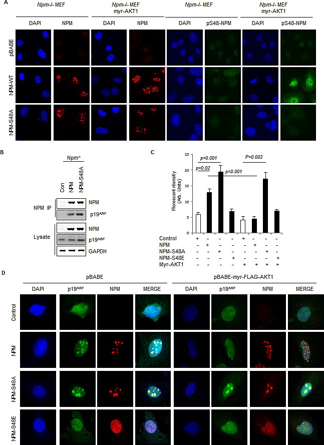
Fig.2. Phosphorylation of NPM-Ser48 regulates the localization of NPM and p19A.

(A) Npm−/−, p53−/−double null MEF were infected with pBabe retrovirus expressing FLAG-tagged-myr-AKT1 in combination with NPM-WT or NPM-S48A as indicated. Cells were fixed and stained with DAPI and anti-NPM (left) or anti-phospho-S48-NPM (pS48-NPM). (B) NPM immunoprecipitates and whole cell lysates from Npm−/−;p53−/− MEFs expressing human NPM or NPM-S48A were probed with the indicated antibodies.(C) Graph, quantification of p19ARF staining intensity in confocal images using ImageJ. (D) Npm−/−, p53−/−double null MEF were infected with pBABE retrovirus expressing FLAG-tagged-myr-AKT1 in combination with NPM-WT, NPM-S48A or S48E as indicated. Cells were fixed and stained with DAPI, anti-NPM and anti p19ARF.
