Abstract
Summary:
Sometimes, after local anesthetic injection for surgical procedures of the upper eyelid, it is possible to observe superficial preseptal hematomas or excessive lid swelling that may distort the tissues and obscure surgical landmarks. We present a technique to perform local anesthesia of the upper eyelids, using a 27-gauge needle and a 26-gauge filling cannula, that may decrease the incidence of hematomas and bruising.
Local anesthesia and intravenous sedation are frequently used for patients undergoing upper eyelid surgery, although general anesthesia may be desirable in some instances.
The local anesthetic is usually administered as a diffuse superficial slowly subcutaneous injection along the upper lid skin crease.1
Risk of hemorrhage during injections may be possible especially when the patient is taking prescribed anticoagulants or when hypertension is present; to provide bleeding control, local anesthetic is associated with epinephrine (1:100,000).2
Sometimes, after local anesthetic injection for surgical procedures of the upper eyelid, it is possible to observe superficial preseptal hematomas or excessive lid swelling. They usually disappear within a few days, but in the absence of appropriate treatment, they may cause severe subcutaneous scars, septal retractions, retractile ectropion, and pigmentation disorders.3,4
Bruising and hematomas are experienced by every patient undergoing surgical procedures of the upper eyelid, so it is not really a complication but an expected side effect that may distort the tissues and obscure surgical landmarks.
METHODS AND RESULTS
We present a technique to perform local anesthesia of the upper eyelids, using a 27-gauge needle and a 26-gauge filling cannula, that may decrease the incidence of hematomas and bruising.
After skin marking, we perform local anesthesia using a mix of lidocaine 2% with epinephrine (1:100,000).
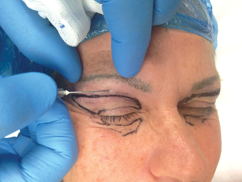
We use the needle to enter the skin of the eyelids, as an “invitation” to the cannula: the entry point is above the upper border of the skin excision on a line running through the center of the eyebrow (Fig. 1).
Fig. 1.

The needle is used to enter the skin of the eyelids, as an “invitation” to the cannula: the entry point is above the upper border of the skin excision on a line running through the center of the eyebrow.
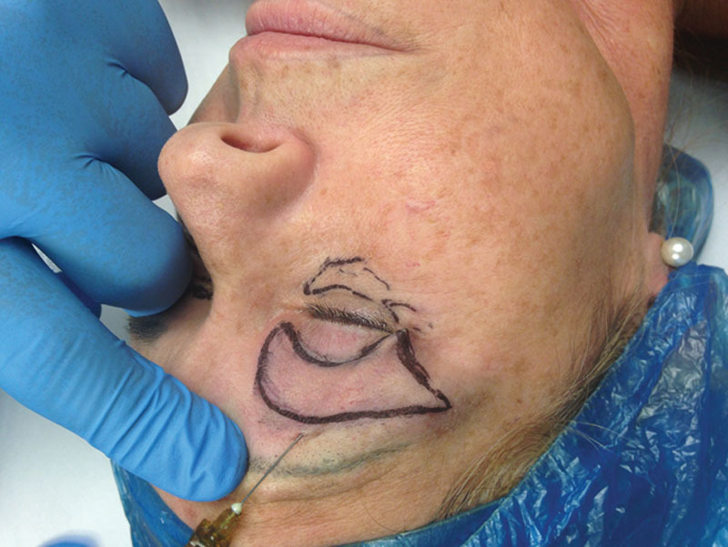
Sometimes, another alternative entry point may be at the level of the lateral part of the eyelid (Fig. 2).
Fig. 2.

Another alternative entry point may be at the level of the lateral part of the eyelid.
The injection of anesthetic with the cannula is performed superficially in a continuous movement while the noninjecting hand spreads the eyelid tissues with traction for better visualization.
When injecting near the eye, surgeon must be very careful to stabilize his or her hand on the patient’s face, so that if there is any surprise movement, the hand will follow and any undesired complication is avoided.2
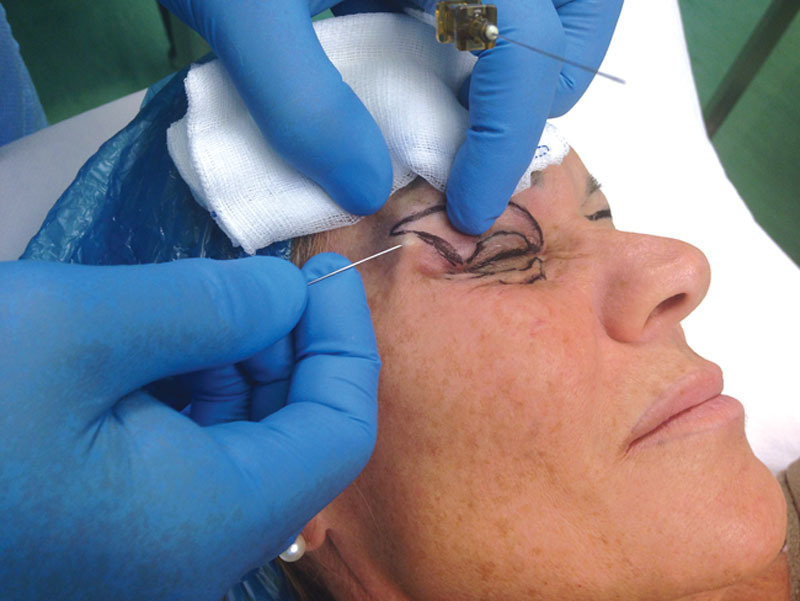
The needle is cautiously advanced in a superior-inferior direction, toward the canthus, and the anesthetic is administrated while the cannula is pulled back, without the need to be retraced. In this way, there is the possibility to perform the anesthesia of the entire eyelid with a single injection (Fig. 3).
Fig. 3.

The needle is cautiously advanced in a superior-inferior direction and the anesthetic is administrated while the cannula is pulled back, without the need to be retraced.
This technique decreases the number of times the tissues are entered, thus causing less pain and bruising.
Postoperative care includes ophthalmic antibiotic ointment applied 3–4 times daily for 2 weeks.
DISCUSSION AND CONCLUSIONS
We think that this technique of using 27-gauge needle and a 26-gauge filling cannula for local anesthesia allows minor incidence of hematomas and bruising after eyelid surgery.
PATIENT CONSENT
The patient provided written consent for the use of her image.
Footnotes
Disclosure: The authors have no financial interest to declare in relation to the content of this article. The Article Processing Charge was paid for by the authors.
REFERENCES
- 1.Vagefi MR, Lin CC, McCann JD, et al. Local anesthesia in oculoplastic surgery: precautions and pitfalls. Arch Facial Plast Surg. 2008;10:246–249. doi: 10.1001/archfaci.10.4.246. [DOI] [PubMed] [Google Scholar]
- 2.Mehta S, Belliveau MJ, Oestreicher JH. Oculoplastic surgery. Clin Plast Surg. 2013;40:631–651. doi: 10.1016/j.cps.2013.08.005. [DOI] [PubMed] [Google Scholar]
- 3.Stephen R, James R. Patrinely Klapper management of cosmetic eyelid surgery complications. Semin Plast Surg. 2007;21:80–93. doi: 10.1055/s-2007-967753. [DOI] [PMC free article] [PubMed] [Google Scholar]
- 4.Lee EJ, Khandwala M, Jones CA. A randomised controlled trial to compare patient satisfaction with two different types of local anaesthesia in ptosis surgery. Orbit. 2009;28:388–391. doi: 10.3109/01676830903071240. [DOI] [PubMed] [Google Scholar]


