Abstract
Affinity purification of Strep-tagged fusion proteins on resins carrying an engineered streptavidin (Strep-Tactin) has become a widely used method for isolation of protein complexes under physiological conditions. Fusion proteins containing two copies of Strep-tag II, designated twin-Strep-tag or SIII-tag, have the advantage of higher affinity for Strep-Tactin compared to those containing only a single Strep-tag, thus allowing more efficient protein purification. However, this advantage is offset by the fact that elution of twin-Strep-tagged proteins with biotin may be incomplete, leading to low protein recovery. The recovery can be dramatically improved by using denaturing elution with sodium dodecyl sulfate (SDS), but this leads to sample contamination with Strep-Tactin released from the resin, making the assay incompatible with downstream proteomic analysis. To overcome this limitation, we have developed a method whereby resin-coupled tetramer of Strep-Tactin is first stabilized by covalent cross-linking with Bis(sulfosuccinimidyl) suberate (BS3) and the resulting cross-linked resin is then used to purify target protein complexes in a single batch purification step. Efficient elution with SDS ensures good protein recovery, while the absence of contaminating Strep-Tactin allows downstream protein analysis by mass spectrometry. As a proof of concept, we describe here a protocol for purification of SIII-tagged viral protein VPg-Pro from nuclei of virus-infected N. benthamiana plants using the Strep-Tactin polymethacrylate resin cross-linked with BS3. The same protocol can be used to purify any twin-Strep-tagged protein of interest and characterize its physiological binding partners.
Keywords: Biochemistry, Issue 86, Strep-tag, fusion protein, Strep-Tactin, protein complex purification, bis(sulfosuccinimidyl) suberate, BS3, protein cross-linking, protein structure stabilization, proteomics, mass spectrometry
Introduction
In recent years, Strep-tag technology has become widely used in many areas of biomedical research, including proteomics and structural biology. This protein purification technology, which relies on the fusion of recombinant proteins to a short Strep-tag peptide, has matured with the advent of affinity matrices carrying Strep-Tactin, a genetically engineered variant of streptavidin with improved peptide-binding capacity.1,2 Fusion proteins containing two copies of Strep-tag II, designated twin-Strep-tag or SIII-tag, exhibit a higher affinity for Strep-Tactin matrices than those containing only a single Strep-tag, ensuring more efficient purification of the recombinant proteins and their associated binding partners. However, the higher affinity of twin-Strep-tagged proteins to Strep-Tactin also has its downside. Competitive elution of such proteins with excess biotin may be incomplete, leading to decreased target protein yield. A more efficient alternative is elution with SDS, but it leads to undesired sample contamination with Strep-Tactin released from the resin, making the assay incompatible with proteomic analysis. This paper presents a technique to overcome this limitation by first stabilizing the resin-coupled tetramer of Strep-Tactin by chemical cross-linking and then using SDS to elute twin-Strep-tagged proteins and their associated complexes from the resulting cross-linked resin. Thus, sufficient protein yield can be achieved without sample contamination with Strep-Tactin, thereby allowing further analysis by mass spectrometry.
The method is suitable for purification of any recombinant fusion protein with a surface-exposed SIII-tag3 or twin-Strep-tag (amino acid sequence WSHPQFEK(GGGS)3 WSHPQFEK and SAWSHPQFEK(GGGS)2 GGSAWSHPQFEK, respectively). The protein can be of animal, plant or bacterial origin and can be isolated from either total cell lysate or enriched organelle fraction. As an example, we describe here the purification of an SIII-tagged protein VPg-Pro of Potato Virus A (PVA)4 from the nuclear fraction of PVA-infected Nicotiana benthamiana plants. The nuclear fraction was isolated as previously described5, with the following modifications: cells were not treated with formaldehyde, sodium butyrate was substituted in all buffers with 5 mM sodium fluoride, complete protease inhibitor was substituted with PMSF, Triton X-100 concentration in extraction buffer #2 was lowered to 0.3% (v/v) and the nuclear pellet obtained by centrifugation through sucrose cushion (extraction buffer #3) was resuspended in 1.45 ml of pre-chilled binding buffer and rotated for 1.5 hours at 4 °C. The resulting nuclear extract containing the SIII-tagged bait protein and associated complexes (bait protein sample) was processed according to the protocol described below (see section 2).
Protocol
1. Cross-linking of Strep-Tactin Polymethacrylate Resin with Bis(sulfosuccinimidyl) Suberate (BS3)
Equilibrate one sealed microtube containing 2 mg of BS3 cross-linker to room temperature. CAUTION: BS3 is a hazardous substance. Wear protective gloves and goggles.
Resuspend Strep-Tactin polymethacrylate resin (50% suspension in 100 mM Tris-HCl, pH 8.0; 1 mM EDTA; 150 mM NaCl) by brief vigorous shaking and immediately transfer 600 µl of the suspension to a spin column using a pipette tip with the end cut off.
Centrifuge at 1,500 x g for 30 sec at room temperature. Discard the flow-through and add 450 µl of phosphate buffered saline (PBS) to the column.
Repeat the previous step 2 more times to completely replace Tris buffer with PBS and leave the resin in 430 µl of PBS adjusted to pH 8.0 after the last centrifugation.
Puncture the foil of the microtube with BS3 with a pipette tip containing 100 µl of ultrapure water. Dissolve the BS3 powder in water by gently pipetting up and down and immediately add 20 µl of the solution to the spin column. The final concentration of BS3 in the cross-linking reaction is ~1.2 mM.
Rotate the column for 30 min at room temperature. Check that the resin is mixed properly with the BS3 solution.
To quench the reaction, add 6 µl of 3M Tris-HCl, pH 7.5 and rotate the column for another 15 min at room temperature.
Centrifuge at 1,500 x g for 30 sec at room temperature. Discard the flow-through and resuspend the cross-linked resin in 450 µl of Tris buffered saline with Tween 20 (TBST). Repeat the centrifugation and wash steps two more times. At the last step, resuspend the resin in 450 µl of TBS.
Transfer the resin suspension to a fresh tube using a pipette tip with the end cut off. Resuspend the resin left in the column with another 450 µl of TBS and transfer to the same tube. Repeat the last step once more to ensure maximum transfer of the resin from the column to the tube.
Let the tube stand for 10 min at room temperature and adjust the volume to 600 µl by removing excess TBS. The resin is ready for immediate use (recommended) or can be stored at 4 °C, without freezing.
2. Binding of the Twin-Strep-tagged Bait Protein and Associated Complexes to the Cross-linked Strep-Tactin Polymethacrylate Resin
Centrifuge 1 ml of the bait protein sample in binding buffer at 17,000 x g for 10 min at 4 °C and transfer the supernatant to a fresh tube.
- To minimize binding of endogenous biotinylated proteins to the Strep-Tactin resin, add avidin to a final concentration of 100 µg/ml and rotate for 15 min at 4 °C.
- If the cross-linked resin from step 1.10 has been stored for an extended period of time at 4 °C, collect the resin by centrifugation at 400 x g for 1 min at 4 °C, discard the supernatant and wash the resin with 1 ml of TBST. Repeat the centrifugation and wash steps two more times, first with TBST and then with TBS. Centrifuge the tube at 400 x g for 1 min at 4 °C and adjust to the original volume by removing excess TBS.
Resuspend the cross-linked Strep-Tactin resin by vortexing. Immediately add 50 µl of the resin suspension to the tube containing the bait protein sample using a cut pipette tip and rotate for another 30 min at 4 °C.
While waiting, set thermomixer to 55 °C and preheat 500 µl of elution buffer for use in step 3.1.
Centrifuge at 400 x g for 1 min at 4 °C. Discard the supernatant and wash the resin on a rotator for 5 min at 4 °C with 1 ml of pre-chilled wash buffer #1. Repeat the centrifugation and wash steps three times. At the last step, resuspend the resin in 250 µl of wash buffer #2.
Transfer the resin suspension to a fresh spin column. Resuspend the resin remaining in the tube in another 250 µl of wash buffer #2 and transfer to the same column.
Centrifuge at 400 x g for 3 min at 4 °C, discard the flow-through and transfer the column to a fresh dolphin-nose 2 ml tube. Proceed immediately to the elution step below.
3. Elution of Specific Protein Complexes
Add 150 µl of preheated elution buffer from step 2.4 to the spin column.
Incubate in thermomixer for 5 min at 55 °C, shaking at 1,400 rpm.
Centrifuge at 1,500 x g for 1 min at room temperature.
Discard the column and store the purified target proteins at ≤ -20 °C.
| Phosphate buffered saline (PBS) | |
| Na2HPO4 | 10 mM |
| KH2PO4 | 2mM |
| NaCl | 137 mM |
| KCl | 2.7 mM |
| pH adjusted to 7.4 unless stated otherwise (pH 8.0 in section 1.4) | |
| Tris buffered saline (TBS) | |
| Tris-HCl, pH 7.4 | 50 mM |
| NaCl | 150 mM |
| Tris buffered saline with Tween 20 (TBST) | |
| Tris-HCl, pH 7.4 | 50 mM |
| NaCl | 150 mM |
| Tween 20 | 0.1% (v/v) |
| Binding buffer | |
| Tris-HCl, pH 8.0 | 25 mM |
| NaCl | 550 mM |
| NaF | 5 mM |
| EDTA | 0.5 mM |
| Glycerol | 10% (v/v) |
| PMSF | 0.1 mM |
| Wash buffer #1 | |
| Tris-HCl, pH 8.0 | 25 mM |
| NaCl | 500 mM |
| NaF | 5 mM |
| EDTA | 0.4 mM |
| Igepal CA-630 | 0.2% (v/v) |
| Glycerol | 5% (v/v) |
| PMSF | 0.1 mM |
| Wash buffer #2 | |
| Tris-HCl, pH 8.0 | 25 mM |
| NaCl | 150 mM |
| Elution buffer | |
| Tris-HCl, pH 8.0 | 25 mM |
| SDS | 1% (w/v) |
Table 1. Buffers used in the present study.
Representative Results
The purification procedure is schematically illustrated in Figure 1, together with a representation of problems associated with other existing purification methods.
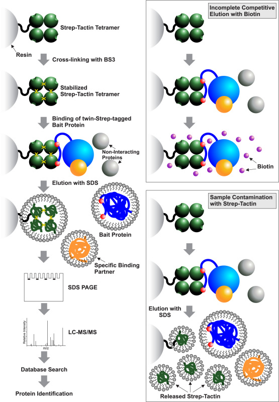 Figure 1. Schematic representation of the purification procedure. The workflow includes stabilization of the resin-coupled Strep-Tactin tetramer via covalent cross-linking with BS3, binding of the twin-Strep-tagged bait protein and associated complexes to the cross-linked resin, washing away of unbound proteins, denaturing elution of the specifically bound protein complexes with 1% SDS and their analysis by liquid chromatography- tandem mass spectrometry (LC-MS/MS). Two red spheres in the bait protein denote the twin-Strep-tag. Limitations of other purification methods are shown in inset boxes on the right. They include incomplete elution of target proteins with biotin and sample contamination with Strep-Tactin following denaturing elution with 1% SDS.
Typical results of the SIII-tagged protein purification using the BS3-cross-linked Strep-Tactin polymethacrylate resin and denaturing elution with 1% SDS are shown in Figure 2. 10% (15 µl) of the spin column eluate containing purified SIII-tagged PVA VPg-Pro and associated proteins was analyzed by SDS-PAGE followed by silver staining. The absence of the 52 kDa band corresponding to PVA VPg-Pro in the negative control lane confirmed the specificity of the pull-down. 40 µl of the remaining spin column eluate were applied to a keratin-free SDS gel and the corresponding lane was cut out and the contained proteins were digested with trypsin and analyzed by liquid chromatography- tandem mass spectrometry (LC-MS/MS). The mass spectrometry analysis identified viral RNA-dependent RNA polymerase (replicase) NIb as the interacting partner of VPg-Pro. Multiple tryptic peptides corresponding to NIb were detected in all four biological replicates of the affinity purification and none in the four controls expressing VPg-Pro without the SIII-tag, confirming the specificity of interaction. Because the molecular weight of NIb (59 kDa) is close to that of SIII-tagged VPg-Pro (52 kDa), the two proteins appeared as a double band during SDS PAGE analysis (Figure 2, arrowhead). Taken together, the results described above provide experimental evidence that affinity purification on Strep-Tactin resins covalently cross-linked with BS3 can be successfully employed to isolate twin-Strep-tagged proteins and identify their physiological binding partners by mass spectrometry.
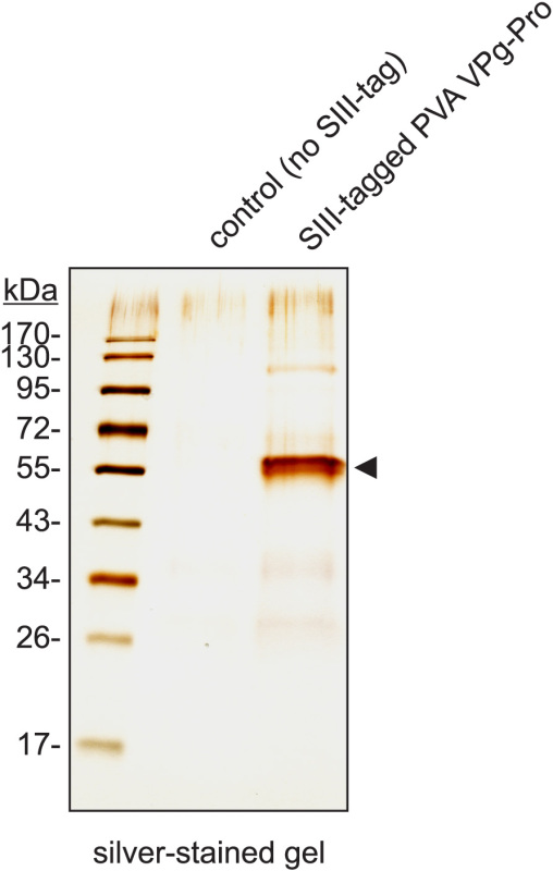 Figure 2. Purification of SIII-tagged PVA VPg-Pro and its binding partner PVA NIb on Strep-Tactin polymethacrylate resin cross-linked with BS3. The figure shows a silver-stained SDS-polyacrylamide gel of eluates from the cross-linked beads. NIb was identified by mass spectrometry as a binding partner of VPg-Pro using an aliquot of the same sample. SIII-tagged PVA VPg-Pro (predicted MW: 52 kDa) and NIb (predicted MW: 59 kDa) migrate as a double band, indicated by an arrowhead. Negative control (central lane) corresponds to purification from cells infected with the virus expressing VPg-Pro without the SIII-tag.
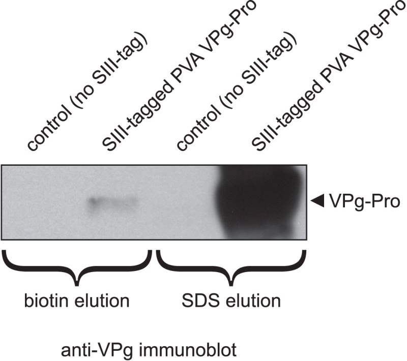 Figure 3. Biotin elution of SIII-tagged PVA VPg-Pro from Strep-Tactin polymethacrylate resin is incomplete compared to elution with 1% SDS. The figure shows an anti-VPg immunoblot of eluates from Strep-Tactin beads. The beads were eluted either with 15 mM biotin or with 1% SDS. The difference in signal intensity reflects different protein yields obtained with the two elution techniques. Negative controls correspond to purifications from cells infected with the virus expressing VPg-Pro without the SIII-tag.
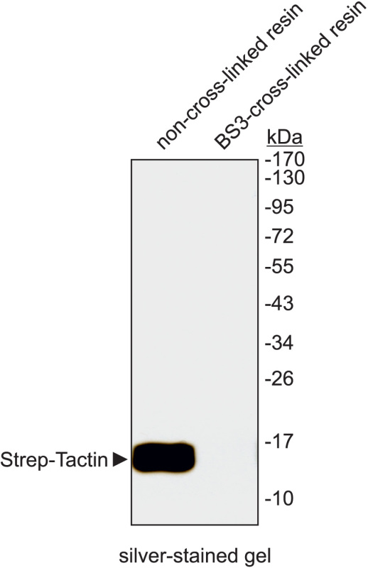 Figure 4. Covalent cross-linking with BS3 prevents the release of Strep-Tactin from the resin during SDS elution. The figure shows a silver-stained gel of SDS eluates from non-cross-linked (left lane) and BS3-cross-linked (right lane) Strep-Tactin polymethacrylate resin. Note the presence of released Strep-Tactin in the left lane but its absence in the right lane.
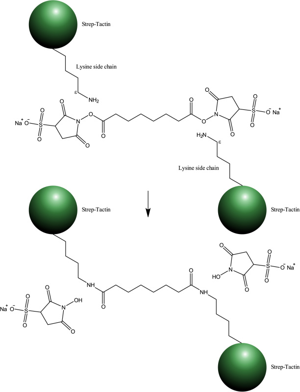 Figure 5. Chemical structure of
Bis(sulfosuccinimidyl) suberate (BS3) and its cross-linking reaction with epsilon amino groups of lysine residues in Strep-Tactin.
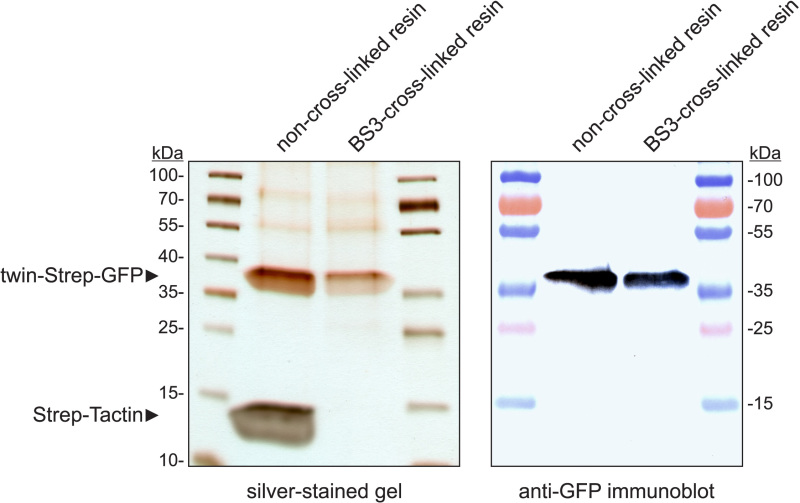 Figure 6. Purification of twin-Strep-tagged green fluorescent protein (GFP) on non-cross-linked and BS3-cross-linked Strep-Tactin polymethacrylate resin. Two equal amounts of human embryonic kidney 293 cells expressing twin-Strep-tagged GFP were lysed and similarly processed using the above protocol, except that in one case the resin has been cross-linked with BS3 and in the other BS3 has been substituted with water in the cross-linking reaction. The figure shows a silver-stained SDS-polyacrylamide gel and anti-GFP immunoblot of purified proteins. Note that some decrease in protein yield is inherent to purification methods involving chemical cross-linking, but this is compensated by the higher sample purity due to the absence of released Strep-Tactin.
Discussion
The above protocol can be used to purify any twin-Strep-tagged bait protein of interest and its associated complexes in any suitable buffer that does not contain biotin or strong denaturants. In the current version of the protocol, binding and washing are performed under relatively stringent conditions in the presence of high salt and non-ionic detergent. Although this results in less background, fragile protein complexes may dissociate under these conditions. To preserve such lower affinity complexes, salt concentration may be lowered and nonionic detergent may be reduced or omitted from binding and wash buffers.
The main advantage of the twin-Strep-tag system is the high affinity of the small (3kDa) tandem tag to engineered streptavidin (Strep-Tactin)1,2, allowing efficient one-step purification of target proteins and their complexes even in batch mode. Because of the strong binding of twin-Strep-tag to Strep-Tactin, the resin can be washed under moderately stringent conditions, such as high detergent and/or salt concentrations, which results in higher target protein purity. However, such strong binding also has its drawbacks. Even in the case of proteins with a single Strep-tag II (amino acid sequence WSHPQFEK), competitive elution from the resin may sometimes be difficult. For example, elution of a Strep (II)-tagged protein kinase, NtCDPK2, from the Strep-Tactin polymethacrylate resin with 10 mM desthiobiotin proved to be unsuccessful, requiring denaturing elution with SDS sample buffer6. The problem was resolved only after desthiobiotin was replaced with stronger competing biotin (10 mM) and elution was carried out for 5 minutes with vigorous shaking7. In the case of the twin-Strep-tag, which has higher affinity for Strep-Tactin, target protein elution may be incomplete even at biotin concentrations as high as 15 mM. As shown in Figure 3, only a small fraction of SIII-tagged PVA VPg-Pro could be eluted from the Strep-Tactin resin with 15 mM biotin compared to the amount eluted with 1% SDS. It is important to note that the efficiency of competitive elution likely also depends on the nature of the eluted protein or protein complex. Larger and more hydrophobic proteins may sterically hinder the accessibility of the biotin binding pocket in Strep-Tactin, thereby making competitive elution less efficient. The above drawbacks are avoided by using elution with SDS, but such elution has its own problems, mainly revolving around the fact that exposure to detergents leads to dissociation of the resin-coupled Strep-Tactin tetramers and sample contamination with released Strep-Tactin8. This is demonstrated in Figure 4 (left lane), which shows a considerable amount of Strep-Tactin released after incubation of the Strep-Tactin resin in elution buffer containing 1% SDS. Such contamination is unacceptable if the sample needs to be further analyzed by LC-MS/MS.
An effective solution to the above contamination problem is the stabilization of the resin-coupled Strep-Tactin tetramer9 by covalent cross-linking. For this purpose, we employed a water-soluble, non-cleavable, homobifunctional amine-reactive cross-linker BS3 with a history of successful use in stabilizing protein structures10-13. Figure 5 shows the chemical formula of BS3 and its cross-linking reaction with free epsilon amino groups of lysine in Strep-Tactin. When the resin-coupled Strep-Tactin tetramer was stabilized by cross-linking with BS3, the problem of sample contamination with released Strep-Tactin was virtually eliminated (Figure 4, right lane). Because the cross-linking with BS3 does not cause extensive protein aggregation14, it does not reduce the accessibility of the biotin binding pocket in streptavidin, making the cross-linked resin suitable for purification of Strep-tagged fusion proteins. It should be noted, however, that chemical cross-linking of proteins with biological activity (antibodies, etc.) stabilizes both active and inactive protein conformations, leading to some activity loss. For example, the well-established immunoprecipitation method using BS3-cross-linked antibodies produces a lower target protein yield compared to that obtained with non-cross-linked antibodies15. Nevertheless, some loss of biological activity is considered to be an acceptable trade-off for the improved sample purity. We have observed similar results with the BS3-cross-linked Strep-Tactin resin. The somewhat lower protein yield obtained with the cross-linked resin was more than compensated by the greatly improved sample purity (Figure 6).
Another potential application of the proposed method is in purification of Strep-tagged membrane proteins solubilized with detergents. Detergents are essential for keeping such hydrophobic proteins and their associated complexes in solution, but their presence may lead to sample contamination with Strep-Tactin released from the affinity resin. A good example of this is the purification of a Strep (II)-tagged membrane protein AtNTT1 on Strep-Tactin beads for the purpose of protein crystallization8. The presence of the detergent laurylamidodimethylpropylaminoxide (LAPAO) in the elution buffer led to the release of Strep-Tactin from the beads and subsequent growth of undesired Strep-Tactin crystals8. Stabilization of the resin-coupled Strep-Tactin tetramer by covalent cross-linking may provide an effective means to overcome this problem.
In summary, chemical cross-linking has been successfully employed to overcome the limitations associated with denaturing elution from the Strep-Tactin resin. This approach helped achieve good target protein recovery without introducing sample contamination. The proposed method can thus be used to identify specific binding partners of twin-Strep-tagged bait proteins by mass spectrometry. Furthermore, the method can be applied to isolate and characterize nucleoprotein complexes, including those reversibly cross-linked with formaldehyde, as well as membrane proteins solubilized with detergents. Finally, chemical cross-linking does not significantly change the physical properties of the Strep-Tactin resin, making the cross-linked resin potentially suitable for high-throughput applications.
Disclosures
No conflicts of interest declared.
Acknowledgments
We gratefully acknowledge the technical support of Sini Miettinen, Minna Pöllänen and Taru Rautavesi. We thank Helka Nurkkala for providing HEK 293 cells expressing twin-Strep-tagged GFP and Pekka Evijärvi for providing sound recording equipment. This work was funded by the Academy of Finland, grant numbers 138329, 134684 and 258978.
References
- Voss S, Skerra A. Mutagenesis of a flexible loop in streptavidin leads to higher affinity for the Strep-tag II peptide and improved performance in recombinant protein purification. Protein Eng. 1997;10(8):975–982. doi: 10.1093/protein/10.8.975. [DOI] [PubMed] [Google Scholar]
- Schmidt TG, Skerra A. The Strep-tag system for one-step purification and high-affinity detection or capturing of proteins. Nat. Protoc. 2007;2(6):1528–1535. doi: 10.1038/nprot.2007.209. [DOI] [PubMed] [Google Scholar]
- Junttila MR, Saarinen S, Schmidt T, Kast J, Westermarck J. Single-step Strep-tag purification for the isolation and identification of protein complexes from mammalian cells. Proteomics. 2005;5(5):1199–1203. doi: 10.1002/pmic.200400991. [DOI] [PubMed] [Google Scholar]
- Hafren A, Hofius D, Ronnholm G, Sonnewald U, Makinen K. HSP70 and its cochaperone CPIP promote potyvirus infection in Nicotiana benthamiana by regulating viral coat protein functions. Plant Cell. 2010;22(2):523–535. doi: 10.1105/tpc.109.072413. [DOI] [PMC free article] [PubMed] [Google Scholar]
- Jaskiewicz M, Peterhansel C, Conrath U. Detection of histone modifications in plant leaves. J. Vis. Exp. 2011. [DOI] [PMC free article] [PubMed]
- Witte CP, Noel LD, Gielbert J, Parker JE, Romeis T. Rapid one-step protein purification from plant material using the eight-amino acid StrepII epitope. Plant Mol. Biol. 2004;55(1):135–147. doi: 10.1007/s11103-004-0501-y. [DOI] [PubMed] [Google Scholar]
- Werner AK, Sparkes IA, Romeis T, Witte CP. Identification, biochemical characterization, and subcellular localization of allantoate amidohydrolases from Arabidopsis and soybean. Plant Physiol. 2008;146(2):418–430. doi: 10.1104/pp.107.110809. [DOI] [PMC free article] [PubMed] [Google Scholar]
- Panwar P, Deniaud A, Pebay-Peyroula E. Contamination from an affinity column: an encounter with a new villain in the world of membrane-protein crystallization. Acta Crystallogr. D. Biol. Crystallogr. 2012;68(10):1272–1277. doi: 10.1107/S090744491202639X. [DOI] [PubMed] [Google Scholar]
- Hendrickson WA, et al. Crystal structure of core streptavidin determined from multiwavelength anomalous diffraction of synchrotron radiation. Proc. Natl. Acad. Sci. U. S. A. 1989;86(7):2190–2194. doi: 10.1073/pnas.86.7.2190. [DOI] [PMC free article] [PubMed] [Google Scholar]
- Bernot KM, Lee CH, Coulombe PA. A small surface hydrophobic stripe in the coiled-coil domain of type I keratins mediates tetramer stability. J. Cell Biol. 2005;168(6):965–974. doi: 10.1083/jcb.200408116. [DOI] [PMC free article] [PubMed] [Google Scholar]
- Singh I, et al. Solution structure of human von Willebrand factor studied using small angle neutron scattering. J. Biol. Chem. 2006;281(50):38266–38275. doi: 10.1074/jbc.M607123200. [DOI] [PubMed] [Google Scholar]
- Weldon WC, et al. Enhanced immunogenicity of stabilized trimeric soluble influenza hemagglutinin. PLoS One. 2010;5(9) doi: 10.1371/journal.pone.0012466. [DOI] [PMC free article] [PubMed] [Google Scholar]
- Wang W, Barger SW. Roles of quaternary structure and cysteine residues in the activity of human serine racemase. BMC Biochem. 2011;12:63. doi: 10.1186/1471-2091-12-63. [DOI] [PMC free article] [PubMed] [Google Scholar]
- Rappsilber J. The beginning of a beautiful friendship: cross-linking/mass spectrometry and modelling of proteins and multi-protein complexes. J. Struct. Biol. 2011;173(3):530–540. doi: 10.1016/j.jsb.2010.10.014. [DOI] [PMC free article] [PubMed] [Google Scholar]
- Sousa MM, Steen KW, Hagen L, Slupphaug G. Antibody cross-linking and target elution protocols used for immunoprecipitation significantly modulate signal-to noise ratio in downstream 2D-PAGE analysis. Proteome Sci. 2011;9:45. doi: 10.1186/1477-5956-9-45. [DOI] [PMC free article] [PubMed] [Google Scholar]


