Abstract
Stretch marks are common skin disorders that are dermal scars with associated epidermal atrophy. They are of significant concern or psychological concern to many. This manuscript describes the use of multipolar radiofrequency with pulsed magnetic fields that was successfully used to diminish these lesions in 16 subjects undergoing a series of treatments. The improvements noted were statistically significant and no serious adverse events were noted.
Striae or stretch marks are a common skin condition occurring in both genders, but are more prevalent among women. These are linear dermal scars accompanied by epidermal atrophy. They usually occur frequently in numerous physiological and pathological conditions, such as adolescent growth spurts, pregnancy, obesity, Cushing's and Marfan syndromes, and long-term systemic or topical steroid use. Decreased expression of collagen and fibronectin genes has also been associated with striae.1
Although they do not cause any significant medical problems, aesthetically striae can be a cause of great concern or psychological stress for many women. Patient demand for nonsurgical, noninvasive, and no-downtime skin rejuvenation procedures has grown dramatically over the past decade as new treatments and technologies have been introduced. During this period, there has been a substantial increase in the utilization of medical prescriptive skin care. The effects of dermal heating are well-recognized to include the modification of collagen structure and stimulation of neocollagenesis (by induction of inflammation that will end in new collagen production by fibroblasts recruited to the heated area). These changes can help improve the appearance of striae due to the increased collagen and elastin. Electrical energy can be advantageous for deep dermal heating as the movement of electrons is not impeded by tissue proteins. Radiofrequency (RF) energy devices have remained the most common and dominant technology in the noninvasive management of skin tightening, wrinkle reduction, cellulite improvement, and body contouring enhancement, as they can treat all these conditions with relatively consistent results. The RF energy is high-frequency alternating electrical current that passes into the dermis and hypodermal tissues without disruption of the epidermal-dermal barrier. The highfrequency oscillating electrical current results in collisions between charged molecules and ions and the micromolecular mechanical energy from these collisions is transformed into heat.1-3
RF devices may be mono-polar, bipolar, or multi-polar. Venus Freeze (Venus Concept, Toronto, Ontario, Canada) is a noninvasive multi-polar (MP2) pulsed electromagnetic field (PEMF) and RF energy generating system with two applicators—Diamondpolar™ (4 RF and PEMF electrodes) for treatment of small areas and Octipolar™ (8 electrodes and 9 PEMF) for treatment of large areas. The treatment applicators transmit bipolar RF energy in a method that creates an organized RF energy matrix, which produces homogeneous heating in the entire treatment area for maximum safety and efficacy, eliminating the need for pre/post cooling mechanisms. The RF energy transmitted mediates thermal stimulation of the extracellular matrix (ECM) in the dermis. This results in an immediate and temporary shrinkage of the collagen triple helix2-4 and subsequently, micro-inflammatory stimulation of the fibroblast, which, in response, produces new collagen (neocollagenesis), new elastin (neoelastogenesis), and ground substances.3,4 This treatment enhances the tensile strength and elasticity of the dermis with the aid of the newly produced proteins and proteoglycans.2,3,5
OBJECTIVES
The objective of this study was to evaluate the safety and effectiveness of using RF and PEMFs for the treatment of striae. The safety of RF and PEMFs for striae treatment was established by physicians’ assessment/observation of adverse events or side effects, such as signs of pain, edema, burn, localized infection, skin pigmentation, and texture alterations. Efficacy of using RF and PEMFs for striae treatment was established by the level of improvement seen visually and by macro-photography.
MATERIALS AND METHODS
Sixteen female subjects between the ages of 30 and 72 (mean age=46.06 years, SD=10.247) with varying degrees of striae participated in this two-center, single-arm pilot study. The subjects were enrolled into the study after meeting all the inclusion/exclusion criteria and providing signed informed consent.
Each subject had a screening assessment and pretreatment photograph (baseline), six treatment visits, which included five measurements of striae bands and pretreatment photographs and two post-treatment visits (1 week and 1 month post-treatments). The treated areas were photographed using high-resolution macro-photography. The pre- and post-treatment photographs were compared by two independent physicians. A sterile six-inch skin ruler was used to measure the length and width of each striae band on the first appointment prior to each treatment and at the follow-up appointments of one week and one month post-treatment series.
Each subject received six treatments using RF and PEMF. Prior to treatment, the treated areas were assessed visually in order to determine skin-relevant parameters. The treated areas were photographed and measured in order to allow comparison and assessment of striae improvement following treatment. The treatment area was cleaned thoroughly with soap and water. The skin surface was dried prior to the treatment.
The treatment parameters, such as time (10 minutes for an area approximately 4x5 inches) and output energy (60-80% with goal being to reach therapeutic in the first minute of treatment) was determined by the physician depending on patient skin type and area of treatment.
For treatment safety evaluation, treated areas were visually assessed for side effects, such as edema, erythema, burn, localized infection, and skin pigmentation, immediately after the treatment. Subjects were also asked questions to assess their willingness to continue with the treatment as well as their rating on observed improvements.
The pretreatment, during treatment, and post-treatment measurement of length and width of each striae bands in the treatment area were recorded per subject. The pretreatment and post-treatment photographs were assessed and graded by two physicians.
RESULTS
All 16 subjects enrolled in the study completed the treatment and the following results were recorded:
No side effects or undesirable safety events were recorded for any subjects throughout the study.
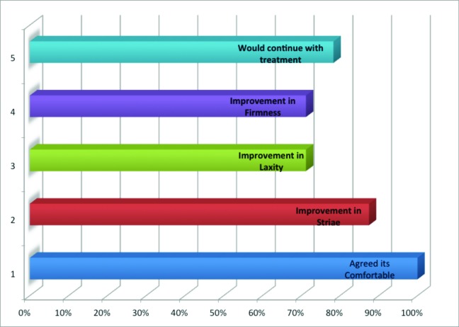
Fourteen out of the 16 subjects agreed that they noticed visible improvement, one was not sure while one did not see any improvement.
All subjects (100%) agreed that the treatment was comfortable. Figure 1 shows the graphical analysis of the outcome of the patient survey conducted during the study.
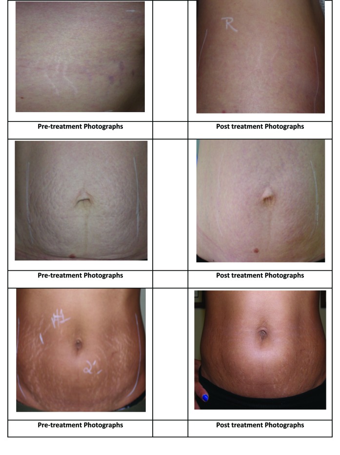
Visual evaluation of pretreatment (baseline) and post-treatment photographs by the two physicians agreed that there was reduction in the visibility of striae after treatment in some of the pairs of photographs reviewed. The Kappa statistical method was used to establish almost perfect agreement on decisions of the two physicians at 95% confidence interval (Kappa value of 0.88, SE(κ)=0.083, 95% CI=0.71 to 1.04). Figure 2 shows reduction in the visibility of striae after six treatments with RF and PEMFs.
Figure 1.
Patient survey results
Figure 2.
Before and after photos showing reduction in the visibility of striae after six treatments with RF and PEMFs
Measurements of length and width of striae bands were statistically analyzed to determine the efficacy of RF and PEMFs in the treatment of striae (Table 1). The mean reduction in length of striae bands measured in the 16 subjects after the treatment was 1.031, SD=0.853. The mean reduction in width of striae bands measured in the 16 subjects after the treatment was 0.160, SD=0.171. Using paired t-test, the reduction in both length and width of striae bands measured at one month post-treatment visit compared to baseline measurements were found to be statistically significant at 95% (p<0.001).
TABLE 1.
Measurements of length and width of striae bands
| SUBJECTS | REDUCTION IN LENGTH (CM) | REDUCTION IN WIDTH (CM) |
|---|---|---|
| 1 | 3.180 | 0.239 |
| 2 | 0.804 | 0.159 |
| 3 | 0.638 | 0.318 |
| 4 | 0.638 | 0.636 |
| 5 | 2.065 | 0.000 |
| 6 | 0.635 | 0.239 |
| 7 | 1.133 | 0.083 |
| 8 | -0.067 | -0.067 |
| 9 | 1.950 | 0.125 |
| 10 | 1.533 | 0.133 |
| 11 | 0.100 | 0.100 |
| 12 | 0.650 | 0.200 |
| 13 | 0.700 | 0.267 |
| 14 | 0.500 | 0.200 |
| 15 | 0.533 | -0.033 |
| 16 | 0.400 | -0.033 |
| Number of subjects | 16.000 | 16.000 |
| Mean reduction | 1.031 | 0.160 |
| Standard deviation | 0.853 | 0.171 |
| Standard error | 0.213 | 0.043 |
| Upper 95% confidence interval | 1.236 | 0.201 |
| Lower 95% confidence interval | 0.826 | 0.119 |
DISCUSSION AND CONCLUSION
All 16 subjects that participated in this study agreed that the treatment was comfortable, and no side effects or undesirable safety events were recorded throughout the six-week treatment period. These results support the safety of RF and PEMFs.
Fourteen (87.5%) out of the 16 subjects agreed that they noticed visible changes. Further, there were statistically significant reductions in both length and width of striae bands measured at one month post-treatment visit compared to baseline measurements in these subjects.
In conclusion, the data generated in this study support the high degree of patient satisfaction and demonstrate the safe, effective use of RF and PEMFs in the treatment of striae.
LIMITATIONS
Although results are promising, the long-term sustainability of the reduced visibility of the striae is not known, and further investigation is required. Patients may achieve a noticeable improvement; however, the complete elimination of the striae is not achieved using this method.
Footnotes
DISCLOSURE:Dr. Dover has a relationship with Allergan, CVS/Skin Effects, Cutera, Cynosure, Kythera, Lumenis, Medicis, Merz, Myoscience, Shaser, Solta, Suneva, Syneron, and Zeltiq. Dr. Rothaus reports no relevant conflicts of interest. Dr. Gold is a consultant and performs research for Venus and is a stockholder in Venus.
REFERENCES
- 1.Lee K, Rho Y, Jang S, et al. Decreased expression of collagen and fibronectin genes in striae distensae tissue. Clin Exp Dermatol. 1994;19:285–288. doi: 10.1111/j.1365-2230.1994.tb01196.x. [DOI] [PubMed] [Google Scholar]
- 2.Alster TS, Lupton JR. Nonablative cutaneous remodeling using radiofrequency devices. Clin Dermatol. 2007;25:487–491. doi: 10.1016/j.clindermatol.2007.05.005. [DOI] [PubMed] [Google Scholar]
- 3.Zelickson BD, Kist D, Bernstein E, et al. Histological and ultrastructural evaluation of the effects of a radiofrequency based non-ablative dermal remodeling device: a pilot study. Arch Dermatol. 2004;140:204–209. doi: 10.1001/archderm.140.2.204. [DOI] [PubMed] [Google Scholar]
- 4.Hantash BM, Ubeid AA, Chang H, et al. Bipolar fractional radiofrequency treatment induces neoelastogenesis and neocollagenesis. Lasers Surg Med. 2009;41:1–9. doi: 10.1002/lsm.20731. [DOI] [PubMed] [Google Scholar]
- 5.Emilia del Pino M, Rosado RH, Azuela A, et al. Effect of controlled volumetric tissue heating with radiofrequency on cellulite and the subcutaneous tissue of the buttocks and thighs. J Drugs Dermatol. 2006;5(8):714–722. [PubMed] [Google Scholar]


