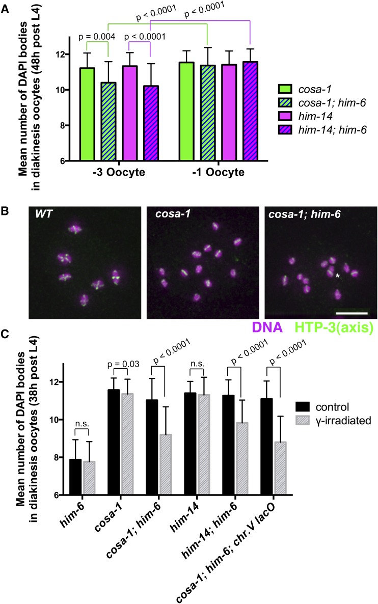
Figure 6.
Quantitation of late-prophase interchromosomal associations in mutants lacking activity of conserved meiotic crossover factors COSA-1 and MutSγ. (A and C) Graphs showing quantitation of DAPI bodies in diakinesis-stage occytes in worms of the indicated genotypes; error bars indicate standard deviation. In this assay, wild-type oocytes have 6 DAPI bodies (reflecting chiasmata connecting all 6 chromosome pairs), and 12 DAPI bodies indicates a complete lack of chiasmata; the assay tends to overestimate the incidence of connections between chromosomes, as some univalents lie too close together to be resolved unambiguously. (A) Worms were fixed for DAPI staining at 48 hr post-L4 stage, and the data for oocytes in the −3 (less mature) and −1 (more mature) positions were graphed separately. For oocytes in the −1 positions, no significant differences were observed between the cosa-1 single mutant and cosa-1; him-6 double mutant or between the him-14 single mutant and him-14; him-6 double mutant, consistent with all six homolog pairs lacking chiasmata and CO at the end of prophase in the double mutants. However, significant differences between the numbers of DAPI bodies detected in the −3 vs. −1 oocytes were observed for the cosa-1; him-6 and him-14; him-6 double mutants (P < 0.0001), but not for the cosa-1 and him-14 single mutants (P = 0.19, P = 0.52). In combination, these data suggest the presence of persistent associations between homologs in the cosa-1; him-6 and him-14; him-6 double mutants that were ultimately resolved prior to ovulation. Numbers of oocyte nuclei scored: him-6, 60; cosa-1; him-6, 193; cosa-1, 71; him-14, 137; him-14; him-6, 129. (B) Immunofluorescence images depicting the full chromosome complements of individual −2 diakinesis oocytes of the indicated genotypes, stained for DNA (purple), and chromosome axis protein HTP-3 (green) following fixation at 38 hr post-L4. HTP-3 cruciform structures (reflecting the presence of chiasmata) are readily detected on bivalents in wild-type oocytes, but infrequent in cosa-1 and cosa-1; him-6 oocytes (none in the images shown). Images are projections of 3-D data stacks encompassing whole nuclei; asterisk indicates univalents that appear to overlap in the projection but are actually from different focal planes. Scale bar, 5 μm. (C) Graph depicting the effect of exposure to 5 krad γ-irradiation on the mean number of DAPI bodies in diakinesis oocytes (fixed 18 hr after IR treatment at 20 hr post-L4). Data for oocytes in the −3, −2, and −1 position were combined. Whereas IR treatment had no detectable impact on the him-6 single mutant (P = 0.41) or the him-14 single mutant (P = 0.91) and only a marginal effect on the cosa-1 single mutant (P = 0.03), IR treatment resulted in highly significant reductions (P < 0.0001) in the number of DAPI bodies detected in cosa-1; him-6 oocytes, him-14; him-6 oocytes, and cosa-1; him-6; syIs44 oocytes (which carry a high-copy transgene array containing multiple copies of the lacO sequence integrated into chromosome V), indicating an increase in the incidence of connections between chromosomes. Numbers of oocyte nuclei scored: him-6 control, 122; him-6 irradiated,140; cosa-1 control, 126; cosa-1 irradiated, 132; cosa-1; him-6 control, 137; cosa-1; him-6 irradiated, 115; him-14 control, 126; him-14 irradiated, 137; him-14; him-6 control, 117; him-14; him-6 irradiated, 139; cosa-1; him-6; syIs44 control, 90; cosa-1; him-6; syIs44 irradiated, 81.

