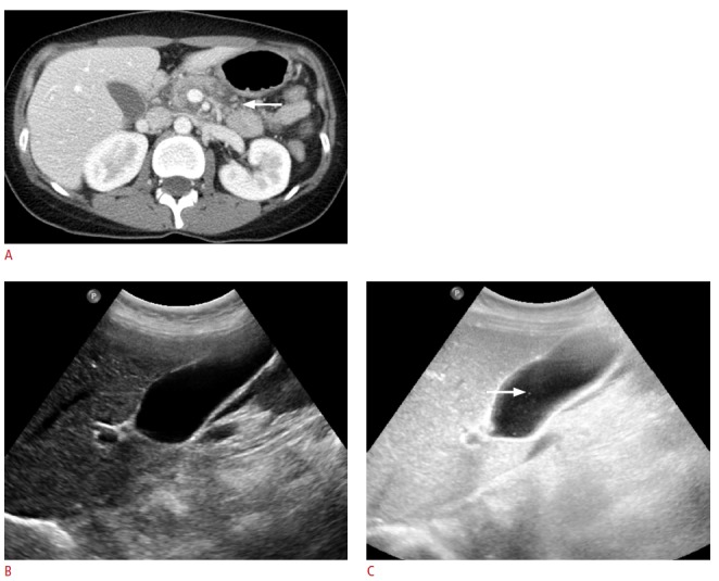
Fig. 4. A 31-year-old woman having 5 episodes of acute recurrent idiopathic pancreatitis.

A. Axial plane of contrast-enhanced abdomen computed tomography shows the swelling of pancreas and peripancreatic fluid (arrow). B. Fundamental ultrasonography shows no stones in the gallbladder lumen. C. Harmonic ultrasonography with a high background noise clearly shows the microliths (arrow) in the gallbladder lumen. Cholecystectomy was recommended, but the patient refused.
