
Figure 1.
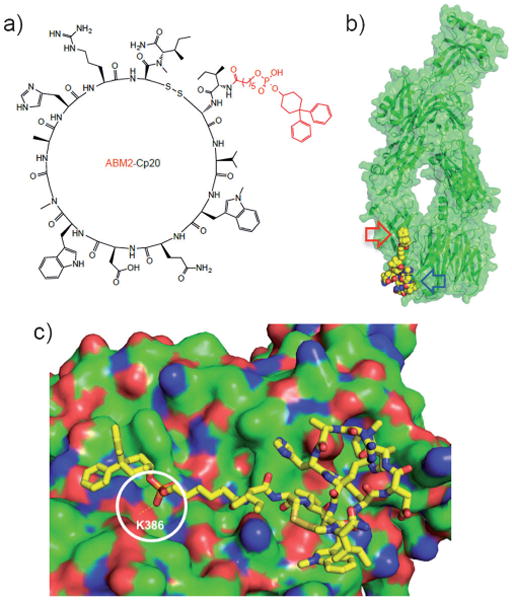
Structure and proposed binding model of ABM2-Cp20. a) Structure of ABM2-Cp20 with the ABM2 tag shown in red. b) Docking of ABM2-Cp20 (yellow spheres) into the compstatin binding site of C3c (green cartoon/surface representation; PDB code: 2QKI); the primary compstatin binding site and the proposed extended contact site for ABM2 are marked with blue and red arrows, respectively. c) Close-up of ABM2-Cp20 (stick representation) docked to C3c (green surface; positive and negative surface charges are shown in red and blue, respectively). The hydrogen bond between ABM2-Cp20 and lysine residue 386 of C3c (K386) predicted from the computational analysis is highlighted by a white circle.