ABSTRACT
Henipaviruses are associated with pteropodid reservoir hosts. The glycoproteins G and F of an African henipavirus (strain M74) have been reported to induce syncytium formation in kidney cells derived from a Hypsignathus monstrosus bat (HypNi/1.1) but not in nonchiropteran BHK-21 and Vero76 cells. Here, we show that syncytia are also induced in two other pteropodid cell lines from Hypsignathus monstrosus and Eidolon helvum bats upon coexpression of the M74 glycoproteins. The G protein was transported to the surface of transfected chiropteran cells, whereas surface expression in the nonchiropteran cells was detectable only in a fraction of cells. In contrast, the G protein of Nipah virus is transported efficiently to the surface of both chiropteran and nonchiropteran cells. Even in chiropteran cells, M74-G was predominantly expressed in the endoplasmic reticulum (ER), as indicated by colocalization with marker proteins. This result is consistent with the finding that all N-glycans of the M74-G proteins are of the mannose-rich type, as indicated by sensitivity to endo H treatment. These data indicate that the surface transport of M74-G is impaired in available cell culture systems, with larger amounts of viral glycoprotein present on chiropteran cells than on nonchiropteran cells. The restricted surface expression of M74-G explains the reduced fusion activity of the glycoproteins of the African henipavirus. Our results suggest strategies for the isolation of infectious viruses, which is necessary to assess the risk of zoonotic virus transmission.
IMPORTANCE Henipaviruses are highly pathogenic zoonotic viruses associated with pteropodid bat hosts. Whether the recently described African bat henipaviruses have a zoonotic potential as high as that of their Asian and Australian relatives is unknown. We show that surface expression of the attachment protein G of an African henipavirus, M74, is restricted in comparison to the G protein expression of the highly pathogenic Nipah virus. Transport to the cell surface is more restricted in nonchiropteran cells than it is in chiropteran cells, explaining the differential fusion activity of the M74 surface proteins in these cells. Our results imply that surface expression of viral glycoproteins may serve as a major marker to assess the zoonotic risk of emerging henipaviruses.
INTRODUCTION
The genus Henipavirus within the Paramyxoviridae family comprises two highly pathogenic members, Hendra virus (HeV) and Nipah virus (NiV), that can cause severe encephalitis in humans, with case fatality rates of 40 to 100%, and have to be dealt with under biosafety level 4 (BSL4) conditions.
HeV was isolated in 1994 from diseased horses in Australia and sporadically spread to persons who had direct contact with infected animals (1). NiV was discovered in 1998 in Malaysia, where it was isolated from pigs and transmitted to pig farmers and abattoir workers (2). Both viruses have their natural reservoir in Asian fruit bats of the genus Pteropus. Infected pteropodid bats do not show clinical signs and can shed the virus via their urine (2–6). In the case of NiV, local outbreaks occurred regularly in India and Bangladesh since 2001 (7), where the virus was transmitted from flying foxes to humans, presumably via consumption of contaminated date palm sap (8–10). Human-to-human transmission also occurs in these countries (11, 12). An additional henipavirus, Cedar virus (CedPV), has been described recently (13).
The detection of cross-neutralizing antibodies and genomic RNA in bats of the species Eidolon helvum indicated that henipaviruses are also present in African fruit bats (14–17). Cross-reacting antibodies were also reported for domestic pig populations in Ghana, suggesting that the occurrence of henipavirus infections may not be restricted to bats (18). So far, all efforts to isolate an African henipavirus have failed, which makes it difficult to assess the zoonotic potential of these viruses (14–18).
The infection of cells by NiV and HeV is initiated by the binding of the viral glycoprotein (G), a type II membrane protein, to the ubiquitously expressed cellular surface receptor ephrin-B2 (EphB2) or EphB3 (19–21). The subsequent release of the viral genome into the cytoplasm is mediated by the action of the viral fusion protein (F), which induces the fusion of the viral envelope with cellular membranes. Coexpression of F and G on the surface of infected or transfected cells results in the fusion of neighboring cells and thus in the formation of syncytia, i.e., multinucleated giant cells (22).
The surface glycoproteins of the African henipavirus M74 share some functional similarities with their counterparts of NiV and HeV. The G protein binds to ephrin-B2, and the F protein is proteolytically cleaved into F1 and F2 in an acidic compartment following internalization from the cell surface (23, 24). There is, however, a major difference in the fusion activity. In the case of NiV and HeV, coexpression of F and G usually results in the formation of multinucleated giant cells. In contrast, the surface glycoproteins of M74 have been found to induce smaller syncytia, and so far they were observed only in a kidney cell line derived from Hypsignathus monstrosus. No syncytia were observed in common nonchiropteran mammalian cells, such as Vero and BHK cells. The inability to induce syncytium formation in nonchiropteran cells has been attributed in part to an inefficient processing of the F protein (24).
To address the question of why the M74 glycoproteins fail to mediate syncytium formation in nonchiropteran cell lines, we characterized the M74-G protein in more detail. We report that surface transport of G is less efficient than that of NiV-G. Only small amounts of M74-G were detected on the surface of nonchiropteran cells. In contrast, surface expression was increased in the three chiropteran cell lines that form syncytia upon coexpression of M74-F and -G. Thus, differences in the surface transport of M74-G are a major determinant for the restricted ability of the surface glycoproteins of the putative African henipavirus M74 to induce syncytium formation.
MATERIALS AND METHODS
Cells.
BHK-21 cells, Vero76 cells, human bronchial epithelial (HBE) cells, a kidney cell line derived from Hypsignathus monstrosus (HypNi/1.1 cells) (23), Hypsignathus monstrosus lung cells (HypLu/2), and Eidolon helvum kidney cells (EidNi/41) were maintained in Dulbecco's minimum essential medium (DMEM; Gibco) supplemented with 5% (BHK-21, Vero76) or 10% (HypNi/1.1, HypLu/2, EidNi/41) fetal calf serum (FCS; Biochrom). HBE cells were maintained in medium containing the same amounts of DMEM (Gibco) and Ham's F-12 medium (PAA) supplemented with 5% FCS. Cells were cultivated in 75-cm2 tissue culture flasks (Greiner Bio-One) at 37°C and 5% CO2.
Plasmids.
The open reading frames (ORF) of M74-F (GenBank accession number AFH96010.1) and NiV-F (GenBank accession number AF212302; kindly provided by A. Maisner) were cloned into the expression vector pCG1 and connected with a sequence coding for a hemagglutinin (HA) epitope at the carboxy-terminal end (M74-F-HA). The ORFs of M74-G (GenBank accession number AFH96011.1) and a synthetic NiV-G (GenBank accession number AF212302; kindly provided by A. Maisner) derived from strain NiV/MY/HU/1999/CDC were cloned into the pCG1 vector, which was kindly provided by R. Cattaneo. The gene constructs were modified such that both glycoproteins contained a carboxy-terminal FLAG epitope (NiV-G-FLAG, M74-G-FLAG). In addition, constructs with an HA epitope at the carboxy-terminal end were generated (NiV-G-HA, M74-G-HA). Furthermore, chimeric G proteins were generated in which the NiV-G ectodomain (ED) was combined with the M74-G transmembrane domain (TD) and cytoplasmic tail (CT), and vice versa. All chimeric proteins contained a carboxy-terminal FLAG epitope (M74-G-FLAG ED, NiV-G-FLAG ED): M74-G ED consists of the amino acid (aa) residues 1 to 70 of NiV-G and aa 88 to 632 of M74-G, whereas NiV-G ED consists of aa 1 to 87 of M74-G and aa 71 to 602 of NiV-G. While the sequence of the NiV-G ED was derived from published data (25, 26), the definitions of the NiV-G TD and CT as well as those of the M74-G ED, TD, and CT were based on the prediction of transmembrane regions by the online tool HMMTOP (www.enzim.hu/hmmtop/).
To determine the intracellular localization of M74-G, cotransfection experiments with fluorescence-labeled cellular compartment markers for endosomes (pEGFP-Endo; Clontech), endoplasmic reticulum (ER) (EGFP-ER, provided by Frank van Kuppeveld), Golgi apparatus (CFP-Golgi; Clontech), and ER-Golgi intermediate compartment (ERGIC) (GFP-ERGIC, provided by Christel Schwegmann-Wessels) were performed (EGFP is enhanced green fluorescent protein, whereas CFP is cyan fluorescent protein).
IFA.
To analyze the cell-to-cell fusion in HBE, HypLu/2, and EidNi/41 cells following coexpression of the NiV- or M74-F and -G proteins, cells were grown on coverslips and transfected with the epitope-labeled constructs using the Lipofectamine 2000 transfection reagent (Life Technologies). At 24 h posttransfection (p.t.), cells were fixed by incubation with phosphate-buffered saline (PBS)-3% paraformaldehyde, permeabilized with 0.2% Triton X-100, and incubated with an antibody against the FLAG epitope (anti-FLAG, mouse; Sigma) and another one against the HA epitope (anti-HA, rabbit; Sigma), followed by an incubation with anti-mouse IgG F(ab′) 2-fragment Cy3 (Sigma) and anti-rabbit IgG fluorescein isothiocyanate (FITC; Sigma), for 1 h and for 30 min, respectively. Immunofluorescence analysis (IFA) was performed using a Nikon Eclipse Ti fluorescence microscope and the NIS Elements AR software (Nikon).
To detect intracellular and surface-expressed G proteins, cells were fixed and, in the case of intracellularly expressed proteins, permeabilized, followed by incubation with the respective antibodies. Finally, the cells were treated with 4′,6-diamidino-2-phenylindole (DAPI; Roth) and mounted in Mowiol.
The intracellular clustering of M74-G, M74-G ED, NiV-G, and NiV-G ED, as well as the coexpression with NiV-G, was investigated by cotransfection of cells with M74-G-FLAG, M74-G-FLAG ED, or NiV-G-FLAG ED and NiV-G-HA, EGFP-Endo, EGFP-ER, CFP-Golgi, or GFP-ERGIC. M74-G-FLAG, NiV-G-FLAG, and the FLAG-labeled ED chimeras were detected by antibody staining as described before. NiV-G-HA was detected by incubation with an antibody directed against the HA epitope (anti-HA, rabbit; Sigma) and an FITC-conjugated anti-rabbit secondary antibody (Sigma). IFA of cotransfected cells was performed using an ApoTome-equipped Axioplan 2 microscope (Carl Zeiss).
Preparation of cell lysates, SDS-PAGE, and Western blot analysis.
Cells were grown in 6-well plates and transfected for expression of epitope-labeled glycoproteins. At 24 h p.t., cells were detached from the bottom of the plates using a cell scraper, collected in reaction tubes, and centrifuged for 15 min at 600 × g and 4°C. After the removal of the supernatants, the cell pellet was resuspended in NP-40 lysis buffer with protease inhibitors and incubated on ice for 2 h. Afterward, the cell lysates were centrifuged for 30 min at 10,000 × g and 4°C, and the supernatant was transferred to a new reaction tube. Subsequently, 4× LDS sample buffer (final concentration, 1×; Life Technologies) was added to the samples, which were then incubated for 10 min at 96°C and were used for SDS-PAGE. To perform SDS-PAGE under reducing conditions, the samples were incubated for 10 min at 96°C in the presence of 0.1 M (final concentration) dithiothreitol (DTT) before they were loaded on the SDS gel (10%). SDS-PAGE and Western blotting were performed. NiV- and M74-G-FLAG were detected by incubation with an anti-FLAG antibody (mouse; Sigma) and anti-mouse horseradish peroxidase (HRP; Dako). Incubation with primary antibodies was performed at 4°C overnight, while secondary antibodies were applied for 1 h at 4°C. For the visualization of protein bands, membranes containing the immobilized proteins were incubated with Super Signal West Dura extended duration substrate (Thermo Scientific), placed in a ChemiDoc imager (Bio-Rad), and analyzed with the Quanti One software (Bio-Rad).
Surface biotinylation.
At 24 h p.t., M74-G-FLAG- or NiV-G-FLAG-transfected cells that had been grown in 6-well plates were washed with ice-cold PBS, followed by incubation with 0.5 mg/ml long-chain (LC) biotin (Thermo Scientific) dissolved in PBS for 20 min at 4°C on a rocking platform shaker. Afterward, the biotin solution was removed, and cells were washed with PBS-0.1 M glycine buffer and further incubated with PBS-glycine for 15 min at 4°C. Next, the cells were scraped from the bottom of the plates, collected in reaction tubes, and centrifuged for 15 min at 600 × g and 4°C. The supernatants were removed by aspiration, and the pellets were resuspended in 500 μl NP-40 lysis buffer with protease inhibitors. Cell lysates were mixed with streptavidin-agarose (Pierce). After overnight incubation at 4°C on an overhead shaker, samples were centrifuged for 5 min at 10,000 × g at 4°C. Next, the supernatants were removed by aspiration, and the agarose-bound proteins were washed with 250 μl NP-40 cell lysis buffer without protease inhibitor and centrifuged again for 5 min at 10,000 × g and 4°C. This step was repeated 3 times, before 50 μl of 2× SDS sample buffer was added to the pellet, and the samples were incubated for 10 min at 96°C to release bound protein from the agarose beads. After a final centrifugation for 5 min at 10,000 × g at room temperature, the resulting supernatants were collected and subjected to SDS-PAGE and Western blotting. Protein detection and visualization of the protein bands were performed as described above.
Flow cytometry.
Cells were grown in 6-well plates and transfected with the empty vector pCG1 or the epitope-labeled NiV- or M74-G-FLAG, as described before. At 24 h p.t., cells were washed and detached by incubation with Accutase (PAA). Next, the cells were transferred to reaction tubes and pelleted by centrifugation at 600 × g and 4°C for 5 min. The cells were subsequently resuspended in PBS without calcium and magnesium ions (PBSM)-1% bovine serum albumin (BSA)-3 mM EDTA and pelleted again. This washing step was repeated three times, before the cells were resuspended in PBSM-1% BSA containing an anti-FLAG antibody (mouse, 1:1,000; Sigma) and incubated for 1 h on an overhead shaker at 4°C. Afterward, the cells were pelleted and washed again (as described above) and resuspended in PBSM-1% BSA containing a biotin-conjugated anti-mouse antibody (1:1,000; Sigma), again for 1 h at 4°C on an overhead shaker. After an additional washing interval, the cells were resuspended in PBSM-1% BSA containing phycoerythrin-conjugated streptavidin (1:1,000; Bio-Rad) for fluorescence labeling and incubated for 30 min at 4°C on an overhead shaker. Next, the cells were washed again and fixed by incubation with PBS-3% paraformaldehyde for 20 min. Finally, the cells were washed again to remove residual paraformaldehyde and resuspended in 500 μl PBSM-1% BSA-3 mM EDTA, and flow cytometry was performed using a Beckman Coulter Epics XL and the Expo 32 ADC XL4 Color software (Beckman Coulter). The experiment was performed three times, with each value determined in quadruplicates.
Endoglycosidase digestion.
Cells grown in 6-well plates were transfected for expression of NiV- or M74-G-FLAG. At 24 h p.t., cell lysates were prepared. For digestion with endoglycosidase H (endo H; New England BioLabs) or N-glycosidase F (PNGase F; New England BioLabs), 9 μl of the cell lysate was combined with 1 μl of 10× glycoprotein denaturing buffer, followed by incubation at 96°C for 10 min. Endoglycosidase digestion was performed according to the manufacturer's protocol. All samples were loaded on an SDS gel (10%), and SDS-PAGE and Western blotting were performed. Proteins were detected by incubation with anti-FLAG antibodies (Sigma) and mouse HRP (Dako).
RESULTS
Syncytium formation induced by coexpression of M74-F and -G in the chiropteran cell lines HypLu/2 and EidNi/41.
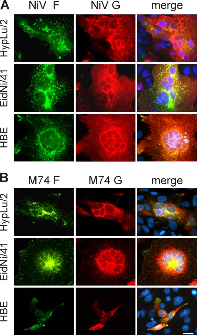
After our recent report that the glycoproteins G and F of a putative African henipavirus are able to induce syncytium formation in the chiropteran HypNi/1.1 cell line, but not in the nonchiropteran BHK-21 and Vero76 cell lines (23), we identified further chiropteran cell lines, which, upon coexpression of M74-F and G, show cell-to-cell fusion. The African henipavirus glycoproteins induced syncytium formation in a lung cell line derived from Hypsignathus monstrosus (HypLu/2) as well as in a kidney cell line derived from Eidolon helvum (EidNi/41) (Fig. 1B). It should be noted that the genetic information for the M74 glycoproteins has been determined by analyzing spleen samples from animals of the latter species (15). As a control for nonchiropteran cells, we included a cell line derived from human bronchial epithelial (HBE) cells. The M74 glycoproteins were unable to induce syncytium formation in HBE cells (Fig. 1B). In contrast, coexpression of the F and G proteins of NiV induced syncytia in all three cell lines (Fig. 1A). Fluorescence signals of M74-F have a more central distribution than those of M74-G; however, this difference was also detected for NiV glycoproteins (compare Fig. 1A and B).
FIG 1.
Coexpression of F and G of NiV (A) and M74 (B) in HypLu/2, EidNi/41, and HBE cells. At 24 h p.t., cells were fixed, permeabilized, and incubated with anti-HA and anti-FLAG antibodies, followed by incubation with secondary Cy3- and FITC-conjugated antibodies. Scale bar indicates 25 μm.
Different expression patterns of M74-G and NiV-G.
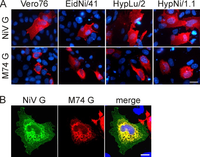
The expression of M74-G was analyzed and compared to that of the NiV-G protein. HypNi/1.1, HypLu/2, EidNi/41, and Vero76 cells were transfected for expression of G protein containing a carboxy-terminal FLAG tag. In permeabilized cells, NiV-G-FLAG was found to be distributed all over the cell (Fig. 2A, top row). In contrast, M74-G-FLAG expression was restricted to central areas of the cell (Fig. 2A, bottom row). The difference between NiV-G and M74-G is most evident in cells coexpressing both glycoproteins (Fig. 2B). The same expression pattern of M74-G-FLAG was observed in BHK-21 cells (data not shown). The intracellular clustering of M74-G appears not to be due to the FLAG tag, because an HA-tagged construct showed the same distribution. Furthermore, the functional activity of M74-G, evaluated by the ability to mediate syncytium formation when coexpressed with M74-F in HypNi/1.1 cells, did not differ between tagged and untagged M74-G protein (24).
FIG 2.
Expression of NiV-G and M74-G in permeabilized cells. (A) Vero76, EidNi/41, HypLu/2, and HypNi/1.1 cells were transfected for expression of FLAG-tagged NiV-G or M74-G. At 24 h p.t., permeabilized cells were immunostained for the presence of FLAG-tagged proteins. Scale bar indicates 25 μm. (B) HypNi/1.1 cells were cotransfected for expression of HA-tagged NiV-G and FLAG-tagged M74-G. At 24 h p.t., permeabilized cells were immunostained for the presence of NiV-G and M74-G. Scale bar indicates 10 μm.
Surface expression of M74-G.
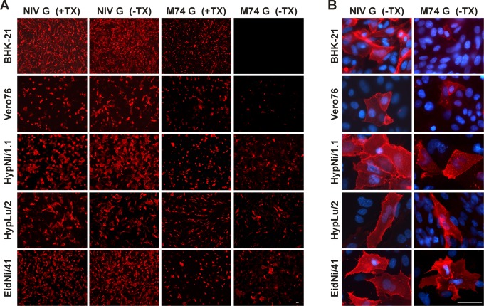
Following the observation that the intracellular expression pattern of M74-G differed from that of NiV-G, we analyzed the surface expression of M74-G. As the G protein is a type II membrane protein, the carboxy-terminal FLAG tag could be used for surface immunofluorescence analysis. IFA of transfected HypNi/1.1, HypLu/2, and EidNi/41 cells revealed that the number of cells which were positive for M74-G surface expression was close to that of antigen-positive permeabilized cells (Fig. 3A, right column). In contrast, surface expression of M74-G in BHK-21 and Vero76 cells was observed only in some of the transfected cells. Nevertheless, the transfection efficiencies of the BHK-21 and Vero76 cell lines were comparable to that of HypNi/1.1 cells, as indicated by IFA of permeabilized cells. This difference between chiropteran and nonchiropteran cells in the surface expression was not observed when the NiV-G protein was analyzed (Fig. 3A and B, left columns). Those few cells among Vero76 and BHK-21 cells that were positive for surface expression of M74-G showed a smaller fluorescence intensity than that of chiropteran cells but a similar surface distribution of the G protein (Fig. 3B, right column).
FIG 3.
Surface expression of M74-G. (A) Cells were transfected for expression of FLAG-tagged NiV-G or M74-G. At 24 h p.t., permeabilized (+TX) and nonpermeabilized (−TX) cells were immunostained by antibodies directed against the FLAG tag. Scale bars indicate 50 μm. (B) Surface expression (−TX) is shown at higher magnification.
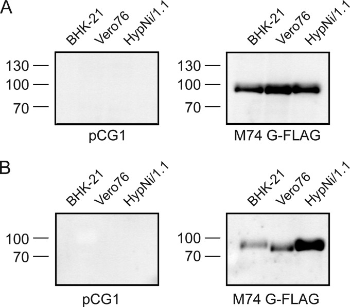
To confirm the results obtained by IFA, we performed an analysis by surface biotinylation and Western blotting. BHK-21, Vero76, and HypNi/1.1 cells were transfected for expression of M74-G and subjected to surface biotinylation. Cell lysates were analyzed by SDS-PAGE and Western blotting. Following SDS-PAGE under reducing conditions, Flag-tagged M74-G was detected in lysates of all three cell lines analyzed as a single band representing the monomeric form of the G protein (Fig. 4A). When SDS-PAGE was performed under nonreducing conditions, two protein bands were detected, representing the dimeric and tetrameric forms of the G protein (23). Expression of M74-G on the cell surface was analyzed by detection of biotinylated proteins. With BHK-21 and Vero76 cells, only weak bands of surface-expressed M74-G were detectable. In contrast, surface biotinylation of HypNi/1.1 cells resulted in a strong G band (Fig. 4B). The different electrophoretic mobility of M74-G from BHK-21 and Vero76 cells may be explained by differences in the glycosylation. The results shown in Fig. 3 and 4 indicate that M74-G is more efficiently expressed on the surface of chiropteran cells than it is on BHK-21 and Vero76 cells.
FIG 4.
Expression of M74-G in cells and on the cell surface. (A) Transfected cells expressing M74-G-FLAG were lysed and subjected to SDS-PAGE under reducing conditions and Western blotting. G proteins were immunostained by antibodies directed against the FLAG tag. Exposure time of 35 s. (B) For detection of G protein on the cell surface, transfected cells were subjected to surface biotinylation. The biotinylated proteins were separated with streptavidin-agarose and analyzed by SDS-PAGE and Western blotting. G proteins were detected by immunostaining. Exposure time of 120 s. Molecular mass markers are indicated on the left (kDa).
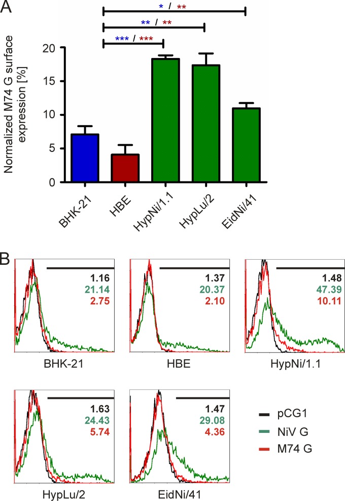
To get quantitative data on the surface expression, an analysis by flow cytometry was performed. Because of the toxic effects of the transfection reagent on Vero76 cells, the procedure was detrimental for these cells, resulting in high background values. Therefore, HBE cells, which do not support syncytium formation by M74 glycoproteins, were included as nonchiropteran cells. As shown in Fig. 5, M74-G was expressed inefficiently on the surface of all cells compared to the expression of NiV-G, with relative expression levels ranging from about four to 18%. The amounts of M74-G on the surface of the chiropteran cells were larger (11%, 17%, and 18%) than those determined for the nonchiropteran cells (4% and 7%). The overall M74-G expression and NiV-G expression were similar.
FIG 5.
Flow cytometry analysis of the surface expression of M74-G. Transfected cells expressing no foreign protein (pCG1) or FLAG-tagged NiV-G or M74-G were incubated with an antibody against the FLAG epitope, followed by incubation with anti-mouse biotin and streptavidin phycoerythrin. The amount of surface-expressing cells was analyzed by flow cytometry. (A) Normalized surface expression of M74-G. To compare the results obtained for the M74-G, the surface expression was normalized to the surface expression of NiV-G, which was set as 100%. The normalized M74-G surface expression is given in percentage of positive cells. Asterisks indicate significance (*, 0.01 < P < 0.05; **, 0.001 < P < 0.01; ***, P < 0.001). (B) Histogram plots of NiV-G and M74-G surface expression in chiropteran and nonchiropteran cells. Black curves represent the histogram plot of pCG1-transfected cells; green and red curves indicate the histogram plots of NiV-G and M74-G surface expression, respectively. The gate setting is indicated by the black line.
Costaining of M74-G and intracellular compartment markers.
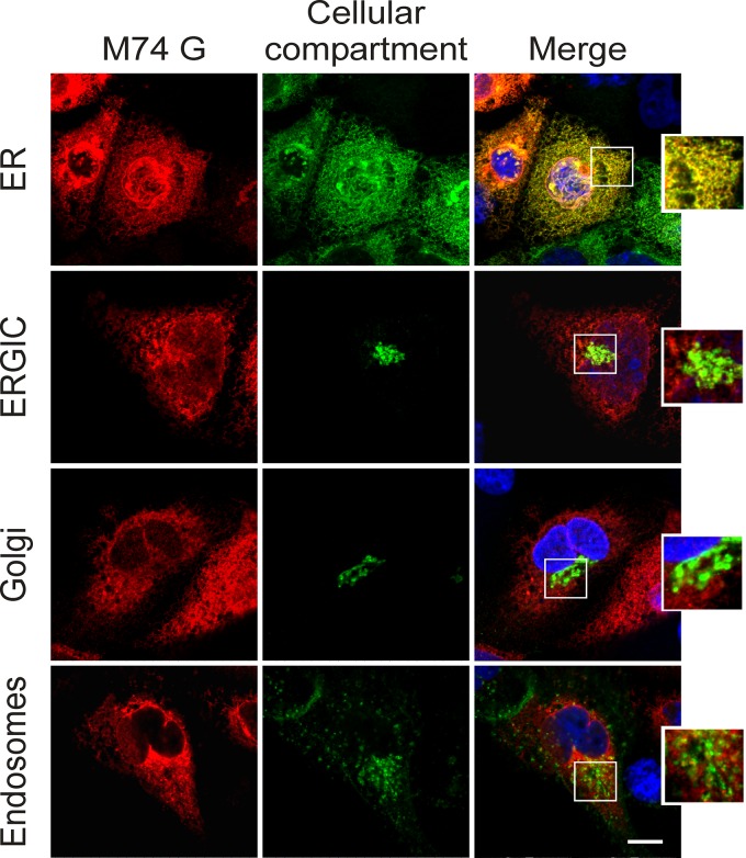
In Fig. 2B, we show that the expression pattern of M74-G is different from that of NiV-G. To get more information about the intracellular localization of M74-G, the henipavirus glycoprotein was coexpressed with marker proteins for different cellular compartments: human RhoB GTPase for endosomes, calreticulin for the ER, β-galactosyltransferase for the Golgi apparatus, or ERGIC53 for the ER-Golgi-intermediate compartment (27–30). When transfected HypNi/1.1 cells were analyzed by immunofluorescence microscopy, colocalization of M74-G was observed only with the ER marker calreticulin but not with any of the other marker proteins (Fig. 6). This result indicates that the majority of the G protein of the African henipavirus accumulates in the ER. The same results were obtained when BHK-21 or Vero76 cells were analyzed (data not shown).
FIG 6.
Clustering of M74-G in intracellular compartments. HypNi/1.1 cells were transfected for coexpression of M74-G-FLAG and a cellular compartment marker for either endosomes, ER, ERGIC, or Golgi. M74-G was detected by immunostaining using antibodies directed against the FLAG tag. Scale bar indicates 10 μm.
Endoglycosidase digestion of M74-G.
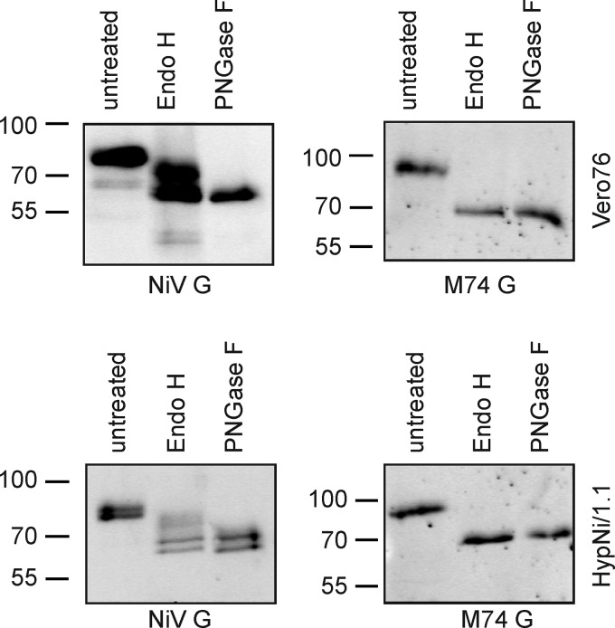
When glycoproteins are transported along the secretory pathway to the cell surface, the mannose-rich N-glycans that are linked to Asn residues in the ER may be converted to complex oligosaccharides in the Golgi apparatus. While both glycan types can be cleaved from the polypeptide backbone by PNGase F, only mannose-rich oligosaccharides are sensitive to the action of endoglycosidase H. Therefore, endoglycosidase treatment provides information about the protein trafficking of M74-G. Transfected Vero76 and HypNi/1.1 cells expressing either NiV-G or M74-G were subjected to endoglycosidase digestion. For NiV-G, treatment with PNGase F resulted in a band with an electrophoretic mobility corresponding to a molecular mass of about 65 kDa, representing the unglycosylated form of the viral protein (Fig. 7). A protein of the same size was also detected after endo H treatment. The latter protein obviously contained only mannose-rich N-glycans and thus may represent molecules from the early secretory pathway. A second band of about 72 kDa corresponds to a protein with partial sensitivity to endo H treatment. Several N-glycans of this protein have been converted to complex oligosaccharides and are thus resistant to endo H, whereas others are still in the mannose-rich form and thus susceptible to cleavage by endo H (Fig. 7). A different picture was obtained when the M74-G protein was subjected to endoglycosidase treatment. With lysates of HypNi/1.1 and Vero76 cells expressing M74-G, the treatment with either endoglycosidase, endo H or PNGase F, resulted in a shift from a protein of approximately 90 kDa to a protein of about 70 kDa, indicating that all N-glycans of M74-G are of the mannose-rich type (Fig. 7). This finding is consistent with the observation that M74-G is predominantly localized in the ER. Even in HypNi/1.1 cells, there was no protein detectable with partial resistance to endo H treatment. Therefore, we assume that the M74-G protein that is transported to the cell surface in HypNi/1.1 cells contains only mannose-rich N-glycans.
FIG 7.
Endoglycosidase digestion of M74-G. Cell lysates of transfected HypNi/1.1 and Vero76 cells were treated with endo H or PNGase F endoglycosidase followed by SDS-PAGE and Western blotting. G proteins were detected by immunostaining using antibodies directed against the FLAG tag. Molecular mass markers are indicated on the left (kDa).
The ectodomain of M74-G is responsible for the restricted surface transport.
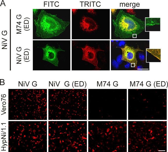
To determine which portion of the G protein is responsible for the observed differences in the surface transport between the NiV and M74 glycoproteins, chimeric proteins were generated. The predicted ectodomain of M74-G was replaced by that of NiV-G and vice versa. The corresponding constructs were analyzed for surface expression and intracellular distribution in BHK-21, Vero76, and HypNi/1.1 cells. Analysis of the expression pattern in permeabilized HypNi/1.1 cells by IFA revealed that the chimeric G protein with the ectodomain (ED) of NiV-G and with the transmembrane domain (TD) and cytoplasmic tail (CT) of M74-G showed an expression comparable to that of the parental NiV-G, whereas the chimeric protein with ED from M74-G clustered in central areas of the cells, as it was observed for the parental M74-G protein. Coexpression studies with NiV-G-HA revealed a complete colocalization with NiV-G-FLAG ED, whereas with M74-G-FLAG ED, colocalization was restricted to central areas of the cell (Fig. 8A). Similar results were obtained when BHK-21 and Vero76 cells were analyzed (data not shown). When the chimeric G proteins were screened for surface expression, we found that NiV G-FLAG ED was expressed on the surface of all cell lines analyzed in amounts that are comparable to those of the parental NiV-G-FLAG. In contrast, the surface expression of M74-G-FLAG ED was very weak in the case of Vero76 cells, as shown above for the parental M74-G-FLAG. For HypNi/1.1 cells, also consistent with parental M74-G, a clear surface expression of M74-G ED was observed (Fig. 8B). Furthermore, coexpression of M74-G-FLAG ED with fluorescent marker proteins in HypNi/1.1 cells indicated a colocalization of M74-G-FLAG ED with the ER but not with marker proteins of endosomes, the Golgi apparatus, or the ERGIC (data not shown). These results indicate that the predicted ectodomain rather than the intracellular or transmembrane region is responsible for the observed expression pattern of M74-G.
FIG 8.
Expression of chimeric G proteins of NiV and M74. (A) HypNi/1.1 cells were transfected for coexpression of NiV-G-HA (green) with either M74-G ED or NiV-G ED (red). Permeabilized cells were immunostained for NiV-G with antibodies against the HA tag (green) and for chimeric proteins with antibodies directed against the FLAG-tag (red). (B) Cells were transfected for expression of the FLAG-tagged native G proteins, NiV-G or M74-G, or either of the corresponding ED chimeras, NiV-G-FLAG ED or M74-G-FLAG ED. The nonpermeabilized cells (−TX) were immunostained for the presence of G proteins by antibodies directed against the FLAG tag. Scale bars indicate 25 μm.
DISCUSSION
Here, we have shown that not only HypNi/1.1 but also HypLu/2 and EidNi/41 cells support syncytium formation by M74 glycoproteins. Syncytium formation in the latter cells has also been reported by Lawrence et al. (31). A negative result was obtained by others (24). An explanation for this difference may be that the authors of the latter work used a subline of these cells (EidNi/43.1) that may be refractory to syncytium formation. We have demonstrated that the M74-G protein shows cell-dependent differences in surface expression. While it was readily detectable on the surface of HypNi/1.1, HypLu/2, and EidNi/41 cells, only few cells showed strong surface fluorescence when BHK-21 or Vero76 cells were analyzed. Surface biotinylation also indicated that the amount of M74-G that is present on the two nonchiropteran cell lines is smaller than that detected on the chiropteran cells. Coexpression of M74-G with NiV-F has been reported to result in the formation of syncytia (24, 31, 32), which were, however, smaller in size and in number than the giant cells obtained by interaction of NiV-F and NiV-G. Analysis by flow cytometry indicated that nonchiropteran cells contain a small amount of M74-G on the surface. This low level of M74-G surface expression that is at or below the level of detection by immunofluorescence microscopy appears to be sufficient for syncytium formation by NiV-F but not by M74-F.
IFA showed that the majority of M74-G was present in central areas of all cells, including HypNi/1.1 cells. This observation is consistent with the finding that M74-G colocalized with a marker protein of the ER but not with markers for ERGIC, Golgi apparatus, or endosomes. The amount of M74-G that is expressed on the surface of HypNi/1.1 cells has to pass the secretory pathway. The inability to detect M74-G in ERGIC and Golgi suggests that the amount of viral protein in these compartments is at or below the detection level.
The sensitivity to endo H treatment appears to point to a location in the ER. NiV-G has been reported to contain six N-linked glycans (33, 34). Sequence analysis predicts six N-glycans also for M74-G (positions 29, 102, 207, 224, 324, and 396). In contrast to NiV-G, which was found to contain some complex oligosaccharides, all N-glycans of M74-G, and also those from HypNi/1.1 cells, were sensitive to endo H treatment. Such a property is usually found in glycoproteins located in a pre-Golgi compartment. Therefore, the amount of M74-G present on the surface of HypNi/1.1 cells is either too low to be detected in the endoglycosidase assay, or the oligosaccharides of M74-G are presented during transport to the cell surface in a way that they are not accessible for conversion into complex N-glycans.
The analysis of chimeric proteins indicated that the ectodomain of M74-G and not the transmembrane domain or the cytoplasmic tail is responsible for the different expression patterns of the G proteins of NiV and M74. Future research has to show which amino acids are crucial for this property.
Though transport of M74-G to the cell surface is inefficient in all cells analyzed, more viral glycoprotein was detected on the surface of the chiropteran cells than was on nonchiropteran cells. This difference can explain the differential ability of the M74 glycoproteins to induce syncytium formation. The expression pattern of M74-F does not explain why the syncytium formation induced by the M74 glycoproteins is restricted to chiropteran cells. No difference was observed in the steady-state distribution of the uncleaved (F0) and cleaved form (F1) of the M74 fusion protein irrespective of the expression in chiropteran or nonchiropteran cells (24) (our unpublished data). However, the kinetics of the proteolytic activation of M74-F was found to be slower than that of NiV-F (24). Though the arbitrary insertion of a nucleotide into the F gene of M74 was reported to result in a protein with enhanced fusion activity (32), the overall activity was still low in comparison to that of NiV-F. Ongoing genomic characterizations of additional African bat henipavirus field strains related to M74 suggest complete conservation of the predicted F open reading frame of M74 without any apparent insertions or deletions (J. F. Drexler, T. Binger, V. M. Corman, and C. Drosten, unpublished data). The study of Pernet et al. (32) thus does not reflect the true biological situation. Because of the inefficient surface expression of M74-G and the impaired processing of M74-F, one should consider the possibility that the replication strategy of the respective bat virus is not aimed at a productive infection with high numbers of progeny virions but rather at a persistent type of infection characterized by cell-to-cell spread. More knowledge about the replication of African henipaviruses is required to understand the biology of these viruses and to evaluate the risk of zoonotic virus transmission. Risk assessment is hampered, however, by the lack infectious virus isolates.
Our findings have implication for attempts to isolate infectious henipaviruses from African bats. Because of the restricted syncytium formation with nonchiropteran cells, chiropteran cells, such as HypNi/1.1, are cells of choice for successful virus isolation. As release of virions may be a limiting factor, it appears promising to look for cell-associated virus transmitted by cell-to-cell spread of infection. In the absence of criteria to judge the zoonotic potential of a growing number of bat viruses, our data suggest that the capacity to efficiently transport glycoproteins to the cell surface may turn out in the future to be a relevant marker for risk assessment. Such analyses can be undertaken even in the absence of virus isolates. This kind of functional data raised for emerging bat viruses may provide more robust information on the pathogenicity of such viruses for nonchiropteran mammals than those based upon sequence similarities alone.
ACKNOWLEDGMENTS
Part of this work was performed by N.K. and M.H. in partial fulfillment of the requirements for the doctoral degree from Stiftung Tierärztliche Hochschule Hannover and Leibniz Universität Hannover, respectively.
We are grateful to Mathias Hornef for his help in microscopy and to Andrea Maisner for valuable advice. We are grateful to Roberto Cattaneo for providing expression plasmids.
N.K. was funded by a fellowship of the Hannover Graduate School for Veterinary Pathobiology, Neuroinfectiology, and Translational Medicine (HGNI). This work was supported by grants to G.H. from DFG (HE 1168/14-1 and SFB621 TP B7) and from Bundesministerium für Bildung und Forschung (Ecology and Pathogenesis of SARS, project code 01Kl1005B; and FluResearchNet, project code 01KI1006D).
Footnotes
Published ahead of print 6 August 2014
REFERENCES
- 1. Murray K, Selleck P, Hooper P, Hyatt A, Gould A, Gleeson L, Westbury H, Hiley L, Selvey L, Rodwell B, et al. 1995. A morbillivirus that caused fatal disease in horses and humans. Science 268:94–97. 10.1126/science.7701348 [DOI] [PubMed] [Google Scholar]
- 2. Chua KB, Koh CL, Hooi PS, Wee KF, Khong JH, Chua BH, Chan YP, Lim ME, Lam SK. 2002. Isolation of Nipah virus from Malaysian Island flying-foxes. Microbes Infect. 4:145–151. 10.1016/S1286-4579(01)01522-2 [DOI] [PubMed] [Google Scholar]
- 3. Yob JM, Field H, Rashdi AM, Morrissy C, van der Heide B, Rota P, bin Adzhar A, White J, Daniels P, Jamaluddin A, Ksiazek T. 2001. Nipah virus infection in bats (order Chiroptera) in peninsular Malaysia. Emerg. Infect. Dis. 7:439–441. 10.3201/eid0703.010312 [DOI] [PMC free article] [PubMed] [Google Scholar]
- 4. Olson JG, Rupprecht C, Rollin PE, An US, Niezgoda M, Clemins T, Walston J, Ksiazek TG. 2002. Antibodies to Nipah-like virus in bats (Pteropus lylei), Cambodia. Emerg. Infect. Dis. 8:987–988. 10.3201/eid0809.010515 [DOI] [PMC free article] [PubMed] [Google Scholar]
- 5. Middleton DJ, Morrissy CJ, van der Heide BM, Russell GM, Braun MA, Westbury HA, Halpin K, Daniels PW. 2007. Experimental Nipah virus infection in pteropid bats (Pteropus poliocephalus). J. Comp. Pathol. 136:266–272. 10.1016/j.jcpa.2007.03.002 [DOI] [PubMed] [Google Scholar]
- 6. Halpin K, Hyatt AD, Fogarty R, Middleton D, Bingham J, Epstein JH, Rahman SA, Hughes T, Smith C, Field HE, Daszak P, Henipavirus Ecology Research Group. 2011. Pteropid bats are confirmed as the reservoir hosts of henipaviruses: a comprehensive experimental study of virus transmission. Am. J. Trop. Med. Hyg. 85:946–951. 10.4269/ajtmh.2011.10-0567 [DOI] [PMC free article] [PubMed] [Google Scholar]
- 7. Luby SP, Hossain MJ, Gurley ES, Ahmed BN, Banu S, Khan SU, Homaira N, Rota PA, Rollin PE, Comer JA, Kenah E, Ksiazek TG, Rahman M. 2009. Recurrent zoonotic transmission of Nipah virus into humans, Bangladesh, 2001–2007. Emerg. Infect. Dis. 15:1229–1235. 10.3201/eid1508.081237 [DOI] [PMC free article] [PubMed] [Google Scholar]
- 8. Rahman MA, Hossain MJ, Sultana S, Homaira N, Khan SU, Rahman M, Gurley ES, Rollin PE, Lo MK, Comer JA, Lowe L, Rota PA, Ksiazek TG, Kenah E, Sharker Y, Luby SP. 2012. Date palm sap linked to Nipah virus outbreak in Bangladesh, 2008. Vector Borne Zoonotic Dis. 12:65–72. 10.1089/vbz.2011.0656 [DOI] [PubMed] [Google Scholar]
- 9. Khan MS, Hossain J, Gurley ES, Nahar N, Sultana R, Luby SP. 2010. Use of infrared camera to understand bats' access to date palm sap: implications for preventing Nipah virus transmission. Ecohealth 7:517–525. 10.1007/s10393-010-0366-2 [DOI] [PubMed] [Google Scholar]
- 10. Luby SP, Rahman M, Hossain MJ, Blum LS, Husain MM, Gurley E, Khan R, Ahmed BN, Rahman S, Nahar N, Kenah E, Comer JA, Ksiazek TG. 2006. Foodborne transmission of Nipah virus, Bangladesh. Emerg. Infect. Dis. 12:1888–1894. 10.3201/eid1212.060732 [DOI] [PMC free article] [PubMed] [Google Scholar]
- 11. Sazzad HM, Hossain MJ, Gurley ES, Ameen KM, Parveen S, Islam MS, Faruque LI, Podder G, Banu SS, Lo MK, Rollin PE, Rota PA, Daszak P, Rahman M, Luby SP. 2013. Nipah virus infection outbreak with nosocomial and corpse-to-human transmission, Bangladesh. Emerg. Infect. Dis. 19:210–217. 10.3201/eid1902.120971 [DOI] [PMC free article] [PubMed] [Google Scholar]
- 12. Luby SP, Gurley ES, Hossain MJ. 2009. Transmission of human infection with Nipah virus. Clin. Infect. Dis. 49:1743–1748. 10.1086/647951 [DOI] [PMC free article] [PubMed] [Google Scholar]
- 13. Marsh GA, de Jong C, Barr JA, Tachedjian M, Smith C, Middleton D, Yu M, Todd S, Foord AJ, Haring V, Payne J, Robinson R, Broz I, Crameri G, Field HE, Wang L-F. 2012. Cedar virus: a novel henipavirus isolated from Australian bats. PLoS Pathog. 8:e1002836. 10.1371/journal.ppat.1002836 [DOI] [PMC free article] [PubMed] [Google Scholar]
- 14. Hayman DT, Suu-Ire R, Breed AC, McEachern JA, Wang L, Wood JL, Cunningham AA. 2008. Evidence of henipavirus infection in West African fruit bats. PLoS One 3:e2739. 10.1371/journal.pone.0002739 [DOI] [PMC free article] [PubMed] [Google Scholar]
- 15. Drexler JF, Corman VM, Gloza-Rausch F, Seebens A, Annan A, Ipsen A, Kruppa T, Muller MA, Kalko EK, Adu-Sarkodie Y, Oppong S, Drosten C. 2009. Henipavirus RNA in African bats. PLoS One 4:e6367. 10.1371/journal.pone.0006367 [DOI] [PMC free article] [PubMed] [Google Scholar]
- 16. Peel AJ, Baker KS, Crameri G, Barr JA, Hayman DT, Wright E, Broder CC, Fernandez-Loras A, Fooks AR, Wang LF, Cunningham AA, Wood JLN. 2012. Henipavirus neutralising antibodies in an isolated island population of African fruit bats. PLoS One 7:e30346. 10.1371/journal.pone.0030346 [DOI] [PMC free article] [PubMed] [Google Scholar]
- 17. Weiss S, Nowak K, Fahr J, Wibbelt G, Mombouli JV, Parra HJ, Wolfe ND, Schneider BS, Leendertz FH. 2012. Henipavirus-related sequences in fruit bat bushmeat, Republic of Congo. Emerg. Infect. Dis. 18:1536–1537. 10.3201/eid1809.111607 [DOI] [PMC free article] [PubMed] [Google Scholar]
- 18. Hayman DT, Wang LF, Barr J, Baker KS, Suu-Ire R, Broder CC, Cunningham AA, Wood JL. 2011. Antibodies to henipavirus or henipa-like viruses in domestic pigs in Ghana, West Africa. PLoS One 6:e25256. 10.1371/journal.pone.0025256 [DOI] [PMC free article] [PubMed] [Google Scholar]
- 19. Negrete OA, Wolf MC, Aguilar HC, Enterlein S, Wang W, Muhlberger E, Su SV, Bertolotti-Ciarlet A, Flick R, Lee B. 2006. Two key residues in ephrinB3 are critical for its use as an alternative receptor for Nipah virus. PLoS Pathog. 2:e7. 10.1371/journal.ppat.0020007 [DOI] [PMC free article] [PubMed] [Google Scholar]
- 20. Negrete OA, Levroney EL, Aguilar HC, Bertolotti-Ciarlet A, Nazarian R, Tajyar S, Lee B. 2005. EphrinB2 is the entry receptor for Nipah virus, an emergent deadly paramyxovirus. Nature 436:401–405. 10.1038/nature03838 [DOI] [PubMed] [Google Scholar]
- 21. Bonaparte MI, Dimitrov AS, Bossart KN, Crameri G, Mungall BA, Bishop KA, Choudhry V, Dimitrov DS, Wang LF, Eaton BT, Broder CC. 2005. Ephrin-B2 ligand is a functional receptor for Hendra virus and Nipah virus. Proc. Natl. Acad. Sci. U. S. A. 102:10652–10657. 10.1073/pnas.0504887102 [DOI] [PMC free article] [PubMed] [Google Scholar]
- 22. Bossart KN, Wang LF, Flora MN, Chua KB, Lam SK, Eaton BT, Broder CC. 2002. Membrane fusion tropism and heterotypic functional activities of the Nipah virus and Hendra virus envelope glycoproteins. J. Virol. 76:11186–11198. 10.1128/JVI.76.22.11186-11198.2002 [DOI] [PMC free article] [PubMed] [Google Scholar]
- 23. Kruger N, Hoffmann M, Weis M, Drexler JF, Muller MA, Winter C, Corman VM, Gutzkow T, Drosten C, Maisner A, Herrler G. 2013. Surface glycoproteins of an African henipavirus induce syncytium formation in a cell line derived from an African fruit bat, Hypsignathus monstrosus. J. Virol. 87:13889–13891. 10.1128/JVI.02458-13 [DOI] [PMC free article] [PubMed] [Google Scholar]
- 24. Weis M, Behner L, Hoffmann M, Kruger N, Herrler G, Drosten C, Drexler JF, Dietzel E, Maisner A. 2014. Characterization of African bat henipavirus GH-M74a glycoproteins. J. Gen. Virol. 95:539–548. 10.1099/vir.0.060632-0 [DOI] [PubMed] [Google Scholar]
- 25. Negrete OA, Chu D, Aguilar HC, Lee B. 2007. Single amino acid changes in the Nipah and Hendra virus attachment glycoproteins distinguish ephrinB2 from ephrinB3 usage. J. Virol. 81:10804–10814. 10.1128/JVI.00999-07 [DOI] [PMC free article] [PubMed] [Google Scholar]
- 26. Zhu Q, Biering SB, Mirza AM, Grasseschi BA, Mahon PJ, Lee B, Aguilar HC, Iorio RM. 2013. Individual N-glycans added at intervals along the stalk of the Nipah virus G protein prevent fusion but do not block the interaction with the homologous F protein. J. Virol. 87:3119–3129. 10.1128/JVI.03084-12 [DOI] [PMC free article] [PubMed] [Google Scholar]
- 27. Ostwald TJ, MacLennan DH. 1974. Isolation of a high affinity calcium-binding protein from sarcoplasmic reticulum. J. Biol. Chem. 249:974–979 [PubMed] [Google Scholar]
- 28. Michalak M, Baksh S, Opas M. 1991. Identification and immunolocalization of calreticulin in pancreatic cells: no evidence for “calciosomes.” Exp. Cell Res. 197:91–99. 10.1016/0014-4827(91)90484-C [DOI] [PubMed] [Google Scholar]
- 29. Tharin S, Dziak E, Michalak M, Opas M. 1992. Widespread tissue distribution of rabbit calreticulin, a nonmuscle functional analogue of calsequestrin. Cell Tissue Res. 269:29–37. 10.1007/BF00384723 [DOI] [PubMed] [Google Scholar]
- 30. Michalak M, Campbell KP, MacLennan DH. 1980. Localization of the high affinity calcium binding protein and an intrinsic glycoprotein in sarcoplasmic reticulum membranes. J. Biol. Chem. 255:1317–1326 [PubMed] [Google Scholar]
- 31. Lawrence P, Escudero Perez B, Drexler JF, Corman VM, Muller MA, Drosten C, Volchkov V. 2014. Surface glycoproteins of the recently identified African henipavirus promote viral entry and cell fusion in a range of human, simian and bat cell lines. Virus Res. 181:77–80. 10.1016/j.virusres.2014.01.003 [DOI] [PubMed] [Google Scholar]
- 32. Pernet O, Beaty S, Lee B. 2014. Functional rectification of the newly described African henipavirus fusion glycoprotein (Gh-M74a). J. Virol. 88:5171–5176. 10.1128/JVI.03655-13 [DOI] [PMC free article] [PubMed] [Google Scholar]
- 33. Biering SB, Huang A, Vu AT, Robinson LR, Bradel-Tretheway B, Choi E, Lee B, Aguilar HC. 2012. N-glycans on the Nipah virus attachment glycoprotein modulate fusion and viral entry as they protect against antibody neutralization. J. Virol. 86:11991–12002. 10.1128/JVI.01304-12 [DOI] [PMC free article] [PubMed] [Google Scholar]
- 34. Xu K, Rajashankar KR, Chan YP, Himanen JP, Broder CC, Nikolov DB. 2008. Host cell recognition by the henipaviruses: crystal structures of the Nipah G attachment glycoprotein and its complex with ephrin-B3. Proc. Natl. Acad. Sci. U. S. A. 105:9953–9958. 10.1073/pnas.0804797105 [DOI] [PMC free article] [PubMed] [Google Scholar]


