ABSTRACT
Amino acid substitutions were introduced into avian influenza virus PB1 in order to characterize the interaction between polymerase activity and pathogenicity. Previously, we used recombinant viruses containing the hemagglutinin (HA) and neuraminidase (NA) genes from the highly pathogenic avian influenza virus (HPAIV) H5N1 strain and other internal genes from two low-pathogenicity avian influenza viruses isolated from chicken and wild-bird hosts (LP and WB, respectively) to demonstrate that the pathogenicity of highly pathogenic avian influenza viruses (HPAIVs) of subtype H5N1 in chickens is regulated by the PB1 gene (Y. Uchida et al., J. Virol. 86:2686–2695, 2012, doi:http://dx.doi.org/10.1128/JVI.06374-11). In the present study, we introduced a C38Y substitution into WB PB1 and demonstrated that this substitution increased both polymerase activity in DF-1 cells in vitro and the pathogenicity of the recombinant viruses in chickens. The V14A substitution in LP PB1 reduced polymerase activity but did not affect pathogenicity in chickens. Interestingly, the V14A substitution reduced viral shedding and transmissibility. These studies demonstrate that increased polymerase activity correlates directly with enhanced pathogenicity, while decreased polymerase activity does not always correlate with pathogenicity and requires further analysis.
IMPORTANCE We identified 2 novel amino acid substitutions in the avian influenza virus PB1 gene that affect the characteristics of highly pathogenic avian influenza viruses (HPAIVs) of the H5N1 subtype, such as viral replication and polymerase activity in vitro and pathogenicity and transmissibly in chickens. An amino acid substitution at residue 38 in PB1 directly affected pathogenicity in chickens and was associated with changes in polymerase activity in vitro. A substitution at residue 14 reduced polymerase activity in vitro, while its effects on pathogenicity and transmissibility depended on the constellation of internal genes.
INTRODUCTION
Influenza A virus is a negative-strand RNA virus with a genome composed of eight segments. Viral subtypes are based on two surface glycoproteins, hemagglutinin (HA) and neuraminidase (NA). To date, 16 HA subtypes (H1 to H16) and 9 NA subtypes (N1 to N9) have been identified in wild birds (1, 2). Wild waterfowl are a natural host of influenza A viruses and serve as an asymptomatic reservoir.
In poultry, only a small proportion of avian influenza viruses (AIVs) are recognized as highly pathogenic avian influenza viruses (HPAIVs) on the basis of their lethality for chickens. Multiple basic amino acids at the HA cleavage sites of the H5 and H7 subtypes represent one of the molecular signatures of HPAIV (3–5). In addition to the HA cleavage site, the pathogenicity of influenza viruses is also affected by components of the viral polymerase complex, consisting of polymerase basic protein 2 (PB2), polymerase basic protein 1 (PB1), and polymerase acidic protein (PA). Reverse genetics of influenza viruses has been used to engineer recombinant viruses with targeted amino acid substitutions and to examine their effects on the pathogenicity of HPAIVs. Amino acid substitutions S224P and N383D in the PA of an H5N1 HPAIV have been found to increase polymerase activity in duck fibroblasts, coinciding with enhanced pathogenicity in ducks (6). The T515A substitution in the PA of another H5N1 HPAIV resulted in a loss of pathogenicity in ducks when the virus was inoculated orally and reduced polymerase activity in vitro (7). The T552S substitution in an H2N1 subtype AIV enhanced viral growth and increased polymerase activity in mammalian cells (8). The amino acid residue at position 627 in PB2 has been recognized as an important determinant of host range for influenza A viruses. Viruses derived from avian hosts have a glutamic acid (627E) at this position, whereas a lysine (627K) is predominant at this position in human isolates (9). Enhanced polymerase activity has been shown to correlate with residue 627K in mammalian cells, and both pathogenicity and mortality were increased in mice (10–12). The Y436H substitution in the PB1 of an H5N1 HPAIV resulted in a loss of pathogenicity in ducks and reduced polymerase activity in vitro (7). Other substitutions in H5N1 HPAIV PB1, such as V473L and P598L, reduced plaque size in Madin-Darby canine kidney (MDCK) cells and decreased viral titers in lungs and tracheas collected from infected mice (13). Both substitutions reduced polymerase activity in vitro (13). The K207R substitution in the PB1 of an H5N1 virus was found to decrease polymerase activity without affecting the mortality of infected mallards and mice (7). The K480R substitution in the PB1 of an A(H1N1)pdm09 virus did not affect viral replication in MDCK cells, although it increased polymerase activity slightly in both mammalian and avian cells in vitro (14).
Previously, we constructed recombinant viruses possessing HA and NA genes from an H5N1 HPAIV strain (15). The internal genes were derived from two different AIVs of low pathogenicity in chickens: A/whistling swan/Shimane/580/2002 (H5N3) (WB) and A/chicken/Yokohama/aq55/2001 (H9N2) (LP) (15). When WB PB1 was replaced with LP PB1 to produce the WB(L/PB1) construct, the mean survival time (MST) was shortened from 3.33 to 2 days postinfection (dpi). WB PB1 showed the opposite effect when it was substituted for LP PB1 to construct LP(W/PB1). The MST with LP(W/PB1) was extended to 7.5 dpi, and the survival rate was increased from 6.7% to 50%. These results indicate that changes in PB1 alter the pathogenicity of the recombinant viruses in chickens, despite the presence of the same surface antigen genes as those in HPAIVs.
In this study, we compared amino acid substitutions in WB PB1 with those in LP PB1 and identified substitutions that affect polymerase activity by performing in vitro polymerase assays. Recombinant viruses with the substitutions described below were constructed for experimental infections in chickens. Through these analyses, we demonstrate that the C38Y substitution in PB1 consistently increased viral replication and polymerase activity in vitro, as well as pathogenicity in chickens. However, the V14A substitution showed variable effects on in vitro polymerase activity and pathogenicity or transmissibility in chickens, depending on the genetic background (Table 1).
TABLE 1.
Functional consequences of amino acid substitutions in this studya
| Substitution | Effecta in: |
|||
|---|---|---|---|---|
| DF-1 cells |
Chickens |
|||
| Viral replication | Polymerase activity | Pathogenicity | Transmissibility | |
| WB → WB(L/PB1) | Up* | Up** | Up | — |
| LP → LP(W/PB1) | Down* | Down** | Down | — |
| WB → WB-PB1-C38Y | No change | Up** | Up* | — |
| LP(W/PB1) → LP(W/PB1-C38Y) | Up* | Up** | Up** | — |
| LP → LP-PB1-Y38C | — | Down** | Down* | Lost |
| LP → LP-PB1-V14A | No change | Down** | No change | Lost |
| WB(L/PB1) → WB(L/PB1-V14A) | Down* | Down** | Downb | No change |
Up, increased by the substitution; Down, decreased by the substitution; —, data not applicable; Lost, lost transmissibility by substitution. For details, see Results. Statistically significant differences were observed in each category (*, P < 0.05; **, P < 0.01).
Viral titers in some organs were significantly decreased.
MATERIALS AND METHODS
Viruses and cells.
In this study, we used three viruses: an H5N1 subtype HPAIV (A/chicken/Yamaguchi/7/2004 [HP]) (16), a low-pathogenicity AIV of the H5N3 subtype (A/whistling swan/Shimane/580/2002; kindly provided by T. Ito, Tottori University) (WB), and an H9N2 subtype virus (A/chicken/Yokohama/aq55/2001) (LP) (17).
293T cells and DF-1 cells (a chicken fibroblast-derived cell line [CRL-12203] obtained from the American Type Culture Collection, Manassas, VA) (18) were cultured in Dulbecco's modified Eagle medium supplemented with 10% fetal calf serum and 1% penicillin-streptomycin (10,000 U/ml penicillin; 10,000 μg/ml streptomycin). MDCK cells were cultured in minimum essential medium (MEM) supplemented with 10% fetal calf serum, 1% penicillin-streptomycin, and amphotericin B (Fungizone; 2.5 μg/ml). DF-1 cells were maintained at 39°C under 5% CO2. MDCK and 293T cells were maintained at 37°C under 5% CO2.
Plasmid construction.
Surface genes (HA and NA) were from HP, and internal genes (PB2, PB1, PA, nucleoprotein [NP], matrix protein [M], and nonstructural protein [NS]) were from A/whistling swan/Shimane/580/2002 or A/chicken/Yokohama/aq55/2001. Segments were inserted into a pHW2000 expression vector (kindly provided by E. Hoffmann, G. Neumann, Y. Kawaoka, G. Hobom, and R. G. Webster, St. Jude Children's Research Hospital, Memphis, TN) (19) as described previously (15). Mutations were introduced by site-directed mutagenesis with the PrimeSTAR mutagenesis basal kit (TaKaRa, Shiga, Japan) according to the manufacturer's protocol and were confirmed by sequencing. The pPolI-Luc-RT plasmid, in which the human polymerase I (Pol I) promoter sequence was replaced with a chicken-derived Pol I promoter, was used for luciferase reporter assays (kindly provided by Leo L. M. Poon, University of Hong Kong, Hong Kong SAR, China) (20).
Luciferase assay of polymerase activity in DF-1 cells.
Plasmid pPolI-Luc-RT and pHW2000 plasmid constructs expressing PB2, PB1, PA, or NP were cotransfected into DF-1 cells with pGL4.74 (Promega, Madison, WI) by using the TransIT-LT1 transfection reagent (Mirus, Madison, WI) as recommended by the manufacturer. Twenty-four hours after transfection at 39°C under 5% CO2, cell extracts were collected, and luciferase activity assays were conducted with the Dual-Luciferase reporter assay system (Promega). The assay results were normalized to the activity of Renilla luciferase, encoded by pGL4.74.
Generation of recombinant viruses.
Recombinant viruses were generated by use of a reverse genetics system (19). Equal amounts of the eight plasmid genes (PB2, PB1, PA, HA, NP, NA, M, and NS) inserted into pHW2000 were transfected into 293T cells by using the TransIT-LT1 transfection reagent (Mirus) as recommended by the manufacturer. After 48 h, supernatants were collected and were used to inoculate MDCK cells in infection medium (MEM supplemented with 0.4% bovine serum albumin, 1% penicillin-streptomycin [10,000 U/ml penicillin and 10,000 μg/ml streptomycin], amphotericin B [2.5 μg/ml], and 3% MEM vitamin solution) in order to propagate the recombinant viruses. Supernatants were harvested following the observation of cytopathic effects and were titrated to determine a 50% egg infective dose (EID50) by the Reed-Muench method (21). Recombinant viruses with the HA gene from HP were handled in the biosafety level-3 facilities at the National Institute of Animal Health, Tsukuba, Japan.
Viral kinetics in vivo.
DF-1 cells in 6-well plates (1 × 10⁶ cells/well) were infected with 0.001 EID50/cell of recombinant viruses, and supernatants were collected at 24, 48, and 72 h postinfection (hpi) and were stored at −80°C. Tenfold serially diluted supernatants were inoculated into embryonated chicken eggs in order to calculate the EID50. Welch's t test was conducted at each time point for statistical analysis of viral titers.
Animal experiments.
Four-week-old specific-pathogen-free White Leghorn L-M-6 chickens were obtained from Nisseiken Co., Ltd. (Tokyo, Japan). To determine pathogenicity, chickens were inoculated intranasally with viruses at 106 EID50s/100 μl, and survival rates were recorded for 10 days. To observe the efficiency of viral transmission, 1 of the inoculated chickens and 3 naïve chickens were placed in the same isolator at 24 hpi. Observation lasted for 10 days from the date of initiation of cohabitation. Inoculated and naïve chickens shared feed and water. All animal experiments were conducted in the biosafety level-3 animal facilities at the National Institute of Animal Health, Tsukuba, Japan. The experimental procedures and care of the animals were approved by the Animal Experiment Committee of the National Institute of Animal Health, Tsukuba, Japan.
Survival analysis.
Survival rates and periods were plotted as Kaplan-Meier survival curves (22). Survival analysis was conducted using the results of this study together with our previous results (15). Differences between recombinant and parental viruses were analyzed by a log rank test.
Titration of viruses in specimens from infected chickens.
Tracheal and cloacal swab specimens were collected at 3, 5, 7, and 10 dpi and upon the death of the chickens. Swabs were dipped into transport medium (MEM containing 0.5% bovine serum-free albumin, 25 μg/ml of amphotericin B, 1,000 U/ml of penicillin, 1,000 μg/ml of streptomycin, 0.01 M HEPES, and 8.8 mg/ml of NaHCO3). Organ, tracheal, and cloacal swab specimens were collected from infected chickens at 24 hpi. Swabs were dipped into transport medium, and organs were homogenized in transport medium using a Precellys 24 tissue homogenizer (Bertin Technologies, Paris, France). Swabs were removed from transport medium, and homogenized specimens were stored at −80°C until titration. Tenfold serially diluted specimens were inoculated into embryonated chicken eggs in order to calculate the EID50. Welch's t tests were conducted for statistical analysis of viral titers at each time point and for the determination of differences in the tissue tropisms of recombinants.
RESULTS
Exchange of the PB1 gene between WB and LP reassortants affected polymerase activity and viral growth in DF-1 cells.
First, to examine whether exchanging the PB1 gene affects the polymerase activity of reassortants, the polymerase activities of recombinant viral ribonucleoproteins (vRNPs) were measured by a luciferase reporter assay in DF-1 cells in vitro (Fig. 1A). The polymerase activity of WB(L/PB1) showed an 8-fold increase over that of WB (P < 0.01), and the MST was shortened by 1.33 days. In contrast, the polymerase activity of LP(W/PB1) showed a 7-fold decrease in activity from that of LP (P < 0.01) and a longer MST than that with LP.
FIG 1.
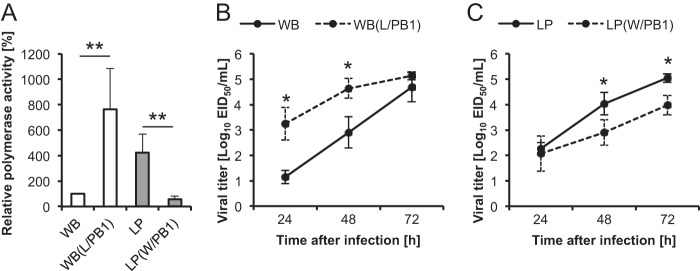
Polymerase activities of recombinant vRNPs generated from DF-1 cells (A) and kinetics of virus multiplication in DF-1 cells (B and C). (A) Polymerase activities (means ± standard deviations from six or more independent experiments) of recombinant vRNPs in DF-1 cells incubated at 39°C were measured in luciferase reporter assays. The polymerase activity of WB was set at 100% as a reference. Double asterisks indicate P values of <0.01. (B and C) Following infection of DF-1 cells with 0.001 EID50/cell, supernatants were collected at 24 hpi, 48 hpi, and 72 hpi, and viral titers of WB and WB(L/PB1) (B) and of LP and LP(W/PB1) (C) were determined. Data are means ± standard deviations at each time point (n, 3 replicates). Values were analyzed by Welch's t test. Single asterisks indicate P values of <0.05.
Next, we examined the growth kinetics of WB and WB(L/PB1) and of LP and LP(W/PB1) in vitro in order to see whether the changes observed in polymerase activity affected the growth of viruses in DF-1 cells (Fig. 1B and C). WB(L/PB1) grew more rapidly than WB until 48 hpi, and the viral titers of WB(L/PB1) were significantly higher than those of WB at 24 hpi and 48 hpi (P < 0.05) (Fig. 1B). There was no significant difference in titers between WB(L/PB1) and WB at 72 hpi. However, growth deceleration of LP(W/PB1) was observed after 48 hpi (Fig. 1C). The viral titers of LP(W/PB1) were significantly lower than those of LP at 48 hpi and 72 hpi (P < 0.05), although there were no significant differences at 24 hpi. These results suggested that the PB1 gene of WB is involved in the lower polymerase activity and lower growth rates of WB and LP(W/PB1) than of LP.
Contribution of the C38Y substitution in WB PB1 to enhanced polymerase activity and viral growth.
Twenty amino acid differences were found between the PB1 sequences of WB and LP (Table 2). To identify the residue(s) responsible for differences in polymerase activity, we selected 7 amino acid residues, at positions 14, 38, 52, 171, 211, 398, and 739, for a site-directed mutagenesis study. The residues at positions 14 and 739 are located in the binding domains for PA and PB2 (23, 24), and the other five substitutions are relatively unique to WB PB1 among AIVs (Table 2). The amino acid residue at each selected site in WB PB1 was changed to the corresponding residue found in LP PB1, and vice versa.
TABLE 2.
Amino acid differences in PB1 between WB and LP, and prevalences of WB PB1 amino acid residues in viruses isolated from avian species and chickens from the NCBI Influenza Virus Resource
| Amino acid position | Residue in: |
No. (%) of strains encoding the same amino acid residue as that in WBa |
|||||
|---|---|---|---|---|---|---|---|
| WB | LP | H5N3 strains (n = 42) | H9N2 strains (n = 501) | H5N1 strains (n = 1,201) | Strains isolated from chickens (n = 1,226) | Strains isolated from avian species (n = 7,762) | |
| 14 | A | V | 39 (92.9) | 269 (53.7) | 144 (12.0) | 445 (36.3) | 6,198 (79.9) |
| 38 | C | Y | 1 (2.4) | 2 (0.4) | 0 (0.0) | 1 (0.1) | 3 (0.0) |
| 52 | R | K | 1 (2.4) | 6 (1.2) | 7 (0.6) | 26 (2.1) | 125 (1.6) |
| 62 | G | K | 40 (95.2) | 385 (76.8) | 1,196 (99.6) | 1,131 (92.3) | 7,590 (97.8) |
| 75 | E | D | 42 (100.0) | 385 (76.8) | 1,185 (98.7) | 1,068 (87.1) | 7,512 (96.8) |
| 76 | D | N | 42 (100.0) | 391 (78.0) | 1,159 (96.5) | 1,118 (91.2) | 7,562 (97.4) |
| 111 | M | I | 42 (100.0) | 403 (80.4) | 1,193 (99.3) | 1,140 (93.0) | 7,636 (98.4) |
| 157 | A | T | 41 (97.6) | 381 (76.0) | 1,195 (99.5) | 1,108 (90.4) | 7,525 (96.9) |
| 171 | L | M | 1 (2.4) | 0 (0.0) | 1 (0.1) | 2 (0.2) | 10 (0.1) |
| 172 | E | D | 41 (97.6) | 304 (60.7) | 1,190 (99.1) | 981 (80.0) | 7,355 (94.8) |
| 175 | D | N | 42 (100.0) | 393 (78.4) | 1,180 (98.3) | 1,108 (90.4) | 7,264 (93.6) |
| 211 | K | R | 1 (2.4) | 25 (5.0) | 22 (1.8) | 38 (3.1) | 173 (2.2) |
| 214 | K | R | 40 (95.2) | 382 (76.2) | 1,194 (99.4) | 1,115 (90.9) | 7,464 (96.2) |
| 215 | K | R | 9 (21.4) | 93 (18.6) | 686 (57.1) | 435 (35.5) | 1,683 (21.7) |
| 317 | M | V | 42 (100.0) | 352 (70.3) | 1,176 (97.9) | 1,050 (85.6) | 7,138 (92.0) |
| 375 | N | T | 29 (69.0) | 325 (64.9) | 1,120 (93.3) | 968 (79.0) | 4,616 (59.5) |
| 387 | K | Q | 39 (92.9) | 263 (52.5) | 1,156 (96.3) | 949 (77.4) | 7,333 (94.5) |
| 398 | N | E | 3 (7.1) | 5 (1.0) | 3 (0.2) | 6 (0.5) | 26 (0.3) |
| 621 | Q | K | 42 (100.0) | 333 (66.5) | 1,184 (98.6) | 1,057 (86.2) | 7,457 (96.1) |
| 739 | D | E | 1 (2.4) | 11 (2.2) | 2 (0.2) | 9 (0.7) | 36 (0.5) |
Shown are the numbers of strains that were categorized into the respective criteria extracted from the NCBI Influenza Virus Resource.
In WB, a cysteine-to-tyrosine substitution at residue 38 (C38Y) of PB1 resulted in higher polymerase activity than that of wild-type WB PB1 (P < 0.01) (Fig. 2A). No significant increase in polymerase activity was observed following substitutions of the other 6 amino acids (Fig. 2A). The effects of C38Y in WB PB1 were also observed when WB-PB1-C38Y was combined with LP PB2, PA, and NP to construct LP(W/PB1-C38Y). The polymerase activity of LP(W/PB1-C38Y) was significantly higher than that of LP(W/PB1) (P < 0.01) (Fig. 2B). Inhibitory effects were observed when the Y38C substitution was introduced into LP PB1. The polymerase activities of WB(L/PB1-Y38C) and LP-PB1-Y38C were significantly lower than those of WB(L/PB1) and LP, respectively (Fig. 2C). Thus, the residue at position 38 is a key amino acid regulating polymerase activity in WB PB1, independently of the other components of the polymerase complex of WB or LP.
FIG 2.
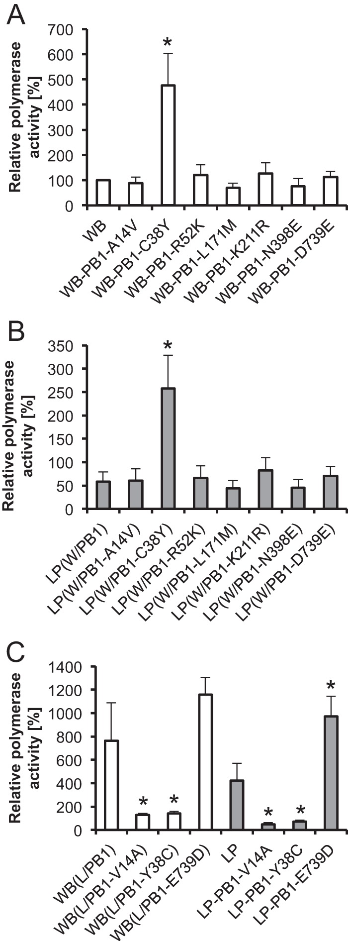
Effects of PB1 amino acid substitutions on polymerase activity. (A and B) The effects of 7 amino acid substitutions in WB PB1 on polymerase activity were analyzed in a WB background (A) and in an LP background (B). Asterisks indicate significant differences (P < 0.01) from WB (A) and from LP(W/PB1) (B). (C) Three amino acid substitutions in LP PB1 were analyzed for their effects on polymerase activity in WB and LP. Asterisks indicate significant differences among WB(L/PB1) variants in the WB background or among LP variants in the LP background (P < 0.01). Values are means ± standard deviations for three or more independent experiments. Statistical differences in mean polymerase activities were assessed with Bonferroni's multiple-comparison test.
In DF-1 cells, the replication of WB-PB1-C38Y, which possesses WB PB1 with the C38Y substitution, did not differ significantly from that of recombinants with wild-type WB PB1 (Fig. 3A). In contrast, the viral titers of LP(W/PB1-C38Y), which possesses WB PB1 with C38Y along with the other internal genes from LP, were significantly higher than those of LP(W/PB1) at 48 hpi and 72 hpi (P < 0.05) (Fig. 3B).
FIG 3.
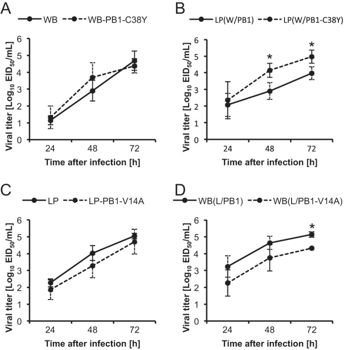
(A and B) Viral titers of recombinants possessing the C38Y substitution in PB1 in a WB background (A) or an LP(W/PB1) background (B). (C and D) Viral titers of recombinants possessing the V14A substitution in PB1 in an LP background (C) or a WB(L/PB1) background (D). Values are means ± standard deviations for three replicates. Viral titers at each time point were analyzed by Welch's t test. Asterisks indicate P values of <0.05.
The V14A substitution in LP PB1 reduced polymerase activity.
We also examined the effects of amino acid alterations from valine to alanine at residue 14 (V14A) and from glutamic acid to aspartic acid at residue 739 (E739D) in LP PB1, because these residues are located in the PA- and PB2-binding domains of PB1. In addition, the frequency of V14 among AIVs was biased in the H5N1 and H9N2 strains (Table 2). V14A caused a significant decrease in polymerase activity when combined with the PB2, PA, and NP of LP or WB (P < 0.01) (Fig. 2C). E739D caused an increase in activity when combined with other components from LP (P < 0.01) but not when combined with other components from WB. No significant differences in viral replication were observed between LP and LP-PB1-V14A (Fig. 3C). The viral titer of WB(L/PB1-V14A) was significantly lower than that of WB(L/PB1) at 72 hpi (P < 0.05) (Fig. 3D).
The substitution at amino acid position 38 in the PB1 gene affected pathogenicity in chickens.
Survival analysis was performed to examine the pathogenicities of recombinant viruses with the PB1 C38Y substitution in chickens. The pathogenicity of WB-PB1-C38Y was found to be higher than that of WB (P = 0.025) (Fig. 4A). The MST of chickens infected with WB was shortened from 3.39 dpi to 2.25 dpi by the C38Y substitution (WB-PB1-C38Y). The effect of C38Y in WB PB1 on survival was greatly enhanced (P = 0.008) by combination with LP PB2, PA, and NP [LP(W/PB1-C38Y)] (Fig. 4B). The survival rate of chickens infected with LP(W/PB1) was decreased from 50% to 0%, and the MST was shortened from 7.50 dpi to 3.00 dpi, by the C38Y substitution. In a comparison of WB with WB-PB1-C38Y, no significant differences were observed between the viral titers in tracheal and cloacal swabs or in organs collected from infected chickens (Fig. 4C and E; Table 3).
FIG 4.
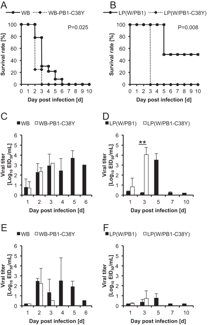
Pathogenicities of recombinant viruses with the C38Y substitution in WB PB1 for chickens. (A and B) Survival plots for chickens infected with WB or WB-PB1-C38Y (A) and for chickens infected with LP(W/PB1) or LP(W/PB1-C38Y) (B). The survivability of chickens (n ≥ 4) was determined, and Kaplan-Meier survival rate curves were plotted and were analyzed by the log rank test. (C to F) Viral titers in tracheal (C and D) and cloacal (E and F) swabs from infected chickens. Swabs were taken from chickens infected with WB or WB-PB1-C38Y (C and E) and from chickens infected with LP(W/PB1) or LP(W/PB1-C38Y) (D and F) at 3, 5, 7, and 10 dpi routinely and upon the deaths of the chickens. Viral titers at 1 dpi were those obtained in the experiment reported in Tables 3 and 4. Viral titers at each time point were analyzed by Welch's t test. Double asterisks indicate a P value of <0.01.
TABLE 3.
Viral titers in organs collected from chickens at 24 h after infection with WB or WB-PB1-C38Y
| Organ or sample | Titer (log10 EID50/ml)a |
|||||
|---|---|---|---|---|---|---|
| WB |
WB-PB1-C38Y |
|||||
| Animal 1 | Animal 2 | Animal 3 | Animal 1 | Animal 2 | Animal 3 | |
| Pancreas | <1.20 | 3.38 | <0.20 | <0.20 | 3.20 | 2.53 |
| Spleen | <1.20 | 3.53 | <0.20 | <0.20 | 4.20 | 3.20 |
| Muscle | <1.20 | 3.30 | <0.20 | <0.20 | 2.20 | 2.20 |
| Liver | <1.20 | 3.92 | <0.20 | 0.53 | 3.07 | 3.20 |
| Bursa of Fabricius | <1.20 | <1.20 | <0.20 | <0.20 | 3.32 | 3.20 |
| Trachea | <1.20 | 2.06 | <0.20 | <0.20 | 3.02 | 2.07 |
| Lung | <1.20 | 5.45 | <0.20 | <0.20 | 3.07 | 2.20 |
| Kidney | <1.20 | 3.32 | <0.20 | <0.20 | 2.53 | 1.70 |
| Heart | <1.20 | 4.87 | <0.20 | <0.20 | 2.38 | 2.53 |
| Comb | <1.20 | 3.38 | <0.20 | <0.20 | 3.32 | 2.87 |
| Wattle | <1.20 | 4.07 | <0.20 | <0.20 | 3.02 | 2.20 |
| Brain | <1.20 | 2.32 | <0.20 | <0.20 | 3.20 | 3.07 |
| Duodenum | — | 3.32 | <0.20 | <0.20 | 3.38 | 2.20 |
| Rectum | <1.20 | 3.92 | <0.20 | <0.20 | 3.53 | 3.53 |
| Blood | <1.20 | 3.30 | <0.20 | <0.20 | 1.20 | 1.38 |
| Tracheal swab | <0.20 | 1.95 | <0.20 | <0.20 | 1.60 | 0.32 |
| Cloacal swab | <0.20 | <0.20 | <0.20 | <0.20 | <0.20 | <0.20 |
The detection limit was either <1.20 or <0.20 log10 EID50/ml. —, sample not available.
The level of viral shedding at 3 dpi in tracheal swabs from LP(W/PB1-C38Y)-infected chickens was significantly higher than that observed with LP(W/PB1) (Fig. 4D). LP(W/PB1) showed the maximum viral titer at 5 dpi; however, the maximum titer in chickens infected with LP(W/PB1-C38Y) was observed at 3 dpi (Fig. 4D). Viral titers in cloacal swabs did not differ significantly between LP(W/PB1-C38Y) and LP(W/PB1) (Fig. 4F). No apparent differences in tissue tropism were observed between LP(W/PB1-C38Y) and LP(W/PB1) (Table 4).
TABLE 4.
Viral titers in organs collected from chickens at 24 h after infection with LP(W/PB1) or LP(W/PB1-C38Y)
| Organ or sample | Titer (log10 EID50/ml)a |
|||||
|---|---|---|---|---|---|---|
| LP(W/PB1) |
LP(W/PB1-C38Y) |
|||||
| Animal 1 | Animal 2 | Animal 3 | Animal 1 | Animal 2 | Animal 3 | |
| Pancreas | <0.20 | <0.20 | <0.20 | <0.20 | <0.20 | <0.20 |
| Spleen | <0.20 | <0.20 | 0.32 | <0.20 | <0.20 | <0.20 |
| Muscle | <0.20 | <0.20 | <0.20 | <0.20 | <0.20 | <0.20 |
| Liver | <0.20 | <0.20 | <0.20 | 0.32 | <0.20 | <0.20 |
| Bursa of Fabricius | <0.20 | <0.20 | <0.20 | <0.20 | <0.20 | <0.20 |
| Trachea | <0.20 | <0.20 | <0.20 | <0.20 | 0.32 | <0.20 |
| Lung | <0.20 | <0.20 | <0.20 | <0.20 | 0.53 | 0.53 |
| Kidney | <0.20 | <0.20 | <0.20 | <0.20 | 0.32 | <0.20 |
| Heart | <0.20 | <0.20 | <0.20 | <0.20 | 0.70 | <0.20 |
| Comb | 0.32 | <0.20 | <0.20 | <0.20 | <0.20 | <0.20 |
| Wattle | <0.20 | <0.20 | <0.20 | <0.20 | <0.20 | <0.20 |
| Brain | 0.32 | <0.20 | <0.20 | <0.20 | <0.20 | <0.20 |
| Duodenum | <0.20 | <0.20 | <0.20 | <0.20 | <0.20 | <0.20 |
| Rectum | <0.20 | <0.20 | <0.20 | <0.20 | <0.20 | <0.20 |
| Blood | <0.20 | <0.20 | <0.20 | <0.20 | <0.20 | <0.20 |
| Tracheal swab | 0.32 | 0.45 | 0.30 | 2.07 | <0.20 | <0.20 |
| Cloacal swab | <0.20 | <0.20 | <0.20 | <0.20 | <0.20 | 0.32 |
The detection limit was <0.20 log10 EID50/ml.
To confirm that the Y residue at position 38 in PB1 regulates H5N1 HPAIV pathogenicity in chickens, we constructed the recombinant virus LP-PB1-Y38C, which possesses the Y38C substitution in LP PB1. We observed by survival analysis that the pathogenicity of LP-PB1-Y38C was reduced from that of LP (P = 0.025) (Fig. 5A). The survival rate of chickens infected with LP-PB1-Y38C was increased from 4.3% to 40%, and the MST was extended from 3.77 dpi to 6.80 dpi (Fig. 5A). Viral titers in tracheal swabs from chickens exposed to LP-PB1-Y38C were significantly lower at 3 dpi than those from chickens exposed to LP (Fig. 5B), while no significant differences were observed in cloacal swabs (Fig. 5C). The transmissibility of LP-PB1-Y38C was examined in order to determine if the decreased pathogenicity resulting from the Y38C substitution is associated with reduced transmissibility of the recombinant virus. LP-PB1-Y38C was not transmitted from infected chickens to any naïve chickens in the same isolator (see Fig. 7), and viral titers in tracheal swabs at 4 dpi did not differ from those observed with LP (Fig. 5B). Thus, both pathogenicity and transmissibility were reduced by the Y38C substitution in LP PB1.
FIG 5.

Pathogenicities of recombinant viruses with the Y38C substitution in LP PB1 for chickens. (A) The survivability of chickens (n ≥ 4) was determined, and Kaplan-Meier survival rate curves were plotted and were analyzed by the log rank test. (B and C) Viral titers in tracheal (B) and cloacal (C) swabs taken from infected chickens at 1, 3, 5, 7, and 10 dpi routinely and upon the deaths of the chickens were determined. Viral titers at each time point were analyzed by Welch's t test. The asterisk indicates a P value of <0.05.
FIG 7.
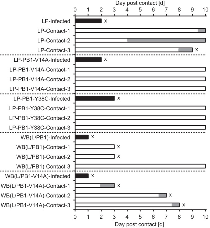
Transmissibilities of recombinant viruses in chickens. Filled and shaded bars indicate durations of detectable virus in tracheal and/or cloacal swabs from infected and contacted chickens, respectively. The letter “x” indicates the time point at which a chicken died from infection. Cohabitation with infected chickens started at 1 dpi.
V14A in LP PB1 reduces transmissibility in chickens.
To investigate the influence of the V14A substitution in LP PB1 on the pathogenicities of recombinants, survival analysis was performed comparing LP with LP-PB1-V14A and WB(LP/PB1) with WB(LP/PB1-V14A). Survival analysis revealed no significant difference between LP and LP-PB1-V14A (P = 0.764) (Fig. 6A). Similarly, there was no significant difference between WB(L/PB1) and WB(L/PB1-V14A) (P = 0.317) (Fig. 6B).
FIG 6.
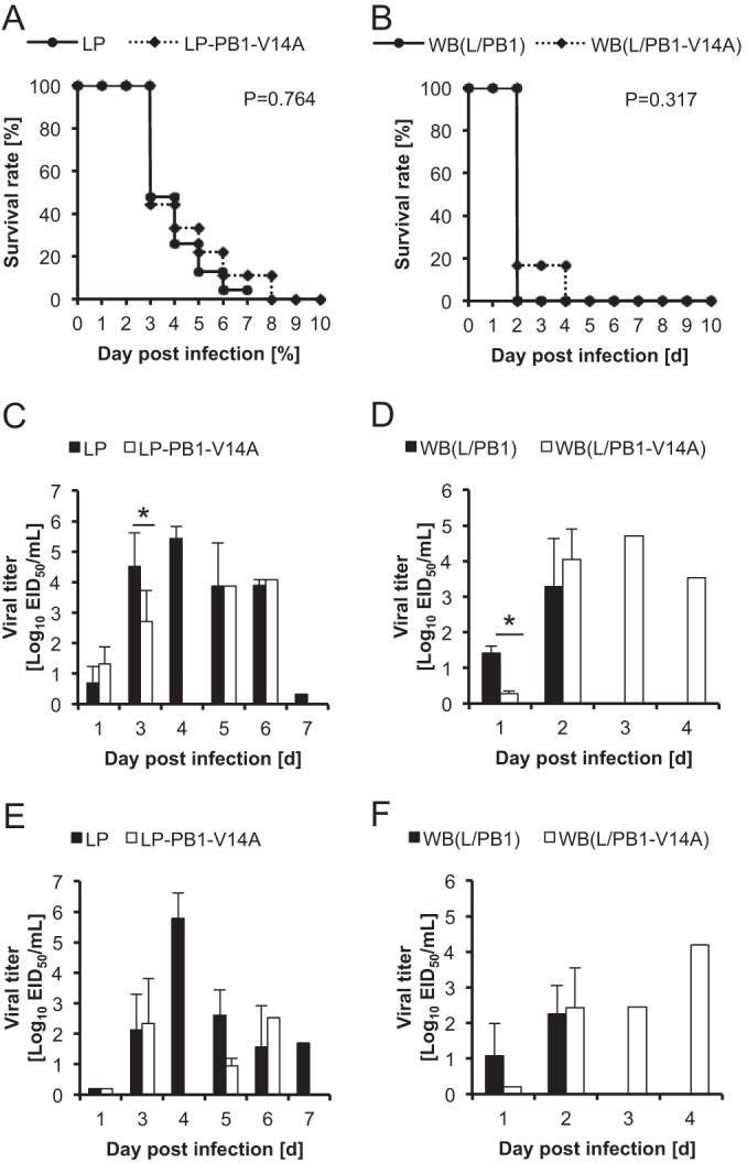
Pathogenicities of recombinant viruses with the V14A substitution in LP PB1 for chickens. (A and B) Survival plots for chickens infected with LP or LP-PB1-V14A (A) and for chickens infected with WB(L/PB1) or WB(L/PB1-V14A) (B). The survivability of chickens (n ≥ 4) was determined, and Kaplan-Meier survival rate curves were plotted and were analyzed by the log rank test. (C to F) Viral titers in tracheal (C and D) and cloacal (E and F) swabs from chickens infected with LP or LP-PB1-V14A (C and E) and from chickens infected with WB(L/PB1) or WB(L/PB1-V14A) (D and F). Swabs were collected at 3, 5, 7, and 10 dpi routinely and upon the deaths of the chickens. Viral titers at 1 dpi were those obtained in the experiment reported in Tables 5 and 6. Viral titers at each time point were analyzed by Welch's t test. Asterisks indicate P values of <0.05.
Next, we compared viral shedding and tissue distribution in chickens infected with a parental virus and those infected with a recombinant, or in chickens infected with different recombinants; LP was compared with LP-PB1-V14A, and WB(L/PB1) was compared with WB(L/PB1-V14A). The viral titers in tracheal swabs from LP-PB1-V14A-infected chickens were significantly lower at 3 dpi than those from LP-infected chickens (Fig. 6C), although no significant difference in viral titers was observed with cloacal swabs (Fig. 6E). The viral titers of LP-PB1-V14A in the lung were decreased (Table 5). A comparison of WB(L/PB1) and WB(L/PB1-V14A) showed that the level of viral shedding from tracheal swabs was significantly lower for WB(L/PB1-V14A) than for WB(L/PB1) at 1 dpi (P < 0.05) (Fig. 6D). There were no significant differences in viral titers in cloacal swabs (Fig. 6F). A comparison of tissue tropism in WB(L/PB1-V14A) and WB(L/PB1) showed a tendency for WB(L/PB1-V14A) titers to decline in most organs; they were significantly lower in the muscle (P < 0.01), liver (P < 0.05), trachea (P < 0.05), and tracheal swabs (P < 0.05) (Table 6) (the same data are shown for tracheal swabs in Fig. 6D).
TABLE 5.
Viral titers in organs collected from chickens at 24 h after infection with LP or LP-PB1-V14A
| Organ or sample | Titer (log10 EID50/ml)a |
|||||
|---|---|---|---|---|---|---|
| LP |
LP-PB1-V14A |
|||||
| Animal 1 | Animal 2 | Animal 3 | Animal 1 | Animal 2 | Animal 3 | |
| Pancreas | <1.20 | <1.20 | <0.20 | <0.20 | <0.20 | 0.87 |
| Spleen | <1.20 | <1.20 | <0.20 | 0.32 | <0.20 | <0.20 |
| Muscle | <1.20 | 1.70 | <0.20 | <0.20 | <0.20 | <0.20 |
| Liver | <1.20 | <1.20 | <0.20 | <0.20 | <0.20 | <0.20 |
| Bursa of Fabricius | <1.20 | 1.32 | 0.32 | <0.20 | <0.20 | <0.20 |
| Trachea | <1.20 | 2.45 | 0.32 | <0.20 | <0.20 | 1.38 |
| Lung | 1.53 | 3.32 | <0.20 | <0.20 | 0.32 | 0.32 |
| Kidney | <1.20 | 2.02 | 0.32 | <0.20 | <0.20 | <0.20 |
| Heart | <1.20 | 2.45 | <0.20 | 0.32 | <0.20 | <0.20 |
| Comb | 1.53 | <1.20 | <0.20 | 0.32 | <0.20 | <0.20 |
| Wattle | <1.20 | 2.07 | <0.20 | <0.20 | <0.20 | <0.20 |
| Brain | <1.20 | 2.02 | <0.20 | <0.20 | <0.20 | <0.20 |
| Duodenum | <1.20 | <1.20 | <0.20 | <0.20 | <0.20 | <0.20 |
| Rectum | <1.20 | <1.20 | <0.20 | <0.20 | <0.20 | <0.20 |
| Blood | <1.20 | <1.20 | <0.20 | <0.20 | <0.20 | <0.20 |
| Tracheal swab | 0.45 | 1.45 | <0.20 | 0.53 | 1.62 | 1.79 |
| Cloacal swab | <0.20 | <0.20 | <0.20 | <0.20 | <0.20 | <0.20 |
The detection limit was either <1.20 or <0.20 log10 EID50/ml.
TABLE 6.
Viral titers in organs collected from chickens at 24 h after infection with WB(L/PB1) or WB(L/PB1-V14A)
| Organ or samplea | Titer (log10 EID50/ml)b |
|||||
|---|---|---|---|---|---|---|
| WB(L/PB1) |
WB(L/PB1-V14A) |
|||||
| Animal 1 | Animal 2 | Animal 3 | Animal 1 | Animal 2 | Animal 3 | |
| Pancreas | 3.32 | 2.53 | 2.53 | 0.53 | 2.20 | <0.20 |
| Spleen | 4.87 | 3.20 | 3.20 | 1.07 | 3.20 | <0.20 |
| Muscle** | 3.20 | 2.32 | 2.38 | <0.20 | 1.20 | <0.20 |
| Liver* | 4.07 | 2.87 | 3.32 | 0.70 | 2.32 | <0.20 |
| Bursa of Fabricius | 3.20 | 2.32 | 1.70 | 0.32 | 2.38 | <0.20 |
| Trachea* | 4.02 | 3.53 | 2.87 | 0.32 | 2.32 | <0.20 |
| Lung | 4.45 | 3.53 | 3.20 | 0.53 | 4.07 | <0.20 |
| Kidney | 4.38 | 2.32 | 2.53 | 0.53 | 2.20 | <0.20 |
| Heart | 4.53 | 3.20 | 3.32 | <0.20 | 3.32 | 0.53 |
| Comb | 3.02 | 2.87 | 2.45 | <0.20 | 3.07 | <0.20 |
| Wattle | 3.20 | 3.20 | 3.20 | <0.20 | 2.53 | <0.20 |
| Brain | 3.87 | 3.87 | 2.32 | <0.20 | 2.32 | <0.20 |
| Duodenum | 3.53 | 2.87 | 2.07 | 0.53 | 2.20 | <0.20 |
| Rectum | 3.87 | 2.20 | 2.20 | <0.20 | 1.87 | <0.20 |
| Blood | 1.32 | 0.87 | 0.32 | <0.20 | 0.53 | <0.20 |
| Tracheal swab* | 1.70 | 1.32 | <0.20 | <0.20 | 0.32 | 0.32 |
| Cloacal swab | 0.70 | 2.32 | <0.20 | <0.20 | <0.20 | <0.20 |
Asterisks indicate statistically significant differences in viral titers between WB(L/PB1) and WB(L/PB1-V14A), as determined by Welch's t test (*, P < 0.05; **, P < 0.01).
The detection limit was <0.20 log10 EID50/ml.
A transmission study was conducted to evaluate the effects of the V14A substitution, which resulted in reduced polymerase activity, while no change in pathogenicity was observed. LP was transmitted to all 3 naïve chickens in the same isolator as an infected chicken by the end of the observation period; in contrast, LP-PB1-V14A was not transmitted to any naïve chickens in the same isolator (Fig. 7). Tracheal and cloacal swabs were collected at 3 dpi from inoculated chickens, and the LP titers from tracheal swabs were significantly higher than the LP-PB1-V14A titers (Fig. 6C). These results were consistent with the differences in transmissibility between LP and the recombinant LP-PB1-V14A virus with reduced polymerase activity. In contrast, WB(L/PB1) was transmitted to 2 of the 3 naïve chickens, and WB(L/PB1-V14A) was transmitted to all 3 naïve chickens, during the observation period (Fig. 7). These two viruses showed nearly identical transmissibility, although the titer of WB(L/PB1-V14A) from tracheal swabs at 1 dpi was significantly lower than that of WB(L/PB1) (Fig. 6D).
DISCUSSION
In this study, we found that the C38Y substitution in WB PB1 increased both polymerase activity in vitro and pathogenicity in chickens. The recombinant viruses with this substitution, WB-PB1-C38Y and LP(W/PB1-C38Y), showed higher pathogenicity in chickens than the parental viruses WB and LP(W/PB1), respectively. Previously, a correlation was reported between enhanced polymerase activity and pathogenicity with substitutions in the PA gene of an H5N1 strain (6). A/duck/Hubei/49/05 (DK/49) showed higher polymerase activity in duck fibroblasts and higher pathogenicity in ducks than A/goose/Hubei/65/05 (GS/65). When the S224P and N383D substitutions in PA were introduced into strain GS/65, polymerase activity and pathogenicity were enhanced (6). The E627K substitution in the PB2 of an AIV was reported to increase polymerase activity in mammalian cells (10), and this substitution in an H5N1 HPAIV enhanced virulence in mice (11, 12). Higher polymerase activity was also attributed to the higher pathogenicity of A/duck/Fujian/01/2002 in mice and chickens than of A/duck/Guangxi/53/2002 (25, 26). In the present study, along with others, demonstrates that enhanced polymerase activity is a major determinant of enhanced pathogenicity.
PB1 serves a central role in the formation of the structural backbone of the viral polymerase complex and has RNA polymerase activity (27–29). In addition, PB1 has viral RNA (vRNA)-binding domains (amino acids 1 to 139, 233 to 249, and 494 to 757) and cRNA-binding domains (amino acids 1 to 139 and 267 to 493) (30, 31). Although C38Y resides in both the vRNA- and cRNA-binding domains, it does not confer binding of PB1 to vRNA. Jung and Brownlee demonstrated that changes from alanine to arginine at positions 233, 238, 239, and 249 impaired vRNA, cRNA, and mRNA synthesis in vitro by disabling the binding activity of PB1 to the vRNA promoter (31), indicating that the essential domains for vRNA binding are located at amino acids 233 to 249. These results are consistent with a previous report demonstrating that hairpin-loop RNA bound to the arginine-rich domain (32). The polymerase function of PB1 resides in four conserved polymerase motifs (amino acids 303 to 306, 403 to 412, 438 to 450, and 474 to 484) (28, 33). V473L and P598L, at positions in motif IV and near the PB2-binding domain, respectively, reduced polymerase activity (13). C38Y does not reside in either the PA-binding domain at the N terminus (amino acids 1 to 15) or the PB2-binding domain at the C terminus (amino acids 685 to 757) of PB1 (23, 24, 34). How C38Y in PB1 affects the polymerase activity needs to be elucidated by further analyses.
The alteration of viral replication by substitutions in the PB1 protein was not always consistent with that of polymerase activities observed in DF-1 cells. Two critical steps must occur with viral transcripts for the replication cycle to be completed: vRNA synthesis and viral protein translation. A temperature-sensitive virus with a C-terminal deletion in the NS1 protein showed reduced vRNA levels and low infectivity at the restrictive temperature (39.5°C), while the accumulation of cRNA and mRNA was not altered (35). In addition, signaling through the nuclear factor κB (NF-κB) pathway, a prerequisite for efficient influenza virus infection, was activated by infection with influenza A virus (36, 37), and an NF-κB inhibitor reduced vRNA production and viral infectivity without influencing viral mRNA levels (38). In the present study, polymerase activities were measured by luciferase assays, which reflect fluctuations in viral mRNA production but not in vRNA levels. A possible explanation of the discrepancy observed between viral replication and polymerase activities is that vRNA production may have been variably affected by mutations in PB1 residue 14 or 38, while mRNA synthesis was consistently affected, though not to an extent that would affect virus production.
LP-PB1-V14A, a recombinant virus with the V14A substitution, displayed a reduced level of viral shedding in infected chickens and lost the ability to be transmitted to naïve chickens. Virus shedding is a key factor in successful transmission between an infected and a naïve chicken, since influenza viruses are transmitted through aerosols, large droplets, or direct contact with secretions (39). Reduced polymerase activity has been shown to be associated with restricted transmissibility of HPAIVs. Hulse-Post et al. reported that reduced polymerase activity with the Y436H substitution in PB1 resulted in lowered transmissibility and pathogenicity in ducks (7). Lyall et al. established transgenic chickens in which the RNA polymerase activity of a polymerase complex from an AIV was hampered by expressing a short hairpin RNA as an RNA decoy (40). When these transgenic chickens were infected with an HPAIV, the efficiency of transmission of the virus to transgenic and nontransgenic chickens was reduced, although transgenic chickens were still susceptible to initial infection. The level of shedding of the virus from the cloacae of transgenic chickens was apparently lower than that in nontransgenic infected chickens. Impaired polymerase activity in LP-PB1-V14A caused reduced transmissibility of the virus without affecting its lethality. Although other causes have not been eliminated, it is likely that the reduced transmissibility of LP-PB1-V14A could be the result of reduced secretions in the trachea. The functional change resulting from the V14A substitution requires further study, considering that Perez and Donis found that A14V does not affect PA binding (41), while Wunderlich et al. demonstrated reduced binding to PA by PB1-derived peptides containing the V14A mutation (42).
It is worth considering whether the V14A substitution may play a role in the dissemination of viruses in the field, since the frequency of valine at residue 14 in PB1 is much higher in H5N1 isolates than in other AIV isolates, a majority of which possess alanine at this position (Table 2). The H5N1 and H9N2 viruses with A14V have emerged since 2001 and 1994, respectively (Fig. 8). During the initial outbreak in 1997, H5N1 viruses possessed A14, but the frequency of V14 suddenly increased in 2003, corresponding with an eruption of the viruses in Asian countries (43). In addition, V14 in H9N2 viruses was confirmed in 1994, and the population has gradually increased.
FIG 8.
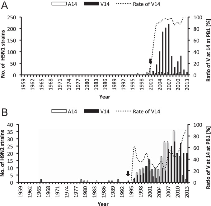
Numbers of strains with alanine or valine at amino acid position 14 in PB1 and percentages of H5N1 (A) and H9N2 (B) strains since 1959. Black arrows indicate when the oldest strain possessing V14 in PB1 appeared. Percentages were calculated when the number of strains of each subtype in the same year was greater than 5.
In conclusion, we found that two novel amino acid substitutions in the PB1 protein, namely, C38Y in WB PB1 and V14A in LP PB1, alter polymerase activity. Altered polymerase activity was shown to be an important factor for viral pathogenesis in chickens. C38Y contributes to an increase in polymerase activity, accompanied by a shortened MST, while V14A affects transmissibility in chickens. Although the molecular bases of the change in polymerase activity and the altered host responses induced by the changes remain to be elucidated, our results demonstrate the involvement of PB1 in the pathogenesis of HPAIV in chickens.
ACKNOWLEDGMENT
This work was supported by a Grant-in-Aid for Scientific Research from the Zoonoses Control Project of the Ministry of Agriculture, Forestry, and Fisheries of Japan.
Footnotes
Published ahead of print 16 July 2014
REFERENCES
- 1.Webster RG, Bean WJ, Gorman OT, Chambers TM, Kawaoka Y. 1992. Evolution and ecology of influenza A viruses. Microbiol. Rev. 56:152–179 [DOI] [PMC free article] [PubMed] [Google Scholar]
- 2.Fouchier RA, Munster V, Wallensten A, Bestebroer TM, Herfst S, Smith D, Rimmelzwaan GF, Olsen B, Osterhaus AD. 2005. Characterization of a novel influenza A virus hemagglutinin subtype (H16) obtained from black-headed gulls. J. Virol. 79:2814–2822. 10.1128/JVI.79.5.2814-2822.2005 [DOI] [PMC free article] [PubMed] [Google Scholar]
- 3.Senne DA, Panigrahy B, Kawaoka Y, Pearson JE, Suss J, Lipkind M, Kida H, Webster RG. 1996. Survey of the hemagglutinin (HA) cleavage site sequence of H5 and H7 avian influenza viruses: amino acid sequence at the HA cleavage site as a marker of pathogenicity potential. Avian Dis. 40:425–437. 10.2307/1592241 [DOI] [PubMed] [Google Scholar]
- 4.Horimoto T, Kawaoka Y. 1994. Reverse genetics provides direct evidence for a correlation of hemagglutinin cleavability and virulence of an avian influenza A virus. J. Virol. 68:3120–3128 [DOI] [PMC free article] [PubMed] [Google Scholar]
- 5.Steinhauer DA. 1999. Role of hemagglutinin cleavage for the pathogenicity of influenza virus. Virology 258:1–20. 10.1006/viro.1999.9716 [DOI] [PubMed] [Google Scholar]
- 6.Song J, Feng H, Xu J, Zhao D, Shi J, Li Y, Deng G, Jiang Y, Li X, Zhu P, Guan Y, Bu Z, Kawaoka Y, Chen H. 2011. The PA protein directly contributes to the virulence of H5N1 avian influenza viruses in domestic ducks. J. Virol. 85:2180–2188. 10.1128/JVI.01975-10 [DOI] [PMC free article] [PubMed] [Google Scholar]
- 7.Hulse-Post DJ, Franks J, Boyd K, Salomon R, Hoffmann E, Yen HL, Webby RJ, Walker D, Nguyen TD, Webster RG. 2007. Molecular changes in the polymerase genes (PA and PB1) associated with high pathogenicity of H5N1 influenza virus in mallard ducks. J. Virol. 81:8515–8524. 10.1128/JVI.00435-07 [DOI] [PMC free article] [PubMed] [Google Scholar]
- 8.Mehle A, Dugan VG, Taubenberger JK, Doudna JA. 2012. Reassortment and mutation of the avian influenza virus polymerase PA subunit overcome species barriers. J. Virol. 86:1750–1757. 10.1128/JVI.06203-11 [DOI] [PMC free article] [PubMed] [Google Scholar]
- 9.Subbarao EK, London W, Murphy BR. 1993. A single amino acid in the PB2 gene of influenza A virus is a determinant of host range. J. Virol. 67:1761–1764 [DOI] [PMC free article] [PubMed] [Google Scholar]
- 10.Massin P, van der Werf S, Naffakh N. 2001. Residue 627 of PB2 is a determinant of cold sensitivity in RNA replication of avian influenza viruses. J. Virol. 75:5398–5404. 10.1128/JVI.75.11.5398-5404.2001 [DOI] [PMC free article] [PubMed] [Google Scholar]
- 11.Hatta M, Gao P, Halfmann P, Kawaoka Y. 2001. Molecular basis for high virulence of Hong Kong H5N1 influenza A viruses. Science 293:1840–1842. 10.1126/science.1062882 [DOI] [PubMed] [Google Scholar]
- 12.Hatta M, Hatta Y, Kim JH, Watanabe S, Shinya K, Nguyen T, Lien PS, Le QM, Kawaoka Y. 2007. Growth of H5N1 influenza A viruses in the upper respiratory tracts of mice. PLoS Pathog. 3:1374–1379. 10.1371/journal.ppat.0030133 [DOI] [PMC free article] [PubMed] [Google Scholar]
- 13.Xu C, Hu WB, Xu K, He YX, Wang TY, Chen Z, Li TX, Liu JH, Buchy P, Sun B. 2012. Amino acids 473V and 598P of PB1 from an avian-origin influenza A virus contribute to polymerase activity, especially in mammalian cells. J. Gen. Virol. 93:531–540. 10.1099/vir.0.036434-0 [DOI] [PubMed] [Google Scholar]
- 14.Chu C, Fan S, Li C, Macken C, Kim JH, Hatta M, Neumann G, Kawaoka Y. 2012. Functional analysis of conserved motifs in influenza virus PB1 protein. PLoS One 7:e36113. 10.1371/journal.pone.0036113 [DOI] [PMC free article] [PubMed] [Google Scholar]
- 15.Uchida Y, Watanabe C, Takemae N, Hayashi T, Oka T, Ito T, Saito T. 2012. Identification of host genes linked with the survivability of chickens infected with recombinant viruses possessing H5N1 surface antigens from a highly pathogenic avian influenza virus. J. Virol. 86:2686–2695. 10.1128/JVI.06374-11 [DOI] [PMC free article] [PubMed] [Google Scholar]
- 16.Mase M, Tsukamoto K, Imada T, Imai K, Tanimura N, Nakamura K, Yamamoto Y, Hitomi T, Kira T, Nakai T, Kiso M, Horimoto T, Kawaoka Y, Yamaguchi S. 2005. Characterization of H5N1 influenza A viruses isolated during the 2003–2004 influenza outbreaks in Japan. Virology 332:167–176. 10.1016/j.virol.2004.11.016 [DOI] [PubMed] [Google Scholar]
- 17.Mase M, Eto M, Imai K, Tsukamoto K, Yamaguchi S. 2007. Characterization of H9N2 influenza A viruses isolated from chicken products imported into Japan from China. Epidemiol. Infect. 135:386–391. 10.1017/S0950268806006728 [DOI] [PMC free article] [PubMed] [Google Scholar]
- 18.Himly M, Foster DN, Bottoli I, Iacovoni JS, Vogt PK. 1998. The DF-1 chicken fibroblast cell line: transformation induced by diverse oncogenes and cell death resulting from infection by avian leukosis viruses. Virology 248:295–304. 10.1006/viro.1998.9290 [DOI] [PubMed] [Google Scholar]
- 19.Hoffmann E, Stech J, Guan Y, Webster RG, Perez DR. 2001. Universal primer set for the full-length amplification of all influenza A viruses. Arch. Virol. 146:2275–2289. 10.1007/s007050170002 [DOI] [PubMed] [Google Scholar]
- 20.Li OT, Chan MC, Leung CS, Chan RW, Guan Y, Nicholls JM, Poon LL. 2009. Full factorial analysis of mammalian and avian influenza polymerase subunits suggests a role of an efficient polymerase for virus adaptation. PLoS One 4:e5658. 10.1371/journal.pone.0005658 [DOI] [PMC free article] [PubMed] [Google Scholar]
- 21.Reed LJ, Muench H. 1938. A simple method of estimating fifty percent endpoints. Am. J. Hyg. 27:493–497 [Google Scholar]
- 22.Kaplan EL, Meier P. 1958. Nonparametric estimation from incomplete observations. J. Am. Stat. Assoc. 53:457–481. 10.1080/01621459.1958.10501452 [DOI] [Google Scholar]
- 23.Obayashi E, Yoshida H, Kawai F, Shibayama N, Kawaguchi A, Nagata K, Tame JR, Park SY. 2008. The structural basis for an essential subunit interaction in influenza virus RNA polymerase. Nature 454:1127–1131. 10.1038/nature07225 [DOI] [PubMed] [Google Scholar]
- 24.Sugiyama K, Obayashi E, Kawaguchi A, Suzuki Y, Tame JR, Nagata K, Park SY. 2009. Structural insight into the essential PB1-PB2 subunit contact of the influenza virus RNA polymerase. EMBO J. 28:1803–1811. 10.1038/emboj.2009.138 [DOI] [PMC free article] [PubMed] [Google Scholar]
- 25.Chen H, Deng G, Li Z, Tian G, Li Y, Jiao P, Zhang L, Liu Z, Webster RG, Yu K. 2004. The evolution of H5N1 influenza viruses in ducks in southern China. Proc. Natl. Acad. Sci. U. S. A. 101:10452–10457. 10.1073/pnas.0403212101 [DOI] [PMC free article] [PubMed] [Google Scholar]
- 26.Leung BW, Chen H, Brownlee GG. 2010. Correlation between polymerase activity and pathogenicity in two duck H5N1 influenza viruses suggests that the polymerase contributes to pathogenicity. Virology 401:96–106. 10.1016/j.virol.2010.01.036 [DOI] [PubMed] [Google Scholar]
- 27.Gonzalez S, Zurcher T, Ortin J. 1996. Identification of two separate domains in the influenza virus PB1 protein involved in the interaction with the PB2 and PA subunits: a model for the viral RNA polymerase structure. Nucleic Acids Res. 24:4456–4463. 10.1093/nar/24.22.4456 [DOI] [PMC free article] [PubMed] [Google Scholar]
- 28.Poch O, Sauvaget I, Delarue M, Tordo N. 1989. Identification of four conserved motifs among the RNA-dependent polymerase encoding elements. EMBO J. 8:3867–3874 [DOI] [PMC free article] [PubMed] [Google Scholar]
- 29.Asano Y, Ishihama A. 1997. Identification of two nucleotide-binding domains on the PB1 subunit of influenza virus RNA polymerase. J. Biochem. 122:627–634. 10.1093/oxfordjournals.jbchem.a021799 [DOI] [PubMed] [Google Scholar]
- 30.Gonzalez S, Ortin J. 1999. Distinct regions of influenza virus PB1 polymerase subunit recognize vRNA and cRNA templates. EMBO J. 18:3767–3775. 10.1093/emboj/18.13.3767 [DOI] [PMC free article] [PubMed] [Google Scholar]
- 31.Jung TE, Brownlee GG. 2006. A new promoter-binding site in the PB1 subunit of the influenza A virus polymerase. J. Gen. Virol. 87:679–688. 10.1099/vir.0.81453-0 [DOI] [PubMed] [Google Scholar]
- 32.Legault P, Li J, Mogridge J, Kay LE, Greenblatt J. 1998. NMR structure of the bacteriophage lambda N peptide/boxB RNA complex: recognition of a GNRA fold by an arginine-rich motif. Cell 93:289–299. 10.1016/S0092-8674(00)81579-2 [DOI] [PubMed] [Google Scholar]
- 33.Biswas SK, Nayak DP. 1994. Mutational analysis of the conserved motifs of influenza A virus polymerase basic protein 1. J. Virol. 68:1819–1826 [DOI] [PMC free article] [PubMed] [Google Scholar]
- 34.He X, Zhou J, Bartlam M, Zhang R, Ma J, Lou Z, Li X, Li J, Joachimiak A, Zeng Z, Ge R, Rao Z, Liu Y. 2008. Crystal structure of the polymerase PAC-PB1N complex from an avian influenza H5N1 virus. Nature 454:1123–1126. 10.1038/nature07120 [DOI] [PubMed] [Google Scholar]
- 35.Falcon AM, Marion RM, Zurcher T, Gomez P, Portela A, Nieto A, Ortin J. 2004. Defective RNA replication and late gene expression in temperature-sensitive influenza viruses expressing deleted forms of the NS1 protein. J. Virol. 78:3880–3888. 10.1128/JVI.78.8.3880-3888.2004 [DOI] [PMC free article] [PubMed] [Google Scholar]
- 36.Ronni T, Matikainen S, Sareneva T, Melen K, Pirhonen J, Keskinen P, Julkunen I. 1997. Regulation of IFN-α/β, MxA, 2′,5′-oligoadenylate synthetase, and HLA gene expression in influenza A-infected human lung epithelial cells. J. Immunol. 158:2363–2374 [PubMed] [Google Scholar]
- 37.Nimmerjahn F, Dudziak D, Dirmeier U, Hobom G, Riedel A, Schlee M, Staudt LM, Rosenwald A, Behrends U, Bornkamm GW, Mautner J. 2004. Active NF-κB signalling is a prerequisite for influenza virus infection. J. Gen. Virol. 85:2347–2356. 10.1099/vir.0.79958-0 [DOI] [PubMed] [Google Scholar]
- 38.Kumar N, Xin ZT, Liang Y, Ly H, Liang Y. 2008. NF-κB signaling differentially regulates influenza virus RNA synthesis. J. Virol. 82:9880–9889. 10.1128/JVI.00909-08 [DOI] [PMC free article] [PubMed] [Google Scholar]
- 39.Tellier R. 2006. Review of aerosol transmission of influenza A virus. Emerg. Infect. Dis. 12:1657–1662. 10.3201/eid1211.060426 [DOI] [PMC free article] [PubMed] [Google Scholar]
- 40.Lyall J, Irvine RM, Sherman A, McKinley TJ, Nunez A, Purdie A, Outtrim L, Brown IH, Rolleston-Smith G, Sang H, Tiley L. 2011. Suppression of avian influenza transmission in genetically modified chickens. Science 331:223–226. 10.1126/science.1198020 [DOI] [PubMed] [Google Scholar]
- 41.Perez DR, Donis RO. 2001. Functional analysis of PA binding by influenza A virus PB1: effects on polymerase activity and viral infectivity. J. Virol. 75:8127–8136. 10.1128/JVI.75.17.8127-8136.2001 [DOI] [PMC free article] [PubMed] [Google Scholar]
- 42.Wunderlich K, Juozapaitis M, Ranadheera C, Kessler U, Martin A, Eisel J, Beutling U, Frank R, Schwemmle M. 2011. Identification of high-affinity PB1-derived peptides with enhanced affinity to the PA protein of influenza A virus polymerase. Antimicrob. Agents Chemother. 55:696–702. 10.1128/AAC.01419-10 [DOI] [PMC free article] [PubMed] [Google Scholar]
- 43.World Health Organization. 2004. Avian influenza A(H5N1). Wkly. Epidemiol. Rec. 79:65–7615024851 [Google Scholar]


