

Abstract
Objective
To test the effectiveness of a high-dose home exercise/telerehabilitation program for manual wheelchair users who have a spinal cord injury (SCI) and determine whether the intervention would reduce pain and increase function, as we hypothesized.
Design
A pre-post trial with outcomes measured at 3 time points: baseline, postintervention (12wk), and follow-up (24+ weeks).
Setting
Subjects performed an exercise program at their homes using telerehabilitation for therapist monitoring of technique and exercise advancement. Baseline and postintervention data were collected at a motion analysis laboratory in a tertiary medical center.
Participants
A convenience sample of manual wheelchair users (N = 16, 3 women; average age, 41y; average time in a wheelchair, 16y) with shoulder pain (average pain duration, 9y) and mechanical impingement signs on physical examination.
Interventions
A 12-week home exercise program of rotator cuff and scapular stabilization exercises was given to each participant. The program included a high dose of 3 sets of 30 repetitions, 3 times weekly, and regular physical therapist supervision via videoconferencing.
Main Outcome Measures
Primary outcomes of pain and function were measured with the Wheelchair User's Shoulder Pain Index (WUSPI), Disabilities of Arm, Shoulder, and Hand (DASH) Index, and Shoulder Rating Questionnaire (SRQ). Secondary outcomes of strength were measured with isometric strength tests of scapulothoracic and glenohumeral muscles, and a static fatigue test of the lower trapezius.
Results
Pain was reduced and function improved after the intervention. There was a significant main effect for pain and function between the 3 time points based on the Friedman signed-ranked test, WUSPI (χ22 = 5.10, P = .014), DASH Index (χ22 = 5.41, P = .012), and SRQ (χ22 = 23.71, P ≤.001). Wilcoxon signed-rank tests demonstrated that isometric strength measurements of the serratus anterior and scapular retractors increased after the exercise intervention ([t = 2.42, P = .04] and [t = 4.67, P = .003], respectively). Muscle impulse produced by the lower trapezius during a fatigue task also improved (t = 2.2, P = .02). No differences were measured in isometric strength for the lower trapezius, glenohumeral rotators, and abductors between the baseline and 12-week time points.
Conclusions
A high-dose scapular stabilizer and rotator cuff strengthening program using telerehabilitation for supervision holds promise for shoulder pain treatment in manual wheelchair users with SCI. Additional work is needed to determine the effectiveness compared with other interventions, as well as the potential for earlier intervention to prevent development of shoulder pain.
Keywords: Exercise therapy, Rehabilitation, Shoulder impingement syndrome, Spinal cord diseases, Telemedicine
People who are dependent on manual wheelchairs for mobility and activities of daily living have their shoulder joints become primary load-bearing structures. The architecture of the shoulder, because of its inherent limited stability and small supporting musculature, is not well designed for tasks required of manual wheelchair users. Thus, a large proportion (30%–75%1-6) of manual wheelchair users report shoulder pain, which is believed to be predominantly from mechanical subacromial impingement (SAI).4,6,7 Exercise interventions to treat SAI syndrome have been shown to successfully reduce pain.1,8-16 A few studies1,17-19 of manual wheel-chair users have documented a reduction in shoulder pain after an exercise intervention. However, the diversity of study designs and interventions has limited the ability to reach a consensus about the most effective exercise prescription.
Previous clinical trials1,12,14,16,18-20 to treat SAI pain with exercise commonly used protocols requiring doses of 3 sets of 10 to 15 repetitions. Literature investigating the dose-response relationship of exercise in treating SAI is minimal. However, Osteras et al21 studied high dose (30 repetitions × 3 sets) versus lower dose (10 repetitions × 2 sets) in a randomized controlled trial in an able-bodied population. The high-dose group had a larger reduction in pain and improvement in function than the low-dose group.
In addition to differences in dosing, assorted environments have been used for exercise interventions. Both clinic-based and home-based exercise programs have proven to reduce SAI pain in clinical trials.1,8-11,13-15,17-19,22 Home-based exercise protocols have commonly included supervisory visits in the clinic during the period of intervention in order to verify correct technique of the exercise performance, and to appropriately progress the participant to more advanced exercises.1,8,9,14,16,17,19 To our knowledge, telerehabilitation has not been used to provide supervision for at-home shoulder rehabilitation programs. However, this approach may be effective for situations in which travel would be a burden.
Therefore, we implemented an exercise program for manual wheelchair users with shoulder pain that varied from previously reported studies by using (1) high repetitions per set of exercise, and (2) remote supervision using videoconferencing software for maintenance of technique and exercise progression. The purpose of our study was to test the effectiveness of this program primarily on altering pain and function, and secondarily strength. We hypothesized that our participants would have reduced pain and improved function, with a secondary hypothesis that our participants would have increased strength after the implementation of this intervention.
Methods
Participants
Participants who use manual wheelchairs (N = 16, 13 men/3 women; 15 who had a spinal cord injury, 1 postpolio) were recruited as a convenience sample from area clinics and organizations that provide treatment and services for individuals with disabilities. A repeated-measures design was used with data collected at 3 time points: (1) baseline; (2) immediate post-intervention (12wk); and (3) follow-up, at least 12 weeks after completion of the intervention (24+ weeks). These time points will be referred to as baseline, 12 weeks, and 24+ weeks. The study protocol was approved by the Mayo Clinic Institutional Review Board, and written informed consent was obtained from all participants before initiating test procedures.
Participants were eligible for the study if they (1) were between 18 and 65 years of age; (2) had used a manual wheelchair as their primary means of mobility for a minimum of 1 year; (3) were able to perform transfers and sit independently; (4) had shoulder pain with a date of onset no sooner than 2 weeks from the date of consent; and (5) had access to high-speed internet. Participants were excluded if they had (1) cognitive impairments that limited their ability to independently follow instructions; or (2) previous significant traumatic injury to the shoulder in which preinjury status was not attained. The aforementioned criteria were screened over the phone. Additional screening was done via physical examination performed in the laboratory by a licensed physical therapist just before the baseline data collection. If shoulder pain was deemed to be of cervical origin, or if there was a presence of adhesive capsulitis (defined as a loss of >25% of range of motion) or gross instability, participants were excluded from the study. Participants were required to have shoulder pain of which a major contributor was believed to be SAI (eg, positive Neer, Hawkins-Kennedy, or Jobe impingement signs, pain with humeral elevation, or complaints of anterior lateral shoulder pain during activity).23-25
Intervention
Twelve weeks of strengthening exercises using resistive bandsa was prescribed for the (A) serratus anterior, (B) scapular re-tractors and depressors, and (C) glenohumeral external rotators (fig 1A, 1B, and 1C, respectively). The target dose used for each strengthening exercise was 3 sets of 30 repetitions with a 30-second rest between sets, 3 times per week. Participants were instructed to hold each end position for 3 seconds and control the speed of the eccentric phase. Emphasis was placed on maintaining a neutral thoracic and cervical posture, and avoiding scapular elevation throughout the exercises. Movement from the scapulothoracic junction, rather than the glenohumeral joint, occurred during correct performance of these scapular exercises. Scapular retraction and depression were emphasized during external rotation exercises. If the participant was unable to perform the entire dose of exercise with the lowest level of resistance using proper technique in sitting, the exercise was modified to be performed in supine, instructed to be isometric rather than isotonic, and/or the number of repetitions per set was reduced. After proper technique was achieved for 3 sets of 30 repetitions, advancement of the exercise was prescribed. Examples of exercise advancement included increasing the resistance to a stiffer band, advancing the position to sitting, and advancing the plane of movement for the external rotators to diagonal movements with the arm abducted. In addition to strengthening exercises, each participant was given an anterior chest “open book” stretch (fig 2A). A stretch of the posterior joint structures was prescribed if isolated glenohumeral passive internal rotation of the shoulder measured <60° (fig 2B).
Fig 1.

Strengthening exercises performed in the sitting position for the serratus anterior (A), scapular retractors and depressors (B), and glenohumeral external rotators (C).
Fig 2.
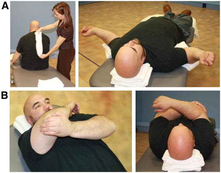
Stretching exercises for the anterior chest (A) and posterior shoulder joint structures (B). Participants lay on a towel roll along their thoracic spine (A) to help stretch their anterior chest. The first image shows where on the participant's anatomy the towel roll is placed. The participant is lying on the towel roll in the second image.
Hands-on instruction of these exercises occurred during the initial laboratory session. During this session, photographs were taken of each participant in order to provide a handout that included participant-specific photographs performing the prescribed exercises (supplemental fig S1, available online only at http://www.archives-pmr.org/). Participants were instructed to exercise free of symptoms. Temporary discomfort in the location of the targeted muscle group was allowed. After initial exercise instruction, regular videoconferencing sessions using the participants' personal computers and free softwareb provided supervision to allow for technique correction and advancement of exercises. Inexpensive web cameras were given to participants who did not have a camera. Weekly videoconferencing sessions were offered to all participants throughout the intervention with the option to videoconference more frequently for the first 2 weeks. When the participants demonstrated good exercise technique independently, a session frequency of every other week was offered.
Outcome measures
To quantify pain and function, participants completed 3 questionnaires specific to the shoulder at baseline, 12-week, and 24+ week time points. The Wheelchair User's Shoulder Pain Index (WUSPI) requires that participants rate their shoulder pain intensity during different activities on visual analog scales anchored by “no pain” and “worst pain ever experienced.”26 Scores range from 0 (no pain) to 150 (severe pain in all activities). The WUSPI has been shown to be both reliable and valid for this study population.27 The Shoulder Rating Questionnaire (SRQ) and the Disabilities of the Arm, Shoulder, and Hand (DASH) Index are assessments of upper extremity function not specific to manual wheelchair users.28,29 Scores for the SRQ range from 17 to 100 with higher scores indicating the least amount of pain and dysfunction.29 Scores for the DASH range from 0 to 100 with low scores indicating the least amount of pain and dysfunction.28 Good reliability and validity of the SRQ and DASH have been reported in the able-bodied population.29,30
Isometric strength measurement was performed using a quantitative muscle testing systemc consisting of load cells and straps. A summary of the positions and stabilizing forces used is presented in table 1. Isometric maximum volitional contractions (MVCs) were held for 3 to 5 seconds. Two trials of each isometric test were performed to ensure that intertrial repeatability was within 10%. A third trial was collected if necessary. The highest MVC (peak force) was used for statistical calculations. Static fatigue testing required participants to hold an MVC of their lower trapezius until task failure (no measurable force output). If the participant did not attain task failure, the task was stopped at 60 seconds. No visual feedback was given during this time; however, continuous and consistent verbal encouragement was given. Muscle impulse was determined by calculating the area under the isometric strength kilogram-force-time curve (kg-f · s) using the trapezoidal numerical integration method. Outcome measures of pain, function, and strength were collected by the same licensed physical therapist who prescribed and supervised the exercise program.
Table 1. Isometric strength testing positions and stabilizations.
| Muscle Group | Joint Positions | Testing Position | Location of Resistance | Stabilization | Image of Test |
|---|---|---|---|---|---|
| Glenohumeral | |||||
| External rotation | SJ: ∼20° abduction, neutral rotation EJ: 90° flexion | Supine | Proximal to wrist | S: Pelvic strap (not pictured) M: Lateral, contralateral ribs |

|
| Internal rotation | SJ: ∼20° abduction, neutral rotation EJ: 90° flexion | Supine | Proximal to wrist | S: Pelvic strap (not pictured) M: Lateral, ipsilateral ribs |

|
| Scapular plane abduction | SJ: ∼80° elevation in the scapular plane | Supine | Proximal to elbow | S: Pelvic strap (not pictured) M: Inferior, bilateral anterior superior iliac spines |

|
| Scapulothoracic | |||||
| Serratus anterior | SJ: 90° flexion EJ: >100° flexion (determined by participant comfort) | Supine | Distal to elbow | S: Pelvic strap M: Anterior ipsilateral ribs |

|
| Scapular retraction | SJ: 90° abduction, neutral rotation EJ: 90° flexion | Prone | Posterior deltoid, on posterior lateral scapula if possible | S: Pelvic strap M: Posterior contralateral ribs |

|
| Lower trapezius* | SJ: ∼120° elevation in the scapular plane, neutral rotation EJ: Extended | Prone | Posterior deltoid, on posterior lateral scapula if possible | S: Pelvic strap M: Posterior contralateral ribs |

|
Abbreviations: EJ, elbow joint; M, manual; S, strap; SJ, shoulder joint.
The same position was used for both the isometric strength and isometric static fatigue tests of the lower trapezius.
Statistical analysis
Since questionnaire data for pain and function were not normally distributed and the sample size was small, data were compared between the 3 time points using the Friedman signed-rank test with a significance level of P<.05 (SAS9.2d). When a significant main effect was detected, post hoc, Bonferroni-adjusted Wilcoxon signed-rank tests were performed. A parametric post hoc power analysis was performed based on an effect-size approach and revealed that with 14 participants, there was 80% power to detect a difference of 17.0, 6.4, and 10.0 on the WUSPI, SRQ, and DASH questionnaires, respectively. The effect size was determined for the nonsignificant primary outcome measures. Strength data for the 2 time points (baseline, postintervention) were compared using a Wilcoxon signed-rank test (JMP9.0.0d).
Results
Our participants ranged in age from 25 to 64 years, with levels of spinal cord injury from C6-7 to L2 (table 2). Three of our 16 participants were women, and 10 were athletes practicing or competing in organized sports (such as wheelchair basketball) at least once a week.
Table 2. Baseline demographic characteristics (N = 16).
| Characteristics | Values |
|---|---|
| Sex: men | 13 (81) |
| Age (y) | 41 (25–64) |
| Weight (kg) | |
| Men | 82.1 (59.3–104.5) |
| Women | 85.9 (45.1–115.0) |
| Level of injury | |
| C6-7 | 1 |
| T2-7 | 5 |
| T8 and below | 9 |
| Postpolio | 1 |
| Time in wheelchair (y) | 16 (3.7–41.8) |
| Duration of shoulder pain (y) | 9 (0.8–22) |
Note. Values are n (%), mean (range), or n.
Two participants did not complete the intervention because of scheduling conflicts. Of those who completed the intervention, 2 participants did not come back to the laboratory for strength testing of their shoulder (illness and lack of transportation); however, they did return pain and function surveys by mail. At the time of the 24+ week follow-up via mail, 2 of 14 who completed the intervention did not return pain and function surveys, and were considered lost to follow-up. Although the completion date of the intended 24-week follow-up survey was not documented, 11 surveys were received between 26 and 30 weeks after the baseline data collection. One participant's survey was lost in the mail, and therefore a second was completed and received at 50 weeks.
Five participants reported exercise program adherence of >75%, 3 reported 75% to 50% adherence, 6 reported 49% to 25% adherence, and no participants had adherence of <25%. The average number of telerehabilitation sessions during the intervention period was .70 sessions per week (range, .50–1.02).
There was a significant main effect for pain and function between the 3 time points based on the Friedman signed-ranked test; WUSPI (χ22 = 5.10, P = .014); DASH Index (χ22 = 5.41, P = .012); and SRQ (χ22 = 23.71, P = <.001). Post hoc Bonferroni-adjusted Wilcoxon signed-rank test analyses revealed statistically significant differences between baseline and 12-week time points for the WUSPI and SRQ, and statistically significant differences between the baseline and 24+ week time points for the WUSPI, DASH Index, and SRQ. No differences in pain or function were found between 12-week and 24+ week time points. After the exercise intervention, the median WUSPI score was reduced from 22.8 (range, 1.2–78.9) to 12.5 (range, 0.0–83.8) (P = .007), and the median SRQ score increased from 81.9 (range, 47.7–95.6) to 90.2 (range, 54.7–99.9) (P<.0001), indicating a reduction in pain and improvement in function. These changes were maintained at the time of follow-up (table 3). Changes in the median DASH Index score were not enough to be statistically significant after the intervention (30.9 [range, 0.8–52.5] to 12.1 [range, 0.8–47.5]) (P = .08); however, 24+ week median DASH Index scores were statistically improved from baseline scores, improving to 7.5 (range, 0.0–29.2) (P = .003). As a result of the nonsignificant finding between baseline and 12-week time points for the DASH Index score, an effect size was calculated (effect size r = .603, d = 1.513). These effect size values indicate that the DASH Index difference between baseline and 12-week time points is large and that the lack of statistical significance was affected by the small sample size.
Table 3. Pain and function scores at baseline, postexercise intervention, and follow-up time points.
| Outcome Measures | Time Point | Freidman Signed-Rank Test P | ||
|---|---|---|---|---|
|
| ||||
| Baseline | Post Intervention (12wk) | Follow-Up (+24wk) | ||
| WUSPI | n = 14 | n = 14 | n = 12* | |
| Median (range) | 22.8 (1.2–78.9) | 12.5† (0.0–83.8) | 10.9† (0.0–31.8) | .014 |
| Median of participants with missing 24-wk data (n = 2) | 39.0 | 28.9 | - | |
| SRQ | n = 14 | n = 14 | n = 11*,‡ | |
| Median (range) | 81.9 (47.7–95.6) | 90.2† (54.7–99.9) | 91.2† (63.6–99.6) | <.001 |
| Median of participants with missing 24-wk data (n = 3) | 72.8 | 73.9 | - | |
| DASH Index | n = 14 | n = 14 | n = 11*,‡ | |
| Median (range) | 30.9 (0.8–52.5) | 12.1 (0.8–47.5) | 7.5† (0.0–29.2) | .012 |
| Median of participants with missing 24-wk data (n = 3) | 35.0 | 25.0 | - | |
Two participants lost to follow-up.
Significantly different than baseline (post hoc Bonferroni-adjusted Wilcoxon signed-rank test; P<.05).
Incomplete completion of survey prevented scoring of 1 participant.
Isometric strength measurements of the serratus anterior and scapular retractors increased after the exercise intervention ([t = 2.42, P = .04] and [t = 4.67, P = .003], respectively). Median serratus anterior measurements improved from 36.4kg (range, 21.4–60.3kg) to 48.0kg (range, 18.5–71.9kg), and scapular retractors improved from 24.6kg (range, 14.5–64.3kg) to 37.4kg (range, 22.2–80.4kg). Muscle impulse produced by the lower trapezius during a fatigue task also improved, from 17.2kg-f • s (range, 3.1–27.9kg-f • s) to 19.1kg-f • s (range, 5.5–38.2kg-f • s) (t = 2.2, P = .02). No differences were measured in isometric strength for the lower trapezius, glenohumeral rotators, and abductors between the baseline and 12-week time points (table 4).
Table 4. Peak isometric strength (kg) and muscle impulse (during static fatigue task [kg-f·s]) at baseline and postexercise intervention time points.
| Measures | n | Time Point | Wilcoxon Signed-Rank Test P | |
|---|---|---|---|---|
|
| ||||
| Baseline | Postintervention (12wks) | |||
| Isometric strength (kg) | ||||
| Glenohumeral | ||||
| External rotation | 12* | 14.2 (5.9–28.7) | 14.5 (9.1–33.7) | .18 |
| Internal rotation | 12* | 24.3 (13.3–43.7) | 28.3 (17.0–47.2) | .13 |
| Scapular plane abduction | 12* | 26.6 (12.3–39.6) | 27.4 (13.5–39.7) | .68 |
| Scapulothoracic | ||||
| Serratus anterior | 12* | 36.4 (21.4–60.3) | 48.0 (18.5–71.9) | .04† |
| Scapular retractors | 11*,‡ | 24.6 (14.5–64.3) | 37.4 (22.2–80.4) | .003† |
| Lower trapezius | 11*,‡ | 24.0 (9.3–45.3) | 27.5 (10.7–52.4) | .17 |
| Static fatigue (kg-f • s) | ||||
| Lower trapezius | 11*,‡ | 17.2 (3.1–27.9) | 19.1 (5.5–38.2) | .02† |
Note. Values are median (range) or as otherwise indicated.
Two participants were unable to travel to laboratory for postintervention strength testing.
Significant change; P<.05.
One participant did not tolerate the prone position for testing.
Discussion
The purpose of this study was to test the effectiveness of a home exercise program, with high-repetition dosing and telerehabilitation, for manual wheelchair users with chronic shoulder pain. Consistent with our hypotheses, participants reported a statistically significant reduction in shoulder pain and improvement in shoulder function after the intervention. This improvement was maintained at the time of follow-up. Additionally, there were statistically significant increases in isometric strength of the serratus anterior and scapular retractors, and in the muscle impulse produced by the lower trapezius during a static fatigue test.
A few studies1,17,19 have also documented a reduction in chronic shoulder pain after a home-based exercise intervention in experienced manual wheelchair users. These studies had baseline WUSPI scores ranging from 23 to 51, and postintervention scores ranging from 14 to 20. These consistent results from differing investigations provide evidence that in this population of long-time wheelchair users, complete symptom elimination is not typically attained. The apparent floor effect in pain reduction may be explained by the presence of underlying soft tissue pathology in the shoulder. As part of a larger study, 10 of our participants received magnetic resonance imaging of the shoulder without contrast, and 7 of them had positive findings of partial or complete tendon tears. The remaining 3 showed signs of tendinopathy. This tendon damage as seen on magnetic resonance imaging may help explain why complete symptom relief was not achieved.
Our program targeted the serratus anterior, scapular retractors, and glenohumeral external rotators. However, a statistically significant increase in strength of the external rotators was not found. There are a few possible explanations for this finding. We chose high exercise doses based on the study by Osteras,21 who found high-repetition doses reduced pain more than lower doses. This loading scheme (lighter resistance, more repetitions) does not favor optimization of hypertrophy gain.31 It is possible that the endurance of this muscle group, which we did not measure, did improve despite the lack of increased MVC force production. Because static fatigue tests are physically demanding, we chose to only measure the endurance of the lower trapezius. In addition, 10 of our 16 participants were athletes. Their baseline strength scores in some muscle groups may have been elevated as a result of their training, creating a ceiling effect and leaving less room for improvement from our intervention.
Strength testing of the scapular muscles is inherently difficult because of the lack of a static axis at the scapulothoracic junction and small segment anatomy on which to place the resistive force. The average changes in strength and impulse values that were observed in this study may be smaller in magnitude than the minimal detectible change (MDC) of the respective measures. Repeatability testing of our methods resulted in MDCs of 12.1kg, 8.6kg, and 4.4kg-f • s for the serratus anterior, scapular retractors, and muscle impulse of the lower trapezius fatigue tests, respectively. When considering these tests in a clinical setting, change beyond the MDC is necessary to determine that an individual has improved beyond test-retest variability of the measure. Strength improvements in 7, 5, and 6 of our participants were equal to or exceeded the MDC of the serratus anterior test, scapular retractor test, and lower trapezius fatigue test, respectively.
Study limitations
Among the limitations to be considered when interpreting these findings is our small sample size. There were 14 participants who completed the exercise intervention after 2 dropped out of the study. We did not have a control group, and therefore we did not compare our high-dose protocol with a low-dose comparison group, nor did we compare supervision via videoconferencing with supervision via an office visit. Therefore, we cannot conclude that our program was more or less effective than previously described exercise interventions. As a result of collecting a convenience sample, 10 of our participants were athletes, and our participants reported low to moderate levels of pain at baseline (median WUSPI score, 22.8). Therefore, the results may vary in individuals with higher levels of pain and dysfunction or in the nonathletic population. Our study may have been biased since the same physical therapist who performed the telerehabilitation also collected the baseline and postintervention data. However, the tester did not have access to the baseline data at the time of retesting, and Mulroy et al19 also found similar improvements in pain, function, and strength with methodology that prevented experimenter-effect bias.
Conclusions
After a conservative, inexpensive, and accessible exercise intervention, shoulder pain was reduced, even in individuals with longstanding symptoms. Our findings indicate that a high-dose shoulder strengthening program of the scapular stabilizers and rotator cuff muscles using telerehabilitation for supervision may be a viable option for the treatment of shoulder pain in manual wheelchair users. Additional work is needed to determine its effectiveness compared with other interventions, as well as the potential for earlier intervention to prevent the development of shoulder pain and tissue pathology.
Supplementary Material
Supplemental Fig 1 (A) Low Row Exercise (Strengthens Lower Trap)
(B) External Rotation Exercise
(C) Shoulder Punch Plus Exercise (for your serratus anterior)
Acknowledgments
We thank Wendy J. Hurd, PhD, PT, Joan Kopacz, MSPT, and Krista Coleman Wood, PhD, PT, for their contribution in the protocol development and for their clinical expertise.
Supported by the Paralyzed Veterans of America and by the National Center for Advancing Translational Science (Clinical and Translational Science Award grant nos. UL1 RR024150, TL1 TR000137). The contents of the article are solely the responsibility of the authors and do not necessarily represent the official views of the National Institutes of Health or the Paralyzed Veterans of America.
List of abbreviations
- DASH
Disabilities of the Arm, Shoulder, and Hand
- MDC
minimal detectible change
- MVC
maximum volitional contraction
- SAI
subacromial impingement
- SRQ
Shoulder Rating Questionnaire
- WUSPI
Wheelchair User's Shoulder Pain Index
Footnotes
The Hygenic Corp, 1245 Home Ave, Akron, OH 44310.
Skype Communications SARL, Luxembourg.
Aeverl Medical LLC, PO Box 170, Gainesville, GA 30503.
SAS Institute Inc, 100 SAS Campus Dr, Cary, NC 27513.
Presented to the American Physical Therapy Association, 2014, and in part to the American Society of Biomechanics, 2013.
Disclosures: none.
References
- 1.Curtis KA, Tyner TM, Zachary L, et al. Effect of a standard exercise protocol on shoulder pain in long-term wheelchair users. Spinal Cord. 1999;37:421–9. doi: 10.1038/sj.sc.3100860. [DOI] [PubMed] [Google Scholar]
- 2.Jain NB, Higgins LD, Katz JN, Garshick E. Association of shoulder pain with the use of mobility devices in persons with chronic spinal cord injury. PM R. 2010;2:896–900. doi: 10.1016/j.pmrj.2010.05.004. [DOI] [PMC free article] [PubMed] [Google Scholar]
- 3.Gellman H, Sie I, Waters RL. Late complications of the weight-bearing upper extremity in the paraplegic patient. Clin Orthop Relat Res. 1988;233:132–5. [PubMed] [Google Scholar]
- 4.Brose SW, Boninger ML, Fullerton B, et al. Shoulder ultrasound abnormalities, physical examination findings, and pain in manual wheelchair users with spinal cord injury. Arch Phys Med Rehabil. 2008;89:2086–93. doi: 10.1016/j.apmr.2008.05.015. [DOI] [PubMed] [Google Scholar]
- 5.Alm M, Saraste H, Norrbrink C. Shoulder pain in persons with thoracic spinal cord injury: prevalence and characteristics. J Rehabil Med. 2008;40:277–83. doi: 10.2340/16501977-0173. [DOI] [PubMed] [Google Scholar]
- 6.Akbar M, Balean G, Brunner M, et al. Prevalence of rotator cuff tear in paraplegic patients compared with controls. J Bone Joint Surg Am. 2010;92:23–30. doi: 10.2106/JBJS.H.01373. [DOI] [PubMed] [Google Scholar]
- 7.Bayley JC, Cochran TP, Sledge CB. The weight-bearing shoulder. The impingement syndrome in paraplegics. J Bone Joint Surg Am. 1987;69:676–8. [PubMed] [Google Scholar]
- 8.Brox JI, Gjengedal E, Uppheim G, et al. Arthroscopic surgery versus supervised exercises in patients with rotator cuff disease (stage II impingement syndrome): a prospective, randomized, controlled study in 125 patients with a 2 1/2-year follow-up. J Shoulder Elbow Surg. 1999;8:102–11. doi: 10.1016/s1058-2746(99)90001-0. [DOI] [PubMed] [Google Scholar]
- 9.Holmgren T, Bjornsson Hallgren H, Oberg B, Adolfsson L, Johansson K. Effect of specific exercise strategy on need for surgery in patients with subacromial impingement syndrome: randomised controlled study. BMJ. 2012;344:e787. doi: 10.1136/bmj.e787. [DOI] [PMC free article] [PubMed] [Google Scholar]
- 10.Kuhn JE. Exercise in the treatment of rotator cuff impingement: a systematic review and a synthesized evidence-based rehabilitation protocol. J Shoulder Elbow Surg. 2009;18:138–60. doi: 10.1016/j.jse.2008.06.004. [DOI] [PubMed] [Google Scholar]
- 11.Littlewood C, Ashton J, Chance-Larsen K, May S, Sturrock B. Exercise for rotator cuff tendinopathy: a systematic review. Physiotherapy. 2012;98:101–9. doi: 10.1016/j.physio.2011.08.002. [DOI] [PubMed] [Google Scholar]
- 12.Lombardi I, Jr, Magri AG, Fleury AM, Da Silva AC, Natour J. Progressive resistance training in patients with shoulder impingement syndrome: a randomized controlled trial. Arthritis Rheum. 2008;59:615–22. doi: 10.1002/art.23576. [DOI] [PubMed] [Google Scholar]
- 13.Ludewig PM, Borstad JD. Effects of a home exercise programme on shoulder pain and functional status in construction workers. Occup Environ Med. 2003;60:841–9. doi: 10.1136/oem.60.11.841. [DOI] [PMC free article] [PubMed] [Google Scholar]
- 14.McClure PW, Bialker J, Neff N, Williams G, Karduna A. Shoulder function and 3-dimensional kinematics in people with shoulder impingement syndrome before and after a 6-week exercise program. Phys Ther. 2004;84:832–48. [PubMed] [Google Scholar]
- 15.Michener LA, Walsworth MK, Burnet EN. Effectiveness of rehabilitation for patients with subacromial impingement syndrome: a systematic review. J Hand Ther. 2004;17:152–64. doi: 10.1197/j.jht.2004.02.004. [DOI] [PubMed] [Google Scholar]
- 16.Morrison DS, Frogameni AD, Woodworth P. Non-operative treatment of subacromial impingement syndrome. J Bone Joint Surg Am. 1997;79:732–7. doi: 10.2106/00004623-199705000-00013. [DOI] [PubMed] [Google Scholar]
- 17.Nawoczenski DA, Ritter-Soronen JM, Wilson CM, Howe BA, Ludewig PM. Clinical trial of exercise for shoulder pain in chronic spinal injury. Phys Ther. 2006;86:1604–18. doi: 10.2522/ptj.20060001. [DOI] [PubMed] [Google Scholar]
- 18.Nash MS, van de Ven I, van Elk N, Johnson BM. Effects of circuit resistance training on fitness attributes and upper-extremity pain in middle-aged men with paraplegia. Arch Phys Med Rehabil. 2007;88:70–5. doi: 10.1016/j.apmr.2006.10.003. [DOI] [PubMed] [Google Scholar]
- 19.Mulroy SJ, Thompson L, Kemp B, et al. Strengthening and optimal movements for painful shoulders (STOMPS) in chronic spinal cord injury: a randomized controlled trial. Phys Ther. 2011;91:305–24. doi: 10.2522/ptj.20100182. [DOI] [PubMed] [Google Scholar]
- 20.Walther M, Werner A, Stahlschmidt T, Woelfel R, Gohlke F. The subacromial impingement syndrome of the shoulder treated by conventional physiotherapy, self-training, and a shoulder brace: results of a prospective, randomized study. J Shoulder Elbow Surg. 2004;13:417–23. doi: 10.1016/j.jse.2004.02.002. [DOI] [PubMed] [Google Scholar]
- 21.Osteras H, Torstensen TA, Osteras B. High-dosage medical exercise therapy in patients with long-term subacromial shoulder pain: a randomized controlled trial. Physiother Res Int. 2010;15:232–42. doi: 10.1002/pri.468. [DOI] [PubMed] [Google Scholar]
- 22.Senbursa G, Baltaci G, Atay A. Comparison of conservative treatment with and without manual physical therapy for patients with shoulder impingement syndrome: a prospective, randomized clinical trial. Knee Surg Sports Traumatol Arthrosc. 2007;15:915–21. doi: 10.1007/s00167-007-0288-x. [DOI] [PubMed] [Google Scholar]
- 23.Hawkins RJ, Kennedy JC. Impingement syndrome in athletes. Am J Sports Med. 1980;8:151–8. doi: 10.1177/036354658000800302. [DOI] [PubMed] [Google Scholar]
- 24.Jobe FW, Moynes DR. Delineation of diagnostic criteria and a rehabilitation program for rotator cuff injuries. Am J Sports Med. 1982;10:336–9. doi: 10.1177/036354658201000602. [DOI] [PubMed] [Google Scholar]
- 25.Neer CS., II Impingement lesions. Clin Orthop Relat Res. 1983;(173):70–7. [PubMed] [Google Scholar]
- 26.Curtis KA, Roach KE, Applegate EB, et al. Development of the Wheelchair User's Shoulder Pain Index (WUSPI) Paraplegia. 1995;33:290–3. doi: 10.1038/sc.1995.65. [DOI] [PubMed] [Google Scholar]
- 27.Curtis KA, Roach KE, Applegate EB, et al. Reliability and validity of the Wheelchair User's Shoulder Pain Index (WUSPI) Paraplegia. 1995;33:595–601. doi: 10.1038/sc.1995.126. [DOI] [PubMed] [Google Scholar]
- 28.Hudak PL, Amadio PC, Bombardier C. Development of an upper extremity outcome measure: the DASH (Disabilities of the Arm, Shoulder and Hand) [corrected]. The Upper Extremity Collaborative Group (UECG) Am J Ind Med. 1996;29:602–8. doi: 10.1002/(SICI)1097-0274(199606)29:6<602::AID-AJIM4>3.0.CO;2-L. [DOI] [PubMed] [Google Scholar]
- 29.L'Insalata JC, Warren RF, Cohen SB, Altchek DW, Peterson MG. A self-administered questionnaire for assessment of symptoms and function of the shoulder. J Bone Joint Surg Am. 1997;79:738–48. [PubMed] [Google Scholar]
- 30.Beaton DE, Katz JN, Fossel AH, Wright JG, Tarasuk V, Bombardier C. Measuring the whole or the parts? Validity, reliability, and responsiveness of the Disabilities of the Arm, Shoulder and Hand outcome measure in different regions of the upper extremity. J Hand Ther. 2001;14:128–46. [PubMed] [Google Scholar]
- 31.Garber CE, Blissmer B, Deschenes MR, et al. American College of Sports Medicine position stand. Quantity and quality of exercise for developing and maintaining cardiorespiratory, musculoskeletal, and neuromotor fitness in apparently healthy adults: guidance for prescribing exercise. Med Sci Sports Exerc. 2011;43:1334–59. doi: 10.1249/MSS.0b013e318213fefb. [DOI] [PubMed] [Google Scholar]
Associated Data
This section collects any data citations, data availability statements, or supplementary materials included in this article.
Supplementary Materials
Supplemental Fig 1 (A) Low Row Exercise (Strengthens Lower Trap)
(B) External Rotation Exercise
(C) Shoulder Punch Plus Exercise (for your serratus anterior)