

Abstract
Objectives
The purpose of this study was to determine if surgeons could reliably predict if patients with tibia fractures treated with intramedullary nails will proceed to nonunion based on their clinical scenario and radiographs at three months.
Design
Blinded randomized questionnaire based on a retrospective cohort
Setting
University level one trauma center.
Patients/Participants
Fifty-six patients who underwent intramedullary fixation for tibia fractures with incomplete healing at three months.
Methods
A questionnaire was applied to 56 consecutive patients treated between 2005–2009 with intramedullary fixation for tibia fractures who had incomplete healing at three months. Each case was developed into a vignette that included the three-month radiographs and detailed clinical histories. The questionnaire was distributed to three fellowship-trained trauma surgeons, who were asked to predict if the fracture would go onto nonunion.
Main outcome measurement
Diagnostic accuracy of predicting nonunion in patients with incomplete healing of their tibia fracture at 3 months.
Results
The combined overall diagnostic accuracy of all three surgeons was 74%. Sensitivity and specificity were 62% and 77% respectively. Radiographic features and injury mechanism were the most commonly cited clinical information used to predict fracture healing. The average positive predictive value was 73%. In 9 patients with diabetes, the diagnostic accuracy was 88%.
Conclusion
Clinical judgment at three months allows for correct prediction of eventual nonunion development in a majority of patients. We suggest that analysis of the entire clinical picture be used to predict fracture healing at 3 months. A protocol of waiting for six months before reoperation in all patients treated with intramedullary nailing for tibia fractures may subject patients to prolonged disability and discomfort.
Keywords: Tibial Nonunion, intramedullary fixation, tibia fracture, Timing of Treatment, diagnostic accuracy
INTRODUCTION
Tibia fracture is the most common long bone fracture [1]. They occur in patients with a variety of demographics and by a variety of mechanisms [2]. Among tibia fractures treated with intramedullary fixation, nonunions can occur in up to 14 percent of patients [3–6]. Although there are some clear indications for early intervention for ununited tibia fractures, such as infection, malalignment, and fixation failure, the optimal timing for treatment of ununited tibia fractures without these complications has not been clearly defined. A recent multi-center prospective study to evaluate reamed and unreamed intramedullary nails in patients with tibial fractures (SPRINT [6]) suggested that delaying any surgical intervention for at least six months post-operatively may decrease the need for reoperation. However, some authors have suggested that nonunion repair be performed as early as three months. [4, 7, 8, 9]. The purpose of this study was to determine if it was possible to reliably predict if a patient would proceed to nonunion based on standard clinical and radiographic features at three months after fracture. A secondary purpose was to determine patient factors leading surgeons to predict nonunion. If surgeons are able to reliably predict, at three months, that a patient will progress to tibial nonunion at six months, prompt treatment can proceed, minimizing patient morbidity, discomfort, and debilitation. Our hypothesis was that clinical judgment, based on clinical data and radiographs at three months, allows for early reliable prediction of eventual tibial nonunion development.
PATIENTS AND METHODS
Patients
The research was conducted at a single level one trauma center after approval from the human subjects committee and the internal review board. Four hundred and sixty-nine patients who underwent intramedullary fixation for tibia shaft fractures (OTA type 42) between 2005–2009 were identified from hospital and department databases. Excluded were pediatric patients with open physes, and adult patients with: nail fracture; segmental bone loss great than 1cm; varus or valgus malalignment greater than 15 degrees; and concomitant tibial plateau (OTA type 41) or pilon (OTA type 43) fractures. Eighty-three patients were excluded based on these criteria and one hundred and twenty-eight patients had incomplete data or were lost to follow-up. Leaving 258 patients that met initial inclusion criteria.
Definitions of Union and Non-union
Nonunion was defined as a combination of radiographic lack of bridging callus on four cortices, clinical tenderness at the fracture site on palpation, and pain with full weightbearing. The fracture was considered healed if there was no tenderness at the fracture site, no pain with full weightbearing, and the radiographs demonstrated the presence of bridging callus on three or more cortices. This “gold standard” was used based on previous reported studies on tibial nonunions [8,10,11]. Of the 258 patients who were not excluded, 202 were clinically healed at 3 months using the definition stated above. The patients who were thought to be completely healed at 3 months had this diagnosis confirmed with follow-up at 6 months. Fifty-six patients had incomplete healing of their tibia fracture at 3 months and were subject to study. The average age of the fifty-six patients was thirty-four years (range 18 – 75). There were fifty-two males and four females [Table 1]. All patients were treated with a reamed intramedullary tibial nail.
Table I.
Demographics of the study population
| All patients | Incompletely healed at 3 months | |
|---|---|---|
| Sample size | 258 | 56 |
| Age | 32 ± 11 | 34 ± 14 |
| Gender | ||
| Male | 234 | 52 |
| Female | 24 | 4 |
| Mechanism of Injury | ||
| Motor vehicle accident | 54 | 11 |
| Motorcycle accident | 48 | 16 |
| Fall from height | 37 | 8 |
| Pedistrian struck | 33 | 9 |
| Same level fall | 45 | 6 |
| Assault | 12 | 2 |
| Gun shot | 24 | 4 |
| Other | 5 | 0 |
| Open injury | 112 | 37 |
| Grade 1 | 39 | 6 |
| Grade 2 | 44 | 17 |
| Grade 3 | 29 | 14 |
| Closed injury | 146 | 21 |
| Biologics used at index surgery | 66 | 15 |
| Diabetic | 74 | 20 |
| Smoker | 65 | 17 |
Using previously stated nonunion criteria, an independent evaluator identified twenty-nine patients who developed a nonunion at six months postoperatively and twenty-seven patients who achieved full union by six months. This stratification was used to define the final outcome for the 56 patients studied. All patients with nonunions underwent surgical repair.
Of the twenty-nine patients who developed nonunion, five patients had positive cultures at the time of nonunion repair but had no clinical signs of infection at the three month visit.
Clinical Vignette
A clinical vignette was constructed for each patient based on their clinical and radiographic findings from the three-month time point. These vignettes were then arranged in random order and compiled into an electronic questionnaire (Microsoft PowerPoint 2007, Microsoft Corporation, Redmond, WA). The vignettes presented radiographic images and clinical data including age, gender, weight, mechanism of injury, Gustilo classification if the fracture was open, medical history, tobacco use, clinical exam findings and if any biologics were used at the time of their initial surgery [Figure 1]. The vignettes were blinded by removing all patient health information identifiers and were distributed to three fellowship-trained trauma surgeons who were asked to predict if the fracture would go onto nonunion at 6 months, and the reasoning for their judgment. For their reasoning, the respondents were given options to choose from which included patient factors, injury factors, surgical or technical factors, and radiographic features. The respondents were not privy to how many vignettes were in each group, union versus nonunion. The range for years in practice among the three surgeons was from one year to fifteen years. Of the 56 patients examined in the vignette, the primary surgery was performed by one of the three surgeons in 24 patients (43%).
Figure 1.


A sample vignette with responses that was used in this study.
Statistical Analysis
Statistical analysis included calculation of the diagnostic accuracy, sensitivity and specificity, and positive and negative predictive values. Further statistical testing included using Fischer exact test and the Chi square test for comparing proportional differences. Statistical analysis was performed using Microsoft Excel (Microsoft Corporation, Redmond, Washington, USA) and SPSS (IBM Corporation, Armonk, New York, USA).
RESULTS
Diagnostic Accuracy
The combined overall diagnostic accuracy of all three surgeons for correctly predicting nonunion was 74% (Surgeon A: 73%, Surgeon B: 73%, Surgeon C: 75%). Sensitivity and specificity for prediction of nonunion were 62% and 77% respectively. Positive (PPV) and negative predictive values (NPV) of nonunion prediction were 73% and 69% respectively [Table 2].
Table 2.
Overall performance of surgeons in nonunion prediction
| Surgeon A | Surgeon B | Surgeon C | Average | |
|---|---|---|---|---|
| Sensitivity | 0.48 | 0.63 | 0.74 | 0.62 |
| Specificity | 0.90 | 0.72 | 0.69 | 0.77 |
| Positive predictive value | 0.81 | 0.68 | 0.69 | 0.73 |
| Negative predictive value | 0.65 | 0.68 | 0.74 | 0.69 |
| Overall accuracy | 0.73 | 0.73 | 0.75 | 0.74 |
When considering the 202 patients that were completely healed at three months with the fifty-six patients that were incompletely healed, the combined overall diagnostic accuracy for identifying or predicting union rises to 94% (243/258).
Callus Formation
Lack of callus formation (70%) and mechanism of injury (73%) were most commonly cited as factors used to predict nonunion. There were 39 patients in which radiographic features were used primarily. Of six patients with no callus formation, the surgeons predicted nonunion 89% of the time and were correct 89% of the time. Of the 10 patients with callus formation on one cortex, the surgeons predicted nonunion 57% of the time and were correct 63% of the time. Of 11 patients with callus formation in two cortices, the surgeons predicted nonunion 42% of the time and were correct 70% of the time. Of 29 patients with callus formation in three cortices, the surgeons predicted nonunion 26% of the time and were correct 75% of the time. The diagnostic accuracy was significantly higher in those patients with no callus formation (p<0.001). The amount of callus formation also had a negative correlation with probability of surgeons predicting nonunion (r=−0.98, p<0.001) [Figure 2].
Figure 2.

Graph depicting the correlation between amount of callus formation, diagnostic accuracy and likelihood of surgeons predicting nonunion.
Mechanism of Injury
There were 41 patients in which mechanism of injury was used as the primary decision factor. Of 36 patients with a high energy mechanism of injury (fall from height, high speed MVC), the surgeons predicted nonunion 42% of the time and were correct 86% of the time. Of 20 patients with a low energy mechanism of injury (fall from standing height, low speed MVC), the surgeons predicted nonunion 25% of the time and were correct 60% of the time. The rate of correct predictions was significantly higher in those patients with higher energy mechanism of injury (p=0.03).
Premorbid Conditions
Of the nine patients with diabetes, the diagnostic accuracy was 89% compared to 70% in those without diabetes (p=0.05). Of the 30 patients who used tobacco, the diagnostic accuracy was 76% compared to 70% in those who did not use tobacco (p=0.3). In 34 patients with open fractures, the diagnostic accuracy was 67% compared to closed injuries 80% (p=0.001).
Agreement between surgeons
Overall diagnostic agreement of surgeons was fair with a kappa value of 0.38. The reason each surgeon gave for their diagnosis correlated well with a kappa value of 0.71.
Combined Predictions of Three Surgeons
In ten patients, all three surgeons correctly predicted nonunion. All ten were males under 60 years old and had no callus formation or signs of radiographic healing at 3 months. 8/10 patients had high energy open fractures, 8/10 patients used tobacco, 4/10 patients had diabetes, and none had biologic adjuvants used at initial surgery.
In 18 patients for whom all three surgeons correctly predicted union, 17/18 patients were under 60 years old, 16/18 patients had callus formation or signs of radiographic healing at months, 8/18 patients used tobacco, 4/18 patients were female, 4/18 patients had biologic adjuvants used at initial surgery, 4/18 patients had high energy open fractures, and only 2/18 patients had diabetes.
There were four patients in whom all three surgeons predicted union when, in fact, the fracture went onto nonunion. None of these patients had callus formation at three months. Of these four patients, none had diabetes, three had open fractures, and three used tobacco. In contrast, there was only one patient for whom all three surgeons predicted nonunion when in fact the fracture was fully healed at six months. He was a 40 year old obese male smoker who sustained a Type IIIA open tibia fracture after being struck by a truck. There was some evidence of primary healing on radiographs at three months [Figure 3]. At the sixth month post-operative visit, he did not have any pain at the fracture site and the radiographs reveal a healed fracture.
Figure 3.


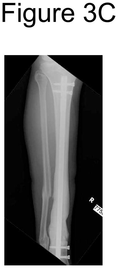

3 month post-operative (A) AP and (B) lateral radiograph of a 40 year old obese male smoker who sustained a Type IIIA open tibia fracture after being struck by a truck. There was some callus formation at this point. 6 month post-operative (C) AP and (D) lateral radiographs shows callus formation and healed fracture with no clinical pain at the fracture site.
DISCUSSION
A lack of consensus exists for timing of intervention for ununited tibial fractures. Our study sought to examine whether clinical judgment based on information available at three months could predict eventual nonunion, in a subset of patients with ununited tibial fractures after intramedullary nailing. The results showed that clinical judgment at three months allowed for correct prediction of eventual nonunion in a substantial percentage (PPV 73%) of patients.
Sensitivity and specificity for prediction of nonunion were 62% and 77%, respectively. To understand this in real-world terms, in 100 patients with nonunion, clinical judgment will correctly predict nonunion in 62 of them. In 100 patients with ultimate union, clinical judgment will correctly predict this outcome in 77. Positive and negative predictive values of nonunion prediction were 73% and 69% respectively. Thus, in 100 patients who are predicted clinically to go onto nonunion, 73 will in fact go onto nonunion. In 100 patients who are predicted clinically to go onto union, 69 will in fact go onto union. Overall accuracy for all three surgeons was similar despite their variability in clinical experience.
The specificity (77%) was higher than the sensitivity (62%) in detecting nonunion, suggesting a conservative mindset to predicting nonunion at three months. Thus, as a corollary, the accuracy rate for predicting union is higher than the rate for predicting nonunion.
We also asked surgeons to specify reasons for predicting nonunion. Lack of callus formation and mechanism of injury were the most common reason for predicting nonunion. This correlates well with previously well-defined risk factors for nonunion in literature [5, 10]. Not surprisingly, the amount of callus formation had a direct correlation with probability of surgeons predicting union. In addition, the surgeons were most accurate in those fractures that had the least amount of callus formation. The surgeons also tended to predict higher nonunion rates and had a higher accuracy rate in patients who sustained a high energy injury compared to those with low energy mechanisms. In addition, predicting nonunion in diabetic patients and patients with closed injuries had a higher rate of success.
A systematic review of the literature identified no other previous studies that have examined diagnostic accuracy of nonunion based on 3 month clinical and radiographic data. The SPRINT [6] study suggested delaying re-operation and allowing increased time for these fractures to heal may prevent unnecessary surgery. In their study, re-operations were disallowed within six months of initial surgery. Exceptions included reoperations performed because of infections, fracture gaps, nail breakage, bone loss, or malalignment. Of the 1226 patients analyzed, reoperation was performed in 106 patients (8%). Approximately 50% of the 106 patients had a re-operation performed prior to six-months. The SPRINT investigators concluded waiting six months allowed for lower reoperation rates compared to previous literature [7, 13–15] where reoperation was performed as early as two months.
The strength of this study includes its similarity to day-to-day clinical decision making. The physicians were given only information available at the three month time point and asked to make a prediction based on this clinical and radiographic information. Also, the consecutive nature of patient selection minimized the selection bias for the vignettes. The blinded and random nature of the vignettes minimized respondent bias secondary to prior knowledge.
There are several limitations to this study. Although the questionnaire itself was blinded and randomized, we could not control for certain patient demographics such as age, gender and weight. Although the predominance of young males in the cohort may limit the applicability of the results to all patients, this cohort represents a typical trauma population. Furthermore, the small number of surgeons surveyed and the academic, tertiary referral study setting may limit the general applicability of this study to the general orthopedic community. In addition, the operating surgeons for these 56 patients as well the three surgeons in the study are diverse in their operating experience and training. This could have exposed the study to confounding variables that are unforeseen. Also, the three surgeons performed the index intramedullary nail procedure in 43% of the patients. Though unlikely due to the long amount of time elapsed between the index procedure and this study, they may have remembered parts of the case while answering the questionnaire despite efforts to blind the questionnaire. We also did not examine whether the patients received physical therapy or early aggressive weightbearing in this study which may affect rate of fracture of healing and callus formation. In addition, our kappa value for interobserver agreement was fair at 0.38 and the surgeons all had similar diagnostic accuracy (~74%). This means although the surgeons were correct most of the time, they sometimes disagreed on the specific vignettes. Finally, we did not limit the study to aseptic nonunions in order to re-create the reality of clinical practice. If a patient did not exhibit clear clinical signs of deep infection at the three months visit (wound dehiscence, drainage, fevers), we did not routinely obtain laboratory infection markers to evaluate for deep infection until at least 6 months when a nonunion repair was planned. None of the patients in our study had laboratory infection markers obtained, nor did they have clear clinical signs of deep infection, at the three month time point. By including cases in our questionnaire that were eventually proven to be septic, we subject the analysis of the results to possible confounding bias, but increase the applicability of this study to day-to-day clinical practice. As a corollary, we feel that real-life predictions would likely yield more accurate results. Though we tried to present the patient in the clinical vignette as detailed as possible, there are often subtle clues on history and exam that can be picked up better in a real-life setting. In addition, the surgeon would also have access to prior radiographs that would show progression of fracture-healing.
In conclusion, we have demonstrated that tibial nonunion can be reliably predicted at three months postoperatively using clinical and radiographic data in a subset of patients. Diagnostic accuracy is higher in patients with less callus formation, high energy mechanisms, closed injuries, and diabetes. A standardized protocol of waiting for six months before reoperation in all patients may subject a large proportion of patients to unnecessary, prolonged disability and discomfort.
Footnotes
This study and the database involved have been approved by the Washington University School of Medicine Human Studies Committee (IRB)
This study was presented in part at the Annual Meeting of the Orthopaedic Trauma Association, San Antonio, Texas, 2011.
Level of Evidence: Diagnostic Level II. See Instructions for Authors for a complete description of levels of evidence.
Conflicts of Interest and Source of Funding: No funding was received for this research. WMR is a consultant and receives royalties from Wright Medical and Smith and Nephew. MJG is a consultant for Synthes, Stryker, Amgen, DGI Med, and RTI biologic. For the remaining authors none were declared.
References
- 1.Howard M, Court-Brown CM. Epidemiology and management of open fractures of the lower limb. Br J Hosp Med. 1997;57(11):582–7. [PubMed] [Google Scholar]
- 2.Weiss RJ, Montgomery SM, Ehlin A, et al. Decreasing incidence of tibial shaft fractures between 1998 and 2004: information based on 10,627 Swedish inpatients. Acta Orthop. 2008;79(4):526–533. doi: 10.1080/17453670710015535. [DOI] [PubMed] [Google Scholar]
- 3.Gustilo RB, Anderson JT. Prevention of infection in the treatment of one thousand and twenty-five open fractures of long bones: retrospective and prospective analyses. J Bone Joint Surg Am. 1976;58(4):453–8. [PubMed] [Google Scholar]
- 4.Chalidis BE, Petsatodis GE, Sachinis NC, et al. Reamed interlocking intramedullary nailing for the treatment of tibial diaphyseal fractures and aseptic nonunions. Can we expect an optimum result? Strategies Trauma Limb Reconstr. 2009;4(2):89–94. doi: 10.1007/s11751-009-0065-0. [DOI] [PMC free article] [PubMed] [Google Scholar]
- 5.Russell TA. Fractures of the tibial diaphysis. In: Levine AM, editor. Orthopaedic knowledge update: trauma. Vol. 1. Rosemont IL: American Academy of Orthopaedic Surgeons; 1996. pp. 171–9. [Google Scholar]
- 6.Bhandari M, Guyatt G, et al. SPRINT Investigators. Randomized trial of reamed and unreamed intramedullary nailing of tibial shaft fractures. J Bone Joint Surg Am. 2008;90(12):2567–78. doi: 10.2106/JBJS.G.01694. [DOI] [PMC free article] [PubMed] [Google Scholar]
- 7.Blachut PA, O’Brien PJ, Meek RN, Broekhuyse HM. Interlocking intramedullary nailing with and without reaming for the treatment of closed fractures of the tibial shaft. A prospective, randomized study. J Bone Joint Surg Am. 1997;79:640–646. doi: 10.2106/00004623-199705000-00002. [DOI] [PubMed] [Google Scholar]
- 8.Brinker MR, O’Connor DP. Exchange nailing of ununited fractures. J Bone Joint Surg Am. 2007;89:177–188. doi: 10.2106/JBJS.F.00742. [DOI] [PubMed] [Google Scholar]
- 9.Finkemeier CG, Schmidt AH, Kyle RF, et al. A prospective, randomized study of intramedullary nails inserted with and without reaming for the treatment of open and closed fractures of the tibial shaft. J Orthop Trauma. 2000;14:187–193. doi: 10.1097/00005131-200003000-00007. [DOI] [PubMed] [Google Scholar]
- 10.Court-Brown CM, Keating JF, Christie J, et al. Exchange intramedullary nailing. J Bone Joint Surg Br. 1995;77-B:407–11. [PubMed] [Google Scholar]
- 11.Oh JK, Bae JH, Oh CW, et al. Treatment of femoral and tibial diaphyseal nonunions using reamed intramedullary nailing without bone graft. Injury. 2009;40:309–14. doi: 10.1016/j.injury.2008.02.024. [DOI] [PubMed] [Google Scholar]
- 12.Malik MH, Harwood P, Diggle P, et al. Factors affecting rates of infection and nonunion in intramedullary nailing. J Bone Joint Surg Br. 2004;86(4):556–60. [PubMed] [Google Scholar]
- 13.Larsen LB, Madsen JE, Høiness PR, et al. Should insertion of intramedullary nails for tibial fractures be with or without reaming? J Orthop Trauma. 2004;18:144–9. doi: 10.1097/00005131-200403000-00003. [DOI] [PubMed] [Google Scholar]
- 14.Keating JF, O’Brien PJ, Blachut PA, et al. Locking intramedullary nailing with and without reaming for open fractures of the tibial shaft. A prospective, randomized study. J Bone Joint Surg Am. 1997;79:334–41. doi: 10.2106/00004623-199703000-00003. [DOI] [PubMed] [Google Scholar]
- 15.Finkemeier CG, Schmidt AH, Kyle RF, et al. A prospective, randomized study of intramedullary nails inserted with and without reaming for the treatment of open and closed fractures of the tibial shaft. J Orthop Trauma. 2000;14:187–93. doi: 10.1097/00005131-200003000-00007. [DOI] [PubMed] [Google Scholar]