Abstract
Background
Cryotherapy has been associated with a significant decrease in nerve conduction velocity and muscle contraction with possible effects on exercise and physical training.
Objectives
To quantify the electromyographic response of the lateral gastrocnemius, tibialis anterior, fibularis longus, rectus femoris and gluteus medius to ankle inversion following cold water immersion.
Method
The peak values of the root mean square (RMS) were obtained from 35 healthy and active university subjects after the use of a tilt platform to force the ankle into 30º of inversion before, immediately after, and 10, 20, and 30 minutes after water immersion at 4±2ºC, for 20 minutes. The Shapiro-Wilk test, repeated measures analysis, Bonferroni's post-hoc, and linear regression analysis provided the results.
Results
Peak RMS was significantly lower at all times after cold water immersion, with residual effect of up to 30 minutes, when compared to pre-immersion for all muscles, except for immediate post-immersion for the gluteus medius.
Conclusions
After cold water immersion of the ankle, special care should be taken in activities that require greater neuromuscular control.
Keywords: cryotherapy, electromyography, inversion platform, ankle, physical therapy
Introduction
Cryotherapy is indicated in the management of acute tissue injuries1. Its effectiveness is well established in pain relief, edema and inflammatory process control, and decreases in blood flow, metabolic rate, temperature, and intramuscular nerve conduction velocity2. In clinical practice, it is used in sports injuries of the ankle/foot and in injuries of physically active individuals during physical therapy3.
Bleakley and Costello2 pointed out that the application of topical cooling prior to exercise has become a popular method; however, the authors report that its benefits should compensate any deleterious physiological effects, considering that it has been associated with a significant decrease in nerve conduction velocity4,5 in muscle contraction6 and with possible effects on exercise and physical training7.
Currently, studies8-10 have shown different results related to the effects of local cooling on proprioception11, motor control12, joint position3,9, and electromyographic (EMG) response13. Despite the frequent use of cryotherapy in the final stages of rehabilitation for foot and ankle injuries in athletes and the early return to sport, there is disagreement about the effect of cooling on peroneal muscle function during weight-bearing exercise13 and on other muscles related to this joint and the lower limbs. It is assumed that, with the decrease in nerve conduction velocity and consequently less recruitment and muscle activity, the ankle is left vulnerable to injury. However, there is limited evidence on the effects of local cooling on motor control8,10, which reinforces the need for further studies on the subject.
Therefore, the present study aimed to determine the effect of cold water immersion of the ankle on the amplitude of the EMG response of the corresponding lower limb muscles after ankle inversion, as well as to detect the possible changes 30 minutes post-immersion.
Method
This study uses an analysis of repeated measures design to determine the peak RMS (Root Mean Square) values of the lateral gastrocnemius (LG), tibialis anterior (TA), fibularis longus (FL), rectus femoris (RF), and gluteus medius (GM) muscles, before (pre), immediately after (post), and 10, 20, and 30 minutes after cold water immersion.
The study was approved by the Research Ethics Committee of Hospital das Clínicas, Faculdade de Medicina de Ribeirão Preto/Universidade de São Paulo (FMRP/USP), Ribeirão Preto, SP, Brazil protocol number 2968/2010, and all volunteers signed an informed consent form. The study was registered as a clinical trial (ClinicalTrials.gov Identifier: NCT01870414) and met the ethical standards in sport and exercise science research14.
The volunteers were recruited by verbal invitation on the university campus and referred to evaluation and screening. The exclusion criteria were as follows: history of muscular or joint injury to the lower limbs in the last six months; diagnosis of metabolic, rheumatic or balance disorders; and cold or pain hypersensitivity during the tests.
A convenience sample of 39 participants fulfilled the required criteria; however, 4 were excluded due to incompatible schedules. Our main focus was to evaluate healthy subjects for the integrity of the sensory and motor system, thus determining the results obtained exclusively from cold water immersion applied to the cooled and non-cooled lower limb muscles.
For the EMG signals, the Myomonitor IV (Delsys, Boston, MA, USA) was used. This module has impedance of 109 Ohms, 16-bit resolution, input band of 1V, sampling frequency of 1000Hz, bandpass filters of 20 to 450Hz, Overall Channel Noise ≤1.2uV RMS, with microcomputer connection and 1000x gain. The single differential surface sensors (Delsys, Boston, MA, USA) had 1-mm wide x 10-mm long silver-bar electrodes, 10 mm apart, Preamplifier Gain 10 V/V±1%, CMRR - 92 dB, and Input Impedance >1015Ώ //0.2pF. Data Acquisition Software was used (Delsys, Boston, MA, USA) for acquisition, storage, and analysis of data.
For fixation of the electrodes, the volunteer was asked to stand up. After the skin was shaved and cleansed, the electrodes were fixed over the muscle bellies following the standards of the international protocol, Surface Electromyography for the Non-Invasive Assessment of Muscles. A reference electrode was positioned on the anterior, ipsilateral tibial tuberosity with double-sided adhesive and elastic bandage.
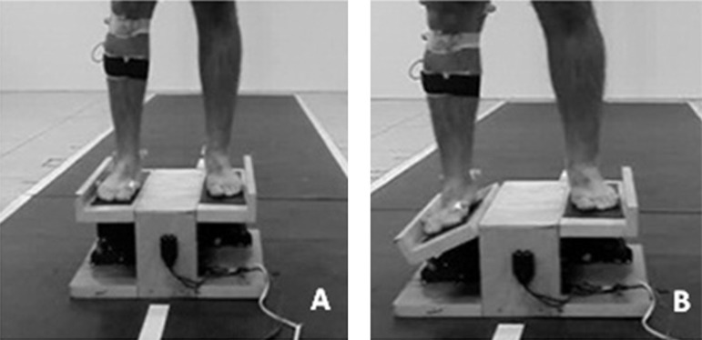
The volunteer was in the orthostatic position on a tilt platform, eyes open and bare feet (Figure 1). The platform was synchronized with the electromyography equipment and activated by the computer. The device specifications and the inversion angle (30º) were based on a systematic review15. Each subject received instructions and was given the opportunity to feel the sudden inversion perturbation so this movement could be learned and recognized. The collection consisted of six tilts, three movements for each side, at random. For the data analysis, the mean value of the three collections was considered after the tilt of the platform for the dominant leg (defined as the leg that the subject would choose to kick a ball).
Figure 1.
Before (A) and after (B) 30º of ankle inversion movement, after the tilt of the platform.
Next, the subjects were placed in a seated position. The dominant leg was immersed for 20 minutes13,16 in cold water at a temperature of 4±2ºC17 and a depth of 20 cm, below the positioning of the electrodes; therefore, there was no need to remove those between trials. The water temperature was controlled using an infrared thermometer (MultTemp®, Porto Alegre, RS, Brazil). The cold water was stirred during the procedure, and whenever necessary, some ice cubes were added to maintain the temperature. After immersion, the lower limb was dried with a towel. The skin surface temperature of the anterior ankle region was measured to confirm cooling.
For the analysis, the peak RMS after the tilt of the inversion platform at 30º was considered. Similarly, Berg et al. considered that higher levels of RMS showed greater muscle activity13. For normalization of the peak RMS signal, pre-cold water immersion was considered as 100% because it is considered a dynamic activity18,19.
Results were verified by applying the Shapiro-Wilk test, repeated measures analysis, Bonferroni's post-hoc test, and linear regression analysis. The statistical analysis was performed using the Statistical Package for Social Sciences (SPSS®), version 15.0. The level of statistical significance was set at 5%.
Results
Thirty-five male subjects, 21.6 (±3.2) yrs of age, 177.9 (±7.9) cm tall, 82.8 (±19.3) Kg in weight and 25.9 (±4.8) Kg.m-2 of body mass index (BMI), were evaluated. Skin surface temperature before and after cold water immersion was 27.7(±3)ºC and 7.1 (±1)ºC, respectively (p=0.0001).
The results in Table 1 show that the peak RMS values were significantly lower for all post-immersion times when compared to those obtained pre-immersion for all muscles, except for immediate post-immersion for GM.
Table 1.
Electromyographic analysis of the peak RMS values of the dominant lower limb muscles after the tilt of the inversion platform at 30º. Mean values (SD) expressed as percentage of pre cold water immersion peak (%). n=35.
| Moment analysis with cold water immersion | |||||
|---|---|---|---|---|---|
| Muscles | Pre | Post | Post 10 min | Post 20 min | Post 30 min |
| Lateral gastrocnemius | 100 (0) | 68.34* (31.30) | 69.14* (30.22) | 66.26* (29.87) | 66.62* 29.83) |
| Tibialis anterior | 100(0) | 53.73* (25.55) | 55.78* (30.24) | 63.74* (50.19) | 50.19* (29.20) |
| Fibularis longus | 100 (0) | 73.65* (43.58) | 74.91* (35.03) | 73.29* (37.84) | 71.10* (39.21) |
| Rectus femoris | 100 (0) | 73.22* (47.00) | 62.77* (32.73) | 65.28* (36.34) | 64.78* (49.29) |
| Gluteus medius | 100 (0) | 86.21 (31.90) | 82.67* (25.02) | 81.23* (24.05) | 75.83* (30.43) |
Mean values (SD) expressed as percentage of pre cold water immersion peak (%)
Statistically significant difference when compared to pre cold water immersion time (p<0.05). Repeated measures analysis and Bonferroni's post-hoc test.
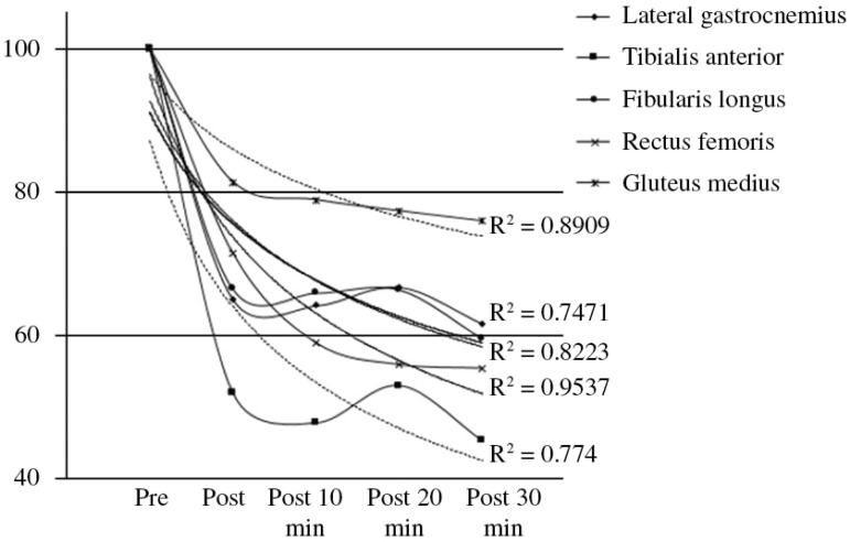
Linear regression analysis was used to determine the decreasing patterns of the peak RMS values. It was observed that the non-cooled muscles (RF and GM) that were not related morphologically to the ankle/foot responded in a more uniform manner to the cooling over time, justifying the use of this experimental model (Figure 2). In contrast, the three other muscles that act on the ankle joint showed a very similar pattern after the cooling treatment.
Figure 2.
Behavior of the peak RMS values of the dominant lower limb muscles, after the tilt of the inversion platform at 30º pre and post cold water immersion. Trend line and R2. n=35.
Discussion
Based on the results, cold water immersion for 20 minutes at 4ºC was effective in reducing skin surface temperature in agreement with Wolf and Basmajian20, who reported decreases in skin and intramuscular temperatures after a 5-minute intervention. These results are consistent with Rupp et al.12, who concluded that cold water immersion was more effective in significantly decreasing intramuscular temperatures in the gastrocnemius when compared to crushed-ice bag during treatment and 90 minutes post-treatment.
Many studies have used electromyography to analyze ankle muscle response10,15 and ankle inversion13,21. In addition, the present study evaluated the effects of cold water immersion on the electromyographic response of muscles that are not mechanically related to the cooled ankle joint. Thus, our study analyzed the RF and GM muscles, which showed a decrease in RMS immediately after the use of cold water immersion, with residual effect that lasted up to 30 min.
The RF is a powerful knee extensor and the GM stabilizes the lateral movement of the pelvis, hip, and knee. Although the ankle inversion causes an anterolateral joint imbalance, consequently it triggers an ascending force to the pelvis, leading to a decrease in activation of the ankle muscles that can change the muscular responses around the hip and knee. This change could be explained in part by the existing modulation response coming from the skin, joint, and muscle receptors, while a reduction in distal afferents would cause an inhibitory effect of the proximal muscle activity. Thus, the cooling of the ankle had a direct reflection on the muscular activity of the non-cooled muscle/joint complexes of the knee and hip.
Following the same pattern, all muscles (LG, TA, and FL) mechanically related to the ankle that received cryotherapy showed significant reduction in RMS. These findings can be explained by the fact that cryotherapy causes a reduction in amplitude and an increase in latency of motor nerve conduction5,22, causing changes in the structures of the axon membrane and in the muscle conductivity of sodium and potassium channels, with a reduction in motor nerve conduction22,23, mainly of the sensorial nerves5. Moreover, Khanmohammadi et al.3 suggested a linear relationship between the rate of muscle spindle discharge and reduced muscle temperature, which can be confirmed by our findings. The results of these studies contrast with those of Berg et al.13 and Cordova et al.21 who observed no differences in FL amplitude or latency during inversion perturbation after cooling the ankle joint. Uchio et al.24 reported some concern for the return of athletes to exercise after cryotherapy because the results from different studies are contradictory, justifying the need for further studies.
There are some limitations that need to be acknowledged and addressed regarding the present study. The sample comprised active and healthy individuals as the authors understand that the tilt platform test could exacerbate pain and/or injury in individuals with ankle disorders. Additionally, the possibility of a control group being submitted to water immersion at room temperature or at rest should also be considered in order to ensure the effects of cold water immersion on ankle muscle activity. Further studies should include the analysis of the non-dominant lower limb that provides support during the tilting.
The results of this study indicate a decrease in the EMG response of the LG, TA, FL, RF, and GM muscles to ankle inversion after the use of cold water immersion in healthy male subjects, with a residual effect that lasts up to 30 minutes. Therefore, after cold water immersion of the ankle, special care should be taken with activities that require greater neuromuscular control.
References
- 1.Collins NC. Is ice right? Does cryotherapy improve outcome for acute soft tissue injury? Emerg Med J. 2008;25:65–68. doi: 10.1136/emj.2007.051664. http://dx.doi.org/10.1136/emj.2007.051664 [DOI] [PubMed] [Google Scholar]
- 2.Bleakley CM, Costello JT. Do Thermal Agents Affect Range of Movement and Mechanical Properties in Soft Tissues? A Systematic Review. Arch Phys Med Rehabil. 2013;94:149–163. doi: 10.1016/j.apmr.2012.07.023. http://dx.doi.org/10.1016/j.apmr.2012.07.023 [DOI] [PubMed] [Google Scholar]
- 3.Khanmohammadi R, Someh M, Ghafarinejad F. The effect of cryotherapy on the normal ankle joint position sense. Asian J Sports Med. 2011;2:91–98. doi: 10.5812/asjsm.34785. http://journals.tums.ac.ir/abs.aspx?tums_id=18302 [DOI] [PMC free article] [PubMed] [Google Scholar]
- 4.Algafly AA, George KP. The effect of cryotherapy on nerve conduction velocity, pain threshold and pain tolerance. Br J Sports Med. 2007;41:365–369. doi: 10.1136/bjsm.2006.031237. http://dx.doi.org/10.1136/bjsm.2006.031237 [DOI] [PMC free article] [PubMed] [Google Scholar]
- 5.Herrera E, Sandoval MC, Camargo DM, Salvini TF. Motor and sensory nerve conduction are affected differently by ice pack, ice massage, and cold water immersion. Phys Ther. 2010;90:581–591. doi: 10.2522/ptj.20090131. http://dx.doi.org/10.2522/ptj.20090131 [DOI] [PubMed] [Google Scholar]
- 6.Bleakley CM, O'Connor S, Tully MA, Rocke LG, Macauley DC, Mcdonough SM. The PRICE study (Protection Rest Ice Compression Elevation): design of a randomised controlled trial comparing standard versus cryokinetic ice applications in the management of acute ankle sprain. BMC Musculoskelet Disord. 2007;19:125–133. doi: 10.1186/1471-2474-8-125. http://dx.doi.org/10.1186/1471-2474-8-125 [DOI] [PMC free article] [PubMed] [Google Scholar]
- 7.Stacey DL, Gibala MJ, Ginis KAM, Timmnos BW. Effects of recovery method after exercise on performance, immune changes, and psychological outcomes. J Orthop Sports Phys Ther. 2010;40:656–665. doi: 10.2519/jospt.2010.3224. http://dx.doi.org/10.2519/jospt.2010.3224 [DOI] [PubMed] [Google Scholar]
- 8.Bleakley CM, Costello JT, Glasgow PD. Should athletes return to sport after applying ice? A systematic review of the effect of local cooling on functional performance. Sports Med. 2012;42:69–87. doi: 10.2165/11595970-000000000-00000. http://dx.doi.org/10.2165/11595970-000000000-00000 [DOI] [PubMed] [Google Scholar]
- 9.Costello JT, Donnelly AE. Cryotherapy and joint position sense in healthy participants: a systematic review. J Athl Train. 2010;45:306–316. doi: 10.4085/1062-6050-45.3.306. http://dx.doi.org/10.4085/1062-6050-45.3.306 [DOI] [PMC free article] [PubMed] [Google Scholar]
- 10.Doeringer JR, Hoch MC, Krause BA. Ice application effects on peroneus longus and tibialis anterior motoneuron excitability in subjects with functional ankle instability. Int J Neurosci. 2010;120:17–22. doi: 10.3109/00207450903337713. http://dx.doi.org/10.3109/00207450903337713 [DOI] [PubMed] [Google Scholar]
- 11.Costello JT, Algar LA, Donnelly AE. Effects of whole-body cryotherapy (-110ºC) on proprioception and indices of muscle damage. Scand J Med Sci Sports. 2012;22:190–198. doi: 10.1111/j.1600-0838.2011.01292.x. http://dx.doi.org/10.1111/j.1600-0838.2011.01292.x [DOI] [PubMed] [Google Scholar]
- 12.Rupp KA, Herman DC, Hertel J, Saliba SA. Intramuscular temperature changes during and after 2 different cryotherapy interventions in healthy individuals. J Orthop Sports Phys Ther. 2012;42:731–737. doi: 10.2519/jospt.2012.4200. http://dx.doi.org/10.2519/jospt.2012.4200 [DOI] [PubMed] [Google Scholar]
- 13.Berg CL, Hart JM, Palmieri-Smith R, Cross KM, Ingersoll CD. Cryotherapy does not affect peroneal reaction following sudden inversion. J Sport Rehabil. 2007;16:285–294. doi: 10.1123/jsr.16.4.285. [DOI] [PubMed] [Google Scholar]
- 14.Harriss DJ, Atkinson G. Update - Ethical standards in sport and exercise science research. Int J Sports Med. 2011;32:819–821. doi: 10.1055/s-0031-1287829. http://dx.doi.org/10.1055/s-0031-1287829 [DOI] [PubMed] [Google Scholar]
- 15.Menacho MO, Pereira HM, Oliveira BIR, Chagas LMPM, Toyohara MT, Cardoso JR. The peroneus reaction time during sudden inversion test: Systematic review. J Electromyogr Kines. 2010;20:559–565. doi: 10.1016/j.jelekin.2009.11.007. http://dx.doi.org/10.1016/j.jelekin.2009.11.007 [DOI] [PubMed] [Google Scholar]
- 16.Hatzel BM, Kaminski TW. The effects of ice immersion on concentric and eccentric muscle performance in the ankle. Isokinet Exerc Sci. 2000;8:103–107. [Google Scholar]
- 17.Kernozek TW, Greany JF, Anderson DR, Van Heel D, Youngdahl RL, Benesh BG. The effect of immersion cryotherapy on medial-lateral postural sway variability in individuals with a lateral ankle sprain. Physiother Res Int. 2008;13:107–118. doi: 10.1002/pri.393. http://dx.doi.org/10.1002/pri.393 [DOI] [PubMed] [Google Scholar]
- 18.Felici F. Neuromuscular responses to exercise investigated through surface EMG. J Electromyogr Kinesiol. 2006;16:578–585. doi: 10.1016/j.jelekin.2006.08.002. http://dx.doi.org/10.1016/j.jelekin.2006.08.002 [DOI] [PubMed] [Google Scholar]
- 19.Murley GS, Menz HB, Landorf KB, Bird AR. Reliability of lower limb electromyography during overground walking: a comparison of maximal- and sub-maximal normalisation techniques. J Biomech. 2010;43:749–756. doi: 10.1016/j.jbiomech.2009.10.014. http://dx.doi.org/10.1016/j.jbiomech.2009.10.014 [DOI] [PubMed] [Google Scholar]
- 20.Wolf SL, Basmajian JV. Intramuscular temperature changes deep to localized cutaneous cold stimulation. Phys Ther. 1973;53:1284–1288. doi: 10.1093/ptj/53.12.1284. [DOI] [PubMed] [Google Scholar]
- 21.Cordova ML, Bernard LW, Au KK, Demchak TJ, Stone MB, Sefton JM. Cryotherapy and ankle bracing effects on peroneus longus response during sudden inversion. J Electromyogr Kines. 2010;20:348–353. doi: 10.1016/j.jelekin.2009.03.012. http://dx.doi.org/10.1016/j.jelekin.2009.03.012 [DOI] [PubMed] [Google Scholar]
- 22.Herrera E, Sandoval MC, Camargo DM, Salvini TF. Effect of walking and resting after three cryotherapy modalities on the recovery of sensory and motor nerve conduction velocity in healthy subjects. Rev Bras Fisioter. 2011;15:233–240. doi: 10.1590/s1413-35552011000300010. http://dx.doi.org/10.1590/S1413-35552011000300010 [DOI] [PubMed] [Google Scholar]
- 23.Luzzati V, Mateu L, Marquez G, Borgo M. Strutural and electrophysiological effects of local anesthetics and of low temperature on myelinated nerves: implication of the lipid chains in nerve excitability. J Mol Biol. 1999;286:1389–1402. doi: 10.1006/jmbi.1998.2587. http://dx.doi.org/10.1006/jmbi.1998.2587 [DOI] [PubMed] [Google Scholar]
- 24.Uchio Y, Ochi M, Fujihara A, Adachi N, Iwasa J, Sakai Y. Cryotherapy influences joint laxity and position sense of the healthy knee joint. Arch Phys Med Rehab. 2003;84:131–135. doi: 10.1053/apmr.2003.50074. http://dx.doi.org/10.1053/apmr.2003.50074 [DOI] [PubMed] [Google Scholar]


