

Genetic factors underlying cognitive performance
Evidence from twin and family studies suggests that individual differences in cognitive performance are partly attributable to genes, but the specific genetic variants remain elusive. Cornelius Rietveld et al. (pp. 13790–13794) identified three genetic variants using a two-step procedure called the proxy-phenotype method that may explain some variation in cognitive performance. The authors performed a genome-wide association study (GWAS) of 106,736 participants and identified 69 SNPs associated with educational attainment. In an independent sample of 24,189 participants, the authors further determined that three of the 69 education-associated SNPs—rs1487441, rs7923609, and rs2721173—were also associated with cognitive performance. In a second independent sample of 8,652 Americans, the authors found that a polygenic score, derived from a weighted sum of the 69 education-associated SNPs, correlated with memory and the absence of dementia. Bioinformatics analyses implicated four genes—KNCMA1, NRXN1, POU2F3, and SCRT—that were associated with a neurotransmitter pathway involved in a cellular mechanism for learning and memory. According to the authors, the results reveal that the phenotype of educational attainment may be used to identify genetic variants that predict the cognitive performance phenotype, suggesting that the proxy-phenotype method improves on existing approaches such as candidate gene analyses and direct GWAS of cognitive performance. — A.G.
Simulations of the Miller experiments

Two dehydroglycine molecules (A) rotate to align their α-carbon atoms (B, indicated by arrows) leading to a proton jumping from one α-carbon to the other, forming glycine (C).
The well-known Miller experiments sought to replicate the chemical conditions that may have existed on a primitive Earth and given rise to the formation of early organic molecules. In 1953, Stanley Miller reported that a mixture of methane, ammonia, water, and hydrogen produced several amino acids after receiving an electric discharge intended to simulate lightning. Antonino Saitta and Franz Saija (pp. 13768–13773) examined whether the electric field in Miller’s experiments merely served as a source of heat energy or exerted a direct influence. Using a computer model simulating the interactions of individual atoms, the authors introduced eight molecules each of water, ammonia, and methane, as well as 10 molecules of carbon monoxide and five of diatomic nitrogen in a condensed state. When the authors applied simulated electric fields above 0.35 V/A, the molecules in the initial system spontaneously formed formic acid molecules within 2 picoseconds and formamide molecules shortly after. Applying fields up to 0.5 V/A to a simulation containing intermediate reaction products led to the formation of a proto-amino acid. The results suggest that the electric field may have played a crucial role in transforming primordial molecules into amino acids, according to the authors. — P.G.
Modeling brain function and disease
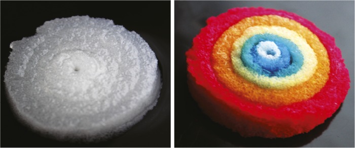
Assembly of six concentric “donuts” of silk scaffold, original color (Left); dyed with food color (Right).
The brain remains one of the least understood human organs, due in part to its complexity as well as technical limitations. Brain-like tissues are critical for understanding brain function and disease, but physiologically relevant models for the brain are elusive. Min Tang-Schomer et al. (pp. 13811–13816) constructed a 3D brain-like tissue that promotes the sustained growth of functional neurons and mimics major structural and mechanical features of the brain. The authors designed stiff and porous scaffolds made of a silk protein called fibroin, and filled the scaffolds with neurons as well as soft extracellular matrix gels that provided structural and biochemical support to the cells. The neurons clustered within the pores, forming long-lasting networks that resembled the brain’s complex circuitry. By contrast, 2D cultures or collagen gels alone did not support the formation of complex neural networks. Moreover, neurons in the 3D brain-like tissue showed realistic responses to damage caused by the impact of a falling weight, resembling cellular response to traumatic brain injury in animal models. According to the authors, the 3D brain-like tissue could help study normal brain function and disorders affecting the central nervous system. — J.W.
Species trait diversity in the Azores

A beetle, Calacalles subcarinatus, endemic to the Azores.
Most island biogeography studies have focused on calculations of species richness and diversity, yet few have examined the diversity of species’ functional traits, which determine how species interact with their environment. Robert Whittaker et al. (pp. 13709–13714) analyzed functional trait data for spiders and beetles on the Azores to quantify the impact of exotic species on island diversity. Both groups of insects play important functional roles: Beetles are numerous and operate at multiple feeding levels, and spiders act as top predators. The authors found that functional diversity of both groups increased in proportion to the number of species on the islands and that these trends did not plateau as island size increased. Moreover, the patterns for exotic species mimicked those of endemic and other native species. As exotic species now outnumber native species on these remote oceanic islands, the results also suggest that the overall array of functional types—known as the functional trait space—on each island has been increased significantly by the anthropogenic introduction of exotic species. The authors add that their data indicate that the islands remain vulnerable to colonization by additional exotic species, urging further research on the interactions between native and exotic species and implications for the persistence of island endemic species. — J.P.J.
Fluorescence imaging of tumors and surgical guidance with carbon nanotubes
Fluorescence imaging of tumors in vivo holds promise for specific, real-time detection and guidance in surgical interventions; yet, current imaging methods are limited by challenges including high background tissue autofluorescence, optical scattering, limited penetration depth, and photobleaching of fluorescent dyes. Debadyuti Ghosh et al. (pp. 13948–13953) developed a fluorescence imaging system that uses virus-assembled single-walled carbon nanotubes (SWNTs) as imaging agents that fluoresce at second-window near-infrared wavelengths (NIR2) between 950 and 1400 nm to penetrate deeply into tissue with minimal optical scattering. SWNTs are resistant to photobleaching, naturally fluoresce under NIR2 wavelengths, and display a large difference between excitation and emission wavelengths, thereby minimizing tissue autofluorescence. Experiments using an M13 virus to stably bind SWNTs demonstrated targeted uptake of the virus-SWNT complex by ovarian tumors and resulted in higher signal-to-noise performance in resolving small tumors, compared with visible and near-infrared dyes. During surgical removal of ovarian tumors from mice, surgeons located and removed more tumor tissue with SWNT-guided fluorescence imaging than without, and the technique enabled detection and removal of submillimeter tumors. The results suggest that fluorescence imaging using SWNTs may be useful as a noninvasive tool for cancer diagnosis, research, and monitoring, and may serve as a surgical guide, according to the authors. — P.G.
Joint protection against Lyme disease
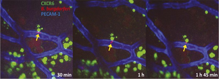
Interaction between extravascular iNKT cell (green) and joint-invading Lyme Borrelia (red).
Lyme disease, caused by the tick-borne bacterium Borrelia burgdorferi, can lead to chronic joint inflammation. Previous studies have shown that mice lacking iNKT cells, a type of natural killer T cell, experience an elevated burden of Borrelia bacteria in their joints. Woo-Yong Lee et al. (pp. 13936–13941) used a technique called spinning disk confocal intravital microscopy to observe the movement of iNKT cells, within the knees of Borrelia-infected mice. The authors found that iNKT cells were distributed outside of knee joint blood vessels and that 90% of the cells were stationary, contrasting with previous studies indicating that iNKT cells travel primarily through blood vessels in the liver. Further, B. burgdorferi bacteria adhered to the inner walls of knee joint blood vessels, across which bacterial cells were inactivated by iNKT cells, and live bacteria that escaped the vasculature were immobilized by iNKT cells. While iNKT cells isolated from joint tissue killed B. burgdorferi bacteria, iNKT cells circulating in the liver demonstrated no such lethality. According to the authors, iNKT cells localized in joints may act as a barrier to invasion by B. burgdorferi, and the relative absence of iNKT cells in human joints may explain why Lyme-related joint inflammation occurs in humans but not rodents. — C.B.