MEDICAL SCIENCES Correction for “Regulation of bone remodeling by vasopressin explains the bone loss in hyponatremia,” by Roberto Tamma, Li Sun, Concetta Cuscito, Ping Lu, Michelangelo Corcelli, Jianhua Li, Graziana Colaianni, Surinder S. Moonga, Adriana Di Benedetto, Maria Grano, Silvia Colucci, Tony Yuen, Maria I. New, Alberta Zallone, and Mone Zaidi, which appeared in issue 46, November 12, 2013, of Proc Natl Acad Sci USA (110:18644–18649; first published October 28, 2013; 10.1073/pnas.1318257110).
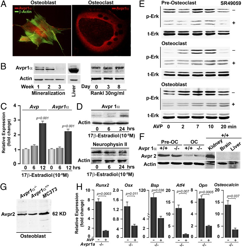
The authors note that Fig. 1 appeared incorrectly. The corrected figure and its legend appear below.
Fig. 1.
Bone cells express Avprs. Immunofluorescence micrographs (A) and Western immunoblotting (B) show the expression of Avpr1α in osteoblasts and osteoclasts, and as a function of osteoblast (mineralization) and osteoclast (with Rankl) differentiation. The expression of Avp (ligand) and Avpr1α (receptor) in osteoblasts is regulated by 17β-estradiol, as determined by quantitative PCR (C) and Western immunoblotting (D). (Magnification: A, 63×.) Because Avp is a small peptide, its precursor neurophysin II is measured. Statistics: Student t test, P values shown compared with 0 h. Stimulation of Erk phosphorylation (p-Erk) as a function of total Erk (t-Erk) by Avp (10−8 M) in osteoclast precursors (preosteoclasts), osteoclasts (OC), and osteoblasts establishes functionality of the Avpr1α in the presence or absence of the receptor inhibitor SR49059 (10−8 M) (E). Western immunoblotting showing the expression of Avpr2 in preosteoclasts, OCs (F), and osteoblasts (G) isolated from Avpr1α−/− mice, as well as in MC3T3.E1 osteoblast precursors (G). Functionality of Avpr2 was confirmed by the demonstration that cells from Avpr1α−/− mice remained responsive to AVP in reducing the expression of osteoblast differentiation genes, namely Runx2, Osx, Bsp, Atf4, Opn, and Osteocalcin (quantitative PCR, P values shown) (H). Only relevant bands from Western blots are shown, with gaps introduced where empty lanes are excised to conserve space.


