Abstract
A meta-analysis of the effects of vector saliva on the immune response and progression of vector-transmitted disease, specifically with regard to pathology, infection level, and host cytokine levels was conducted. Infection in the absence or presence of saliva in naïve mice was compared. In addition, infection in mice pre-exposed to uninfected vector saliva was compared to infection in unexposed mice. To control for differences in vector and pathogen species, mouse strain, and experimental design, a random effects model was used to compare the ratio of the natural log of the experimental to the control means of the studies. Saliva was demonstrated to enhance pathology, infection level, and the production of Th2 cytokines (IL-4 and IL-10) in naïve mice. This effect was observed across vector/pathogen pairings, whether natural or unnatural, and with single salivary proteins used as a proxy for whole saliva. Saliva pre-exposure was determined to result in less severe leishmaniasis pathology when compared with unexposed mice infected either in the presence or absence of sand fly saliva. The results of further analyses were not significant, but demonstrated trends toward protection and IFN-γ elevation for pre-exposed mice.
Author Summary
Arthropod vectors transmit a wide variety of diseases resulting in substantial human morbidity and economic costs worldwide. When hematophagous arthropods blood feed, they release saliva into the host. This saliva elicits a strong immune response and has recently been a focus for vaccine research. There is evidence that the saliva enhances infection in naïve hosts, but that prior exposure to saliva results in less severe infection. This analysis endeavored to determine whether there was a statistically significant enhancement or protective effect with regard to saliva exposure and the progression of disease, and to determine the underlying immune mechanism driving these effects. We found that saliva does indeed enhance infection levels of vector-transmitted pathogens and leishmaniasis pathology in naïve mice and elevates Th2 cytokine levels (IL-4 and IL-10). We also determined that pre-exposure to saliva results in less severe pathology of experimental leishmaniasis in mice. These results are important for vaccine trials and vector control programs, though more studies are needed with regard to pre-exposure.
Introduction
Vector-borne diseases are a major cause of morbidity and mortality in many areas of the world. In addition to their cost to human health, vector-borne diseases can have a high economic cost primarily affecting impoverished nations and the people with the least resources. While there have been efforts to control or eradicate certain vector-borne diseases, these goals have proved frustratingly elusive and the incidence of some vector-borne infections, such as leishmaniasis, is rising [1]. Emerging and reemerging diseases such as Chikungunya threaten to become major public health concerns. More familiar diseases, like malaria and dengue fever, are infecting new populations due to lapses in vector control programs, human migration and increasing vector habitat due to climate change and other human activities [1]–[8]. Although vaccines have been developed for some vector-borne diseases (e.g. yellow fever,) the vast majority and the most problematic still lack vaccines and viable treatment options. The quest for vaccine development has included assessing the potential protective effect of long-term exposure to insect vector saliva. Results have been mixed at best, and there is some controversy as to whether saliva exacerbates disease or protects against its more severe manifestations.
When an arthropod vector bites a host and transmits a pathogen, it releases some of its own saliva into the bite site as well as the pathogen. It is well established that this saliva is highly immunogenic, containing vasodilatory and immunosuppressive compounds [9]. Perhaps the most studied vectors in this regard have been that of sand fly vectors of leishmaniasis. A landmark study in 1988 by Titus and Ribeiro demonstrated that Lutzomyia longipalpis saliva exacerbates Leishmania major infection in naïve mice [10]. Many similar studies have followed, consistently demonstrating that naïve animals either infected via sand fly or coinoculation with salivary gland homogenate along with Leishmania parasites have generally developed larger, longer lasting lesions than animals inoculated with parasites alone [11]–[25]. Furthermore, these effects appear to be consistent across all sand flies, though the salivary composition differs widely between species. In Lutzomyia species, the vasodilatory peptide maxadilan has been implicated in upregulating Th2 cytokines (e.g. IL-4 and IL-10) and down-regulating Th1 cytokines (e.g. IFN- γ) in vitro and in vivo, presenting a potential mechanism for the observed differences in disease progression [18], [26]–[31]. Belkaid et al. further demonstrated that disease enhancement is IL-4 driven, as Phlebotomus papatasi saliva did not enhance disease in IL-4 deficient mice. Furthermore, disease enhancement was even greater in IL-12p40 deficient mice [12]. The infection-enhancing effects of saliva, however, have been demonstrated to be negated by prior exposure to uninfected sand fly saliva [11], [13], [18], [24], [32]–[45]. Immunity to the salivary peptides is theorized to elicit a strong Th1 response in the host, which adversely affects Leishmania parasites. This effect appears to apply to immunization with uninfected sand fly bites and with individual salivary proteins, though laboratory-colonized sand fly saliva is much more effective than wild-caught in providing protection against disease [11], [13].
Studies assessing the effects of mosquito saliva began soon after those of sand flies, with Bissonnette et al and Cross et al demonstrating that Aedes aegypti saliva inhibits IFN-γ, TNF-α, and IL-2 release from murine cells [46], [47]. As with the sand fly studies, the reports that followed have consistently demonstrated that mosquito saliva from all genera also up-regulates Th2 cytokines and down-regulates Th1 cytokines [46]–[56]. Mosquito saliva has also been shown to increase infectivity of various viruses [57]–[62], as well as enhancing viral replication [63], mortality [64]–[66] and even being necessary for infection [57]. However, there has been some controversy with regard to its effect on malaria, with some studies claiming exacerbation of disease and others claiming no effect or even protection from prior immunization [67]–[70].
Hard ticks are a third group of well-studied arthropod vectors with immunomodulatory saliva. These ticks can take up to two weeks to take a complete bloodmeal, so it is necessary for them to secrete these compounds to avoid rejection from the host. Tick saliva has been demonstrated to inhibit pro-inflammatory (Th1) cytokine production [71]–[79], T cell proliferation [71] and neutrophil activity [80]. Accordingly, it has also been implicated in increasing Borrelia and viral infectivity, and immunity against tick saliva may also correspond to decreased effectiveness of the pathogen [81]–[90].
While several review papers on this topic have been published, to date there has not been an analytical comparison of these studies. Here we present a meta-analysis of the effects of vector saliva on disease progression as it applies to three outcomes: pathology, pathogen load, and cytokine levels. Only transient-feeding vectors were included (i.e. sand flies and mosquitoes), as long-term feeding results in a more complicated and not directly comparable interaction. The proportion of mosquito experiments included in each of the analyses varied (22–52% for pathogen load, 25–37.5% for cytokine levels, and 0% for pathology) due to the limited number of published studies including these parameters. Also as a result of paucity, human studies and research on trypanosomes and their vectors were also excluded. For comparability, only in vivo infection, as opposed to macrophage and other in vitro cell studies, and quantification by ELISA and PCR were used for the cytokine evaluation. Furthermore, only IFN-γ, IL-4, and IL-10 were included, as they were the most often studied.
Experiments were placed into two groups: naïve animals exposed to saliva during infection compared with a control group exposed to only pathogens, and animals pre-exposed to saliva before infection compared with a control group of naïve animals exposed to saliva only during infection. A third group, pre-exposed animals compared with those that were needle inoculated and not exposed to saliva at all, was included in the leishmaniasis pathology evaluation. Other than expanding our knowledge of the biology of infection, the results of the analyses concerning the first group could have ramifications for vector control programs and vaccine studies. If control programs are allowed to lapse, newly naïve populations could end up with more severe disease. As for vaccine trials, it would be important to test against vector-borne infection as opposed to needle inoculation. This has been a problem especially with vaccines against leishmaniasis; they may work for needle-inoculated mice but fail to protect against infection via sand fly [91], [92]. The second group mimics natural conditions for endemic populations and naïve ones such as travelers and deployed military service members. It is important to understand potential elevated risks in these populations, as well as potential for vaccine studies. The third group assesses whether immunity induced by saliva pre-exposure just negates the exacerbative effect of saliva, or if there is an added protective effect. While there are certainly limits to this type of analysis, it can be very useful in determining whether observed trends across the published literature are statistically significant effects.
Methods
Data Sources and Extraction
For consistency and comparability, this analysis included only murine studies concerning transient-feeding vectors. A thorough literature search was performed using Pubmed (http://www.ncbi.nlm.nih.gov/pubmed/) for papers published until May 2013. Search terms combined vector saliva, immune response, and specific vectors and diseases such as leishmaniasis, malaria, dengue, sand fly saliva, and mosquito saliva. Other papers were found using the references in previously located articles. Criteria for inclusion were studies using wild-type mouse strains (as opposed to certain immunodeficient) that contained information on one of three outcomes: pathology (leishmaniasis papers only), cytokine levels in vivo (ELISA or PCR), and infection level (parasite or viral load in tissues or parasitemia/viremia). Due to the constraints inherent in a meta-analysis, we limited our focus on pathology to studies evaluating leishmaniasis. The statistical analyses required a certain number of data points and we were unable to find enough studies assessing other pathogens to make comparisons on their own and we could not combine the studies with leishmaniasis pathology studies because the types of assays were not comparable (e.g. lesion size compared to mortality analysis). Similarly, flow cytometric analysis of cytokine expression was excluded because there was not enough conformity across studies in the experimental set up, cells assessed, and gating strategies to be controlled properly.
The Studies were organized into three broad categories of experiments for analysis:
Studies comparing a control group of naïve mice inoculated with the pathogen only to an experimental group of naïve mice infected with the pathogen either by vector feeding or by co-inoculation with the pathogen and a vector salivary gland extract (Saliva vs Control).
Studies comparing a control group of naïve mice infected with the pathogen by vector feeding or by co-inoculation with the pathogen and saliva to an experimental group of mice pre-exposed to vector saliva then infected with the pathogen along with saliva (Pre-exposed vs Saliva).
Studies comparing a control group of naïve mice inoculated with the pathogen only to an experimental group of mice pre-exposed to vector saliva then infected with the pathogen along with saliva (Pre-exposed vs Control).
Cytokines were divided into IFN-γ, IL-4, and IL-10 groups, as these proteins were most commonly measured. Mean values and standard deviations of the mean for each outcome were extracted from either data reported in the papers or from figures using the “grabit” function in MATLAB (Mathworks). When standard errors were reported, they were converted to standard deviations with the formula SE = SD/√N. When no standard deviation or error was reported, standard deviation was calculated as 1/N. The mean was taken for multiple measurements over time.
Data Analysis
The data were analyzed with the metafor package in R (r-project.org). A random effects model was used as there is considerable variation in both mouse strain and vector and pathogen species. The natural log of the ratio of the experimental mean to the control mean was taken [Yi = ln(Xe/Xc)]. Using a ratio allowed us to directly compare studies and provided a means of controlling for differences in experimental design. Variance was calculated by the formula Vi = [SDe2/(Ne*Xe2)]+[SDc2/(Nc*Xc2)]. The code used in R was as follows:
dat1<-read.csv(“[file name]”, header = TRUE)
res1<- rma(Yi,Vi, data = dat1)
summary(res1)
forest(res1, slab = paste(dat1$Author, dat1$Year, sep = “,”))
This code sequence provided a summary of the analysis (most importantly overall effect and p value) and a forest plot of the data for each group.
Results
Infection Level
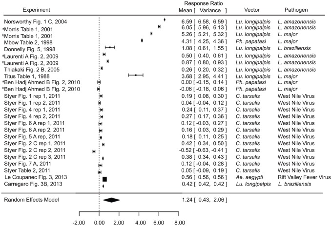
The infection level analysis combined measurements of parasite and viral load in tissues and parasitemia or viremia. Various sand fly and mosquito vectors were included, as were various pathogen species (namely Leishmania species, Plasmodium species, and West Nile virus) (Table S1). Infection of naïve mice in the presence of vector saliva was found to significantly increase infection level (estimate 1.2440, p value 0.0029) compared to pathogen alone (Fig. 1). These results are broadly applicable, considering the variation in vectors (Lu. longipalpis, Ph. papatasi, Culex tarsalis, and Ae. aegypti) and pathogens (L. amazonensis, L. major, L. braziliensis, West Nile Virus, and Rift Valley Fever Virus).
Figure 1. Forest plots of the relationship of vector saliva and infection level in naïve mice (Category 1).
Symbols represent the mean response ratio of the individual studies (squares) and of the entire analysis (diamond) using a Random Effects Model; the size of the square is proportional to the weight of an individual study. Error bars represent 95% Confidence Interval (CI). Squares to the right of the dotted line indicate larger measurements in the experimental (saliva) group, while those on the left indicate larger measurements in the control group. Those that cross the center indicate no significant difference.
Pre-exposure to saliva, however, was not demonstrated to significantly decrease infection level across all vectors and pathogens (estimate −0.6266, p value 0.0868, Table S2) or even across just the sand fly vectors and leishmaniases (estimate −0.8063, p value 0.0865, Table S2). The general trend, however, did indicate protection. There was unfortunately not enough information available to perform an analysis of the third group, that of pre-exposed mice compared with control mice infected without saliva.
In some of the studies, a salivary protein was used as a proxy for saliva as a whole (maxadilan [18] and rLMJ11 [38]). The analysis was conducted both including these studies (Fig. 1; Table S2) and excluding them (Table S2), and the results were not significantly different from each other. Although saliva is a complex cocktail of proteins and the protocols utilized for vaccination utilize greater amounts of a single protein than is found in salivary extracts, this result indicates that maxadilan and LmJ11 are both likely major factors in saliva's immunogenic properties, and that they alone have nearly the same effect as the entire salivary gland homogenate.
Pathology
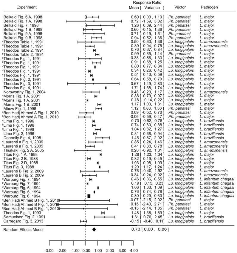
Due to the low number of studies concerning other aspects of pathology, here pathology is synonymous with the size of Leishmania induced lesions. Though all of the studies in this analysis concerned sand flies and Leishmania, they varied considerably with regard to mouse strain, sand fly species, Leishmania species, and experimental design (e.g. infected ear or footpad, experimental group infection by vector feeding or inoculation, amount and times of pre-exposure, etc). Consistent with the infection level results, naïve mice infected in the presence of saliva had significantly larger lesions than those in the control group (estimate 0.319, p value<0.001) (Fig. 2). These results were consistent regardless of whether the saliva came from the natural vector or another species of sand fly (natural vector estimate 0.6183, p value<0.0001; other vector estimate 0.7837, p value<0.0001; Table S2), or even whether the vector was of the natural genus (natural genus estimate = 0.6388, p = <0.0001, other genus estimate 0.8644, p value<0.0001) (Table S2). Saliva also appeared to increase the duration of the lesions, though this factor was not included in the analysis due to inconsistencies in the lengths and intervals of time measured between studies.
Figure 2. Forest plots of the relationship of exposure to vector saliva and Leishmania lesion size in naïve mice (Category 1).
Symbols represent the mean response ratio of the individual studies (squares) and of the entire analysis (diamond) using a Random Effects Model; the size of the square is proportional to the weight of an individual study. Error bars represent 95% Confidence Interval (CI). Squares to the right of the dotted line indicate larger measurements in the experimental (saliva) group, while those on the left indicate larger measurements in the control group. Those that cross the center indicate no significant difference.
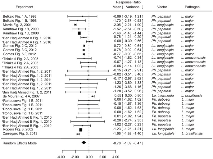
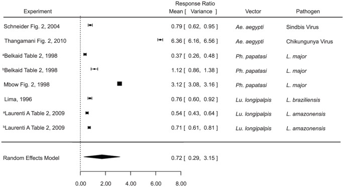
Pre-exposure to saliva, however, was shown to significantly decrease lesion size (estimate −0.7781, p value<0.0001) when compared with naïve mice infected in the presence of the same saliva (Fig. 3). Interestingly, the only study to show the opposite [35] was also the only study conducted on the natural pairing of L. braziliensis and L. intermedia. However, the overall results again remained significant regardless of whether the saliva came from the natural vector or even the natural genus (natural vector estimate −.5839, p value 0.0074, other vector estimate −1.0174, p value<0.0001, natural genus estimate −0.6909, p value 0.0008, other genus estimate −09326, p value 0.0010) (Table S2). It is noteworthy that two of the experiments (bThiakaki Fig. 2A and cThiakaki Fig. 2A [24]) included, trend more toward enhancement, though not statistically significant. The mice in these studies were pre-exposed to Ph. papatasi and Ph. sergenti saliva, respectively, and subsequently exposed to Lu. longipalpis saliva upon infection. The third experiment by the same authors(aThiakaki Fig. 2A [24]), where mice were pre-exposed to Lu. longipalpis saliva, demonstrated protection. These results imply that the protection gained by prior exposure may be somewhat species (or at least genus)- specific.
Figure 3. Forest plots of the relationship of exposure to vector saliva before infection and Leishmania lesion size (Category 2).
Symbols represent the mean response ratio of the individual studies (squares) and of the entire analysis (diamond) using a Random Effects Model; the size of the square is proportional to the weight of an individual study. Error bars represent 95% Confidence Interval (CI). Squares to the right of the dotted line indicate larger measurements in the experimental (pre-exposed) group, while those on the left indicate larger measurements in the control group. Those that cross the center indicate no significant difference.
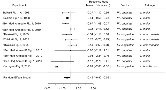
When compared with a control group infected without any saliva at all, mice pre-exposed to saliva developed smaller lesions (estimate −0.4889, p value 0.0254) (Fig. 4). As with the infection level studies, the analysis did not vary by excluding the studies using only maxadilan or rLMJ11 (Table S2).
Figure 4. Forest plots comparing pre-exposure to vector saliva to control groups infected in the absence of saliva on leishmaniasis pathology (Category 3).
Symbols represent the mean response ratio of the individual studies (squares) and of the entire analysis (diamond) using a Random Effects Model; the size of the square is proportional to the weight of an individual study. Error bars represent 95% Confidence Interval (CI). Squares to the right of the dotted line indicate larger measurements in the experimental (pre-exposed) group, while those on the left indicate larger measurements in the control group. Those that cross the center indicate no significant difference.
Cytokines
Studies included in the cytokine analysis were those that measured IFN-γ, IL-4, or IL-10 by either ELISA or PCR. Other cytokines and those measured via flow cytometry were excluded for consistency and due to low numbers and only measurements from in vivo infections were included. IFN-γ analysis of pre-exposed versus naïve mice (n = 5), as well as, of naïve mice infected in the presence versus absence of saliva (n = 9) were inconclusive (Table S2). Though general trends were observed, they were not significant (IFN-γ levels were lower in naïve mice exposed to saliva than in control mice and higher in pre-exposed mice than in the control group, Table S2). Naïve mice exposed to saliva during infection, however, had significantly higher IL-4 levels than control mice exposed only to the pathogen (estimate 1.7196, p value 0.0185) (Fig. 5).
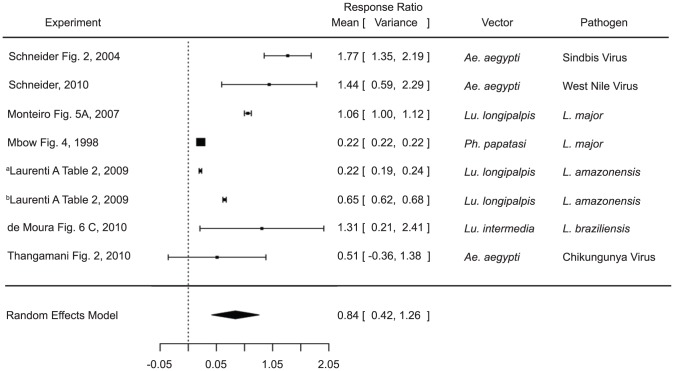
Figure 5. Forest plots of the relationship of vector saliva and IL-4 levels in naïve mice.
Symbols represent the mean response ratio of the individual studies (squares) and of the entire analysis (diamond) using a Random Effects Model; the size of the square is proportional to the weight of an individual study. Error bars represent 95% Confidence Interval (CI). Squares to the right of the dotted line indicate larger measurements in the experimental (saliva) group, while those on the left indicate larger measurements in the control group. Those that cross the center indicate no significant difference.
Likewise IL-10 levels were shown to be significantly higher in naïve mice exposed to saliva during infection (estimate 0.8398, p value<0.001) (Fig. 6). Unfortunately there were not enough measurements of IL-4 or IL-10 in mice pre-exposed to saliva versus naïve mice to conduct an analysis. Both of the cytokine findings were consistent across studies using both mosquito and sand fly vectors and various pathogens (parasitic and viral), suggesting a common mechanism of disease enhancement in the saliva of diverse vectors.
Figure 6. Forest plots of the relationship of vector saliva and IL-10 levels in naïve mice.
Symbols represent the mean response ratio of the individual studies (squares) and of the entire analysis (diamond) using a Random Effects Model; the size of the square is proportional to the weight of an individual study. Error bars represent 95% Confidence Interval (CI). Squares to the right of the dotted line indicate larger measurements in the experimental (saliva) group, while those on the left indicate larger measurements in the control group. Those that cross the center indicate no significant difference.
Discussion
Here we performed a meta-analysis of available data concerning the effects of vector saliva on host immunity. While a vast amount of heterogeneity existed between studies, the use of a ratio allowed us to control for the variability. Overall, our study indicates that saliva enhances infection in naïve mice. Pathogen levels in host blood and tissues are consistently higher in those mice exposed to saliva during infection and this effect holds true for both sand fly and mosquito vectors and for different pathogen species. As such, one would imagine that these results could be extended to other transient-feeding vectors as well, and indeed that has been demonstrated to be the case with Glossina morsitans morsitans/Trypanosoma brucei brucei [93], [94] and Rhodnius prolixus/Trypanosoma cruzi [95]. Both of these studies report higher parasitemia in naïve mice infected in the presence of saliva. More studies need to be performed on these and other vectors, however, to see if the findings are truly consistent. In addition to its effects on infection level, vector saliva also influences leishmaniasis pathology. Here we demonstrated that sand fly saliva enhances Leishmania-induced lesion size. Furthermore, higher levels of morbidity and mortality in mice infected with West Nile virus in the presence of mosquito saliva have been reported [61], [64]–[66]. Thus, the higher infection levels that result from saliva exposure have a demonstrable effect on the disease pathology.
An important consideration in this analysis was whether the vector/pathogen pairing was natural or unnatural. Several sand fly/Leishmania studies used an unnatural combination of Lu. longipalpis saliva with L. major, where the natural vector is one of several Phlebotomus species [10], [18], [22], [23], [38], [41], [96]. Likewise there have been studies using Lu. longipalpis saliva paired with L. amazonensis or e. braziliensis [14]–[16], [18], [19], [21], [22], [24], [97], [98]. While the genus is correct, these Leishmania species are naturally transmitted by different sand fly species [99]. These studies have the potential be both helpful and misleading. What is true for an unnatural pairing may not hold for a natural combination and thus the results may have little practical application. On the other hand, if the effects are similar regardless of the pairing, there may be potential for a more comprehensive vaccine, or at the least important implications for travelers already exposed to different sand fly species. We found that including or excluding the unnatural pairings made no difference in the overall results of the analyses, and that both natural and unnatural pairings (species and genus) generally demonstrated the same results in all categories.
A proposed mechanism for the salivary enhancement of infection has been the up-regulation of host Th2 cytokines. Indeed our analysis demonstrates a marked increase in IL-4 and IL-10 levels in groups exposed to saliva, both sand fly and mosquito, suggesting a strong Th2 response. These results, taken with the lack of enhancement in IL-4 deficient mice [12], strongly imply that the proposed Th2 driven mechanism is in fact correct. In vivo, cytokines function in a milieu of other cytokines and factors and it is the relative balance (or ratio) or these proteins that set the tone of a particular immune response. Whether Th1 cytokines are were regulated in response to saliva exposure is another question we investigated. While IFN- γ levels were generally lower in mice exposed to saliva, the results were not significant. However, upon further examination of the data, the only study to report the opposite also contained the only unnatural vector/pathogen pairing ([96], Lu. longipalpis/L. major). Eliminating this study lowered the p value, but not enough that the results were significant.
The potential for vaccines developed from vector saliva has been an important research topic in recent years. Therefore, a major aim of this study was to determine whether pre-exposure to vector saliva results in less severe infection. In the infection level analysis, while the trend was toward lower levels in pre-exposed mice, the results were not significant and therefore inconclusive. However, the leishmaniasis pathology analysis demonstrated less severe lesions in pre-exposed mice, and this result holds true even when compared with mice unexposed to saliva even during infection. Therefore, with respect to leishmaniasis pathology, pre-exposure does not just negate the infection-enhancing effects of saliva in naïve mice, it actually confers a significant protective effect compared to infection in the absence of saliva. It is interesting to note, however, that while pre-exposure to Lu. intermedia saliva does decrease infection level, it appears to have the opposite effect on lesion size [35]. More studies are necessary to investigate this phenomenon.
While a comprehensive cytokine analysis would be extremely informative with regard to the mechanism of the demonstrated protective effect, unfortunately there were not enough pertinent studies to conduct an analysis on IL-4 or IL-10 levels, and the IFN- γ analysis results were inconclusive. Kamhawi et al. found little change in the level of IL-4 producing cells in pre-exposed mice compared with naïve mice [39], though IFN- γ levels were elevated in pre-exposed mice. Interestingly, all of the studies reported much higher IFN- γ levels in pre-exposed mice except for one using a natural pairing of Ph. papatasi/L. major in BALB/c mice [12]. The same pairing with C57BL/6 mice indicated elevated IFN- γ in pre-exposed mice. This study is the only incidence in the analyses where mouse strain makes a difference, but it illustrates that while these results may be true for some mouse strains and vector/pathogen combinations, they may not be true for other strains or indeed other animals or humans.
Not surprisingly, studies assessing human immune responses to insect bites in disease settings are few and do not present a unifying theme for all vector-transmitted diseases or for a single disease or vector. Some studies suggest that saliva exposure skews the human immune response toward Th2-type immunity [100]–[103] and others suggest a more mixed response [104]–[106].
This meta-analysis has demonstrated conclusively the infection-enhancing effect of transient-feeding vector saliva in murine models of infection and the Th2 driven mechanism behind it; however, more studies need to be conducted on the effects of pre-exposure. A significant protective effect exists with regard to sand fly saliva and leishmaniases, but the mechanism still needs to be clarified. More cytokine studies are needed, as well as additional studies with other. Overall, the vaccine potential of saliva needs to be further investigated. There are many important considerations in the potential development of vaccines, not least that humans may be affected very differently than specific mouse strains, saliva differs widely between vectors, and immunity to saliva has only been demonstrated to result in less severe disease, not prevent infection entirely. Indeed, while the human response to vector saliva has been demonstrated to be similar to the murine one in that saliva enhances infection in naïve human cells [107]–[109], the effects of pre-exposure have been more controversial and appear to be more complicated than in mice [100]–[102], [105], [106].
This study, perhaps most importantly, emphasizes the importance of maintaining vector control programs once started. If allowed to lapse, not only will the protective immunity be lost when vector populations rebuild, but disease may be much more severe in newly naïve populations.
Supporting Information
Metadata associated with each experiment included in analysis. Excel file containing metadata of each experiment. Metadata includes, figure, vector, saliva component, pathogen, natural/unnatural pairing, mouse strain.
(XLSX)
Statistical analysis of each test. Response ration and P-values for each test are reported.
(XLSX)
Acknowledgments
The authors would like to thank Michelle Ngai for her advice regarding statistical analysis, and Matthew Leming for his help with data extraction, and the Eck Institute for Global Health of the University of Notre Dame for its support.
Data Availability
The authors confirm that all data underlying the findings are fully available without restriction. All manuscripts used for the meta-analysis are published. Supporting information contains a table with p-values and the metadata associated with each analyzed paper.
Funding Statement
The authors received no specific funding for this work.
References
- 1. Desjeux P (2004) Leishmaniasis: current situation and new perspectives. Comparative immunology, microbiology and infectious diseases 27: 305–318. [DOI] [PubMed] [Google Scholar]
- 2. Alvar J, Yactayo S, Bern C (2006) Leishmaniasis and poverty. Trends Parasitol 22: 552–557. [DOI] [PubMed] [Google Scholar]
- 3. Bai L, Morton LC, Liu Q (2013) Climate change and mosquito-borne diseases in China: a review. Globalization and health 9: 10. [DOI] [PMC free article] [PubMed] [Google Scholar]
- 4. Bern C, Courtenay O, Alvar J (2010) Of cattle, sand flies and men: a systematic review of risk factor analyses for South Asian visceral leishmaniasis and implications for elimination. PLoS neglected tropical diseases 4: e599. [DOI] [PMC free article] [PubMed] [Google Scholar]
- 5. Estrada-Pena A, Ayllon N, de la Fuente J (2012) Impact of climate trends on tick-borne pathogen transmission. Frontiers in physiology 3: 64. [DOI] [PMC free article] [PubMed] [Google Scholar]
- 6. Medlock JM, Hansford KM, Bormane A, Derdakova M, Estrada-Pena A, et al. (2013) Driving forces for changes in geographical distribution of Ixodes ricinus ticks in Europe. Parasites & vectors 6: 1. [DOI] [PMC free article] [PubMed] [Google Scholar]
- 7. Pradier S, Lecollinet S, Leblond A (2012) West Nile virus epidemiology and factors triggering change in its distribution in Europe. Revue scientifique et technique 31: 829–844. [DOI] [PubMed] [Google Scholar]
- 8. Singh BB, Sharma R, Gill JP, Aulakh RS, Banga HS (2011) Climate change, zoonoses and India. Revue scientifique et technique 30: 779–788. [DOI] [PubMed] [Google Scholar]
- 9. Oliveira F, de Carvalho AM, de Oliveira CI (2013) Sand-Fly Saliva–Man: The Trigger Trio. Frontiers in immunology 4: 375. [DOI] [PMC free article] [PubMed] [Google Scholar]
- 10. Titus RG, Ribeiro JM (1988) Salivary gland lysates from the sand fly Lutzomyia longipalpis enhance Leishmania infectivity. Science (New York, NY 239: 1306–1308. [DOI] [PubMed] [Google Scholar]
- 11. Ahmed SB, Kaabi B, Chelbi I, Derbali M, Cherni S, et al. (2010) Lack of protection of pre-immunization with saliva of long-term colonized Phlebotomus papatasi against experimental challenge with Leishmania major and saliva of wild-caught P. papatasi. The American journal of tropical medicine and hygiene 83: 512–514. [DOI] [PMC free article] [PubMed] [Google Scholar]
- 12. Belkaid Y, Kamhawi S, Modi G, Valenzuela J, Noben-Trauth N, et al. (1998) Development of a natural model of cutaneous leishmaniasis: powerful effects of vector saliva and saliva preexposure on the long-term outcome of Leishmania major infection in the mouse ear dermis. J Exp Med 188: 1941–1953. [DOI] [PMC free article] [PubMed] [Google Scholar]
- 13. Ben Hadj Ahmed S, Chelbi I, Kaabi B, Cherni S, Derbali M, et al. (2010) Differences in the salivary effects of wild-caught versus colonized Phlebotomus papatasi (Diptera: Psychodidae) on the development of zoonotic cutaneous leishmaniasis in BALB/c mice. Journal of medical entomology 47: 74–79. [DOI] [PubMed] [Google Scholar]
- 14. Laurenti MD, da Matta VL, Pernichelli T, Secundino NF, Pinto LC, et al. (2009) Effects of salivary gland homogenate from wild-caught and laboratory-reared Lutzomyia longipalpis on the evolution and immunomodulation of Leishmania (Leishmania) amazonensis infection. Scandinavian journal of immunology 70: 389–395. [DOI] [PubMed] [Google Scholar]
- 15. Laurenti MD, Silveira VM, Secundino NF, Corbett CE, Pimenta PP (2009) Saliva of laboratory-reared Lutzomyia longipalpis exacerbates Leishmania (Leishmania) amazonensis infection more potently than saliva of wild-caught Lutzomyia longipalpis. Parasitology international 58: 220–226. [DOI] [PubMed] [Google Scholar]
- 16. Lima HC, Titus RG (1996) Effects of sand fly vector saliva on development of cutaneous lesions and the immune response to Leishmania braziliensis in BALB/c mice. Infect Immun 64: 5442–5445. [DOI] [PMC free article] [PubMed] [Google Scholar]
- 17. Mbow ML, Bleyenberg JA, Hall LR, Titus RG (1998) Phlebotomus papatasi sand fly salivary gland lysate down-regulates a Th1, but up-regulates a Th2, response in mice infected with Leishmania major. J Immunol 161: 5571–5577. [PubMed] [Google Scholar]
- 18. Morris RV, Shoemaker CB, David JR, Lanzaro GC, Titus RG (2001) Sandfly maxadilan exacerbates infection with Leishmania major and vaccinating against it protects against L. major infection. J Immunol 167: 5226–5230. [DOI] [PubMed] [Google Scholar]
- 19. Norsworthy NB, Sun J, Elnaiem D, Lanzaro G, Soong L (2004) Sand fly saliva enhances Leishmania amazonensis infection by modulating interleukin-10 production. Infect Immun 72: 1240–1247. [DOI] [PMC free article] [PubMed] [Google Scholar]
- 20. Rohousova I, Volf P, Lipoldova M (2005) Modulation of murine cellular immune response and cytokine production by salivary gland lysate of three sand fly species. Parasite immunology 27: 469–473. [DOI] [PubMed] [Google Scholar]
- 21. Samuelson J, Lerner E, Tesh R, Titus R (1991) A mouse model of Leishmania braziliensis braziliensis infection produced by coinjection with sand fly saliva. J Exp Med 173: 49–54. [DOI] [PMC free article] [PubMed] [Google Scholar]
- 22. Theodos CM, Ribeiro JM, Titus RG (1991) Analysis of enhancing effect of sand fly saliva on Leishmania infection in mice. Infect Immun 59: 1592–1598. [DOI] [PMC free article] [PubMed] [Google Scholar]
- 23. Theodos CM, Titus RG (1993) Salivary gland material from the sand fly Lutzomyia longipalpis has an inhibitory effect on macrophage function in vitro. Parasite Immunol 15: 481–487. [DOI] [PubMed] [Google Scholar]
- 24. Thiakaki M, Rohousova I, Volfova V, Volf P, Chang KP, et al. (2005) Sand fly specificity of saliva-mediated protective immunity in Leishmania amazonensis-BALB/c mouse model. Microbes Infect 7: 760–766. [DOI] [PubMed] [Google Scholar]
- 25. Warburg A, Saraiva E, Lanzaro GC, Titus RG, Neva F (1994) Saliva of Lutzomyia longipalpis sibling species differs in its composition and capacity to enhance leishmaniasis. Philosophical transactions of the Royal Society of London Series B, Biological sciences 345: 223–230. [DOI] [PubMed] [Google Scholar]
- 26. Araujo-Santos T, Prates DB, Andrade BB, Nascimento DO, Clarencio J, et al. (2010) Lutzomyia longipalpis saliva triggers lipid body formation and prostaglandin E(2) production in murine macrophages. PLoS neglected tropical diseases 4: e873. [DOI] [PMC free article] [PubMed] [Google Scholar]
- 27. Brodie TM, Smith MC, Morris RV, Titus RG (2007) Immunomodulatory effects of the Lutzomyia longipalpis salivary gland protein maxadilan on mouse macrophages. Infection and immunity 75: 2359–2365. [DOI] [PMC free article] [PubMed] [Google Scholar]
- 28. Gillespie RD, Mbow ML, Titus RG (2000) The immunomodulatory factors of bloodfeeding arthropod saliva. Parasite immunology 22: 319–331. [DOI] [PubMed] [Google Scholar]
- 29. Lerner EA, Ribeiro JM, Nelson RJ, Lerner MR (1991) Isolation of maxadilan, a potent vasodilatory peptide from the salivary glands of the sand fly Lutzomyia longipalpis. J Biol Chem 266: 11234–11236. [PubMed] [Google Scholar]
- 30. Soares MB, Titus RG, Shoemaker CB, David JR, Bozza M (1998) The vasoactive peptide maxadilan from sand fly saliva inhibits TNF-alpha and induces IL-6 by mouse macrophages through interaction with the pituitary adenylate cyclase-activating polypeptide (PACAP) receptor. J Immunol 160: 1811–1816. [PubMed] [Google Scholar]
- 31. Wheat WH, Pauken KE, Morris RV, Titus RG (2008) Lutzomyia longipalpis salivary peptide maxadilan alters murine dendritic cell expression of CD80/86, CCR7, and cytokine secretion and reprograms dendritic cell-mediated cytokine release from cultures containing allogeneic T cells. J Immunol 180: 8286–8298. [DOI] [PMC free article] [PubMed] [Google Scholar]
- 32. Belkaid Y, Butcher B, Sacks DL (1998) Analysis of cytokine production by inflammatory mouse macrophages at the single-cell level: selective impairment of IL-12 induction in Leishmania-infected cells. Eur J Immunol 28: 1389–1400. [DOI] [PubMed] [Google Scholar]
- 33. Ben Hadj Ahmed S, Kaabi B, Chelbi I, Cherni S, Derbali M, et al. (2011) Colonization of Phlebotomus papatasi changes the effect of pre-immunization with saliva from lack of protection towards protection against experimental challenge with Leishmania major and saliva. Parasites & vectors 4: 126. [DOI] [PMC free article] [PubMed] [Google Scholar]
- 34. Collin N, Gomes R, Teixeira C, Cheng L, Laughinghouse A, et al. (2009) Sand fly salivary proteins induce strong cellular immunity in a natural reservoir of visceral leishmaniasis with adverse consequences for Leishmania. PLoS Pathog 5: e1000441. [DOI] [PMC free article] [PubMed] [Google Scholar]
- 35. de Moura TR, Oliveira F, Novais FO, Miranda JC, Clarencio J, et al. (2007) Enhanced Leishmania braziliensis infection following pre-exposure to sandfly saliva. PLoS neglected tropical diseases 1: e84. [DOI] [PMC free article] [PubMed] [Google Scholar]
- 36. de Moura TR, Oliveira F, Rodrigues GC, Carneiro MW, Fukutani KF, et al. (2010) Immunity to Lutzomyia intermedia saliva modulates the inflammatory environment induced by Leishmania braziliensis. PLoS neglected tropical diseases 4: e712. [DOI] [PMC free article] [PubMed] [Google Scholar]
- 37. Ghosh KN, Mukhopadhyay J (1998) The effect of anti-sandfly saliva antibodies on Phlebotomus argentipes and Leishmania donovani. International journal for parasitology 28: 275–281. [DOI] [PubMed] [Google Scholar]
- 38. Gomes R, Oliveira F, Teixeira C, Meneses C, Gilmore DC, et al. (2012) Immunity to sand fly salivary protein LJM11 modulates host response to vector-transmitted leishmania conferring ulcer-free protection. The Journal of investigative dermatology 132: 2735–2743. [DOI] [PMC free article] [PubMed] [Google Scholar]
- 39. Kamhawi S, Belkaid Y, Modi G, Rowton E, Sacks D (2000) Protection against cutaneous leishmaniasis resulting from bites of uninfected sand flies. Science (New York, NY 290: 1351–1354. [DOI] [PubMed] [Google Scholar]
- 40. Oliveira F, Lawyer PG, Kamhawi S, Valenzuela JG (2008) Immunity to Distinct Sand Fly Salivary Proteins Primes the Anti-Leishmania Immune Response towards Protection or Exacerbation of Disease. PLoS neglected tropical diseases 2: e226. [DOI] [PMC free article] [PubMed] [Google Scholar]
- 41. Rogers KA, DeKrey GK, Mbow ML, Gillespie RD, Brodskyn CI, et al. (2002) Type 1 and type 2 responses to Leishmania major. FEMS microbiology letters 209: 1–7. [DOI] [PubMed] [Google Scholar]
- 42. Rohousova I, Hostomska J, Vlkova M, Kobets T, Lipoldova M, et al. (2011) The protective effect against Leishmania infection conferred by sand fly bites is limited to short-term exposure. International journal for parasitology 41: 481–485. [DOI] [PubMed] [Google Scholar]
- 43. Silva F, Gomes R, Prates D, Miranda JC, Andrade B, et al. (2005) Inflammatory cell infiltration and high antibody production in BALB/c mice caused by natural exposure to Lutzomyia longipalpis bites. The American journal of tropical medicine and hygiene 72: 94–98. [PubMed] [Google Scholar]
- 44. Valenzuela JG, Belkaid Y, Garfield MK, Mendez S, Kamhawi S, et al. (2001) Toward a defined anti-Leishmania vaccine targeting vector antigens: characterization of a protective salivary protein. J Exp Med 194: 331–342. [DOI] [PMC free article] [PubMed] [Google Scholar]
- 45. Vlkova M, Rohousova I, Drahota J, Stanneck D, Kruedewagen EM, et al. (2011) Canine antibody response to Phlebotomus perniciosus bites negatively correlates with the risk of Leishmania infantum transmission. PLoS neglected tropical diseases 5: e1344. [DOI] [PMC free article] [PubMed] [Google Scholar]
- 46. Bissonnette EY, Rossignol PA, Befus AD (1993) Extracts of mosquito salivary gland inhibit tumour necrosis factor alpha release from mast cells. Parasite immunology 15: 27–33. [DOI] [PubMed] [Google Scholar]
- 47. Cross ML, Cupp EW, Enriquez FJ (1994) Differential modulation of murine cellular immune responses by salivary gland extract of Aedes aegypti. The American journal of tropical medicine and hygiene 51: 690–696. [DOI] [PubMed] [Google Scholar]
- 48. Boppana VD, Thangamani S, Adler AJ, Wikel SK (2009) SAAG-4 is a novel mosquito salivary protein that programmes host CD4 T cells to express IL-4. Parasite immunology 31: 287–295. [DOI] [PMC free article] [PubMed] [Google Scholar]
- 49. Chen YL, Simons FE, Peng Z (1998) A mouse model of mosquito allergy for study of antigen-specific IgE and IgG subclass responses, lymphocyte proliferation, and IL-4 and IFN-gamma production. International archives of allergy and immunology 116: 269–277. [DOI] [PubMed] [Google Scholar]
- 50. Depinay N, Hacini F, Beghdadi W, Peronet R, Mecheri S (2006) Mast cell-dependent down-regulation of antigen-specific immune responses by mosquito bites. J Immunol 176: 4141–4146. [DOI] [PubMed] [Google Scholar]
- 51. King JG, Vernick KD, Hillyer JF (2011) Members of the salivary gland surface protein (SGS) family are major immunogenic components of mosquito saliva. The Journal of biological chemistry 286: 40824–40834. [DOI] [PMC free article] [PubMed] [Google Scholar]
- 52. Schneider BS, Soong L, Coffey LL, Stevenson HL, McGee CE, et al. (2010) Aedes aegypti saliva alters leukocyte recruitment and cytokine signaling by antigen-presenting cells during West Nile virus infection. PloS one 5: e11704. [DOI] [PMC free article] [PubMed] [Google Scholar]
- 53. Schneider BS, Soong L, Zeidner NS, Higgs S (2004) Aedes aegypti salivary gland extracts modulate anti-viral and TH1/TH2 cytokine responses to sindbis virus infection. Viral immunology 17: 565–573. [DOI] [PubMed] [Google Scholar]
- 54. Wanasen N, Nussenzveig RH, Champagne DE, Soong L, Higgs S (2004) Differential modulation of murine host immune response by salivary gland extracts from the mosquitoes Aedes aegypti and Culex quinquefasciatus. Medical and veterinary entomology 18: 191–199. [DOI] [PubMed] [Google Scholar]
- 55. Wasserman HA, Singh S, Champagne DE (2004) Saliva of the Yellow Fever mosquito, Aedes aegypti, modulates murine lymphocyte function. Parasite immunology 26: 295–306. [DOI] [PubMed] [Google Scholar]
- 56. Zeidner NS, Higgs S, Happ CM, Beaty BJ, Miller BR (1999) Mosquito feeding modulates Th1 and Th2 cytokines in flavivirus susceptible mice: an effect mimicked by injection of sialokinins, but not demonstrated in flavivirus resistant mice. Parasite immunology 21: 35–44. [DOI] [PubMed] [Google Scholar]
- 57. Edwards JF, Higgs S, Beaty BJ (1998) Mosquito feeding-induced enhancement of Cache Valley Virus (Bunyaviridae) infection in mice. Journal of medical entomology 35: 261–265. [DOI] [PubMed] [Google Scholar]
- 58. Limesand KH, Higgs S, Pearson LD, Beaty BJ (2000) Potentiation of vesicular stomatitis New Jersey virus infection in mice by mosquito saliva. Parasite Immunol 22: 461–467. [DOI] [PubMed] [Google Scholar]
- 59. Osorio JE, Godsey MS, Defoliart GR, Yuill TM (1996) La Crosse viremias in white-tailed deer and chipmunks exposed by injection or mosquito bite. The American journal of tropical medicine and hygiene 54: 338–342. [DOI] [PubMed] [Google Scholar]
- 60. Schneider BS, Soong L, Girard YA, Campbell G, Mason P, et al. (2006) Potentiation of West Nile encephalitis by mosquito feeding. Viral immunology 19: 74–82. [DOI] [PubMed] [Google Scholar]
- 61. Styer LM, Lim PY, Louie KL, Albright RG, Kramer LD, et al. (2011) Mosquito saliva causes enhancement of West Nile virus infection in mice. Journal of virology 85: 1517–1527. [DOI] [PMC free article] [PubMed] [Google Scholar]
- 62. Vaughan JA, Scheller LF, Wirtz RA, Azad AF (1999) Infectivity of Plasmodium berghei sporozoites delivered by intravenous inoculation versus mosquito bite: implications for sporozoite vaccine trials. Infection and immunity 67: 4285–4289. [DOI] [PMC free article] [PubMed] [Google Scholar]
- 63. Limesand KH, Higgs S, Pearson LD, Beaty BJ (2003) Effect of mosquito salivary gland treatment on vesicular stomatitis New Jersey virus replication and interferon alpha/beta expression in vitro. J Med Entomol 40: 199–205. [DOI] [PubMed] [Google Scholar]
- 64. Le Coupanec A, Babin D, Fiette L, Jouvion G, Ave P, et al. (2013) Aedes mosquito saliva modulates Rift Valley fever virus pathogenicity. PLoS neglected tropical diseases 7: e2237. [DOI] [PMC free article] [PubMed] [Google Scholar]
- 65. Reagan KL, Machain-Williams C, Wang T, Blair CD (2012) Immunization of mice with recombinant mosquito salivary protein D7 enhances mortality from subsequent West Nile virus infection via mosquito bite. PLoS neglected tropical diseases 6: e1935. [DOI] [PMC free article] [PubMed] [Google Scholar]
- 66. Schneider BS, McGee CE, Jordan JM, Stevenson HL, Soong L, et al. (2007) Prior exposure to uninfected mosquitoes enhances mortality in naturally-transmitted West Nile virus infection. PloS one 2: e1171. [DOI] [PMC free article] [PubMed] [Google Scholar]
- 67. Donovan MJ, Messmore AS, Scrafford DA, Sacks DL, Kamhawi S, et al. (2007) Uninfected mosquito bites confer protection against infection with malaria parasites. Infect Immun 75: 2523–2530. [DOI] [PMC free article] [PubMed] [Google Scholar]
- 68. Kebaier C, Voza T, Vanderberg J (2010) Neither mosquito saliva nor immunity to saliva has a detectable effect on the infectivity of Plasmodium sporozoites injected into mice. Infection and immunity 78: 545–551. [DOI] [PMC free article] [PubMed] [Google Scholar]
- 69. Ramasamy MS, Ramasamy R (1990) Effect of anti-mosquito antibodies on the infectivity of the rodent malaria parasite Plasmodium berghei to Anopheles farauti. Med Vet Entomol 4: 161–166. [DOI] [PubMed] [Google Scholar]
- 70. Schneider BS, Mathieu C, Peronet R, Mecheri S (2011) Anopheles stephensi saliva enhances progression of cerebral malaria in a murine model. Vector borne and zoonotic diseases 11: 423–432. [DOI] [PubMed] [Google Scholar]
- 71. Ferreira BR, Silva JS (1998) Saliva of Rhipicephalus sanguineus tick impairs T cell proliferation and IFN-gamma-induced macrophage microbicidal activity. Veterinary immunology and immunopathology 64: 279–293. [DOI] [PubMed] [Google Scholar]
- 72. Fuchsberger N, Kita M, Hajnicka V, Imanishi J, Labuda M, et al. (1995) Ixodid tick salivary gland extracts inhibit production of lipopolysaccharide-induced mRNA of several different human cytokines. Experimental & applied acarology 19: 671–676. [DOI] [PubMed] [Google Scholar]
- 73. Kopecky J, Kuthejlova M, Pechova J (1999) Salivary gland extract from Ixodes ricinus ticks inhibits production of interferon-gamma by the upregulation of interleukin-10. Parasite immunology 21: 351–356. [DOI] [PubMed] [Google Scholar]
- 74. Kovar L, Kopecky J, Rihova B (2001) Salivary gland extract from Ixodes ricinus tick polarizes the cytokine profile toward Th2 and suppresses proliferation of T lymphocytes in human PBMC culture. The Journal of parasitology 87: 1342–1348. [DOI] [PubMed] [Google Scholar]
- 75. Kovar L, Kopecky J, Rihova B (2002) Salivary gland extract from Ixodes ricinus tick modulates the host immune response towards the Th2 cytokine profile. Parasitology research 88: 1066–1072. [DOI] [PubMed] [Google Scholar]
- 76. Macaluso KR, Wikel SK (2001) Dermacentor andersoni: effects of repeated infestations on lymphocyte proliferation, cytokine production, and adhesion-molecule expression by BALB/c mice. Annals of tropical medicine and parasitology 95: 413–427. [DOI] [PubMed] [Google Scholar]
- 77. Ramachandra RN, Wikel SK (1992) Modulation of host-immune responses by ticks (Acari: Ixodidae): effect of salivary gland extracts on host macrophages and lymphocyte cytokine production. Journal of medical entomology 29: 818–826. [DOI] [PubMed] [Google Scholar]
- 78. Ramachandra RN, Wikel SK (1995) Effects of Dermacentor andersoni (Acari: Ixodidae) salivary gland extracts on Bos indicus and B. taurus lymphocytes and macrophages: in vitro cytokine elaboration and lymphocyte blastogenesis. Journal of medical entomology 32: 338–345. [DOI] [PubMed] [Google Scholar]
- 79. Ribeiro JM, Makoul GT, Levine J, Robinson DR, Spielman A (1985) Antihemostatic, antiinflammatory, and immunosuppressive properties of the saliva of a tick, Ixodes dammini. J Exp Med 161: 332–344. [DOI] [PMC free article] [PubMed] [Google Scholar]
- 80. Montgomery RR, Lusitani D, De Boisfleury Chevance A, Malawista SE (2004) Tick saliva reduces adherence and area of human neutrophils. Infection and immunity 72: 2989–2994. [DOI] [PMC free article] [PubMed] [Google Scholar]
- 81. Chen G, Severo MS, Sohail M, Sakhon OS, Wikel SK, et al. (2012) Ixodes scapularis saliva mitigates inflammatory cytokine secretion during Anaplasma phagocytophilum stimulation of immune cells. Parasites & vectors 5: 229. [DOI] [PMC free article] [PubMed] [Google Scholar]
- 82. Hajnicka V, Fuchsberger N, Slovak M, Kocakova P, Labuda M, et al. (1998) Tick salivary gland extracts promote virus growth in vitro. Parasitology 116 Pt 6: 533–538. [DOI] [PubMed] [Google Scholar]
- 83. Hajnicka V, Kocakova P, Slovak M, Labuda M, Fuchsberger N, et al. (2000) Inhibition of the antiviral action of interferon by tick salivary gland extract. Parasite immunology 22: 201–206. [DOI] [PubMed] [Google Scholar]
- 84. Hannier S, Liversidge J, Sternberg JM, Bowman AS (2004) Characterization of the B-cell inhibitory protein factor in Ixodes ricinus tick saliva: a potential role in enhanced Borrelia burgdoferi transmission. Immunology 113: 401–408. [DOI] [PMC free article] [PubMed] [Google Scholar]
- 85. Kocakova P, Hajnicka V, Slovak M, Nuttall PA, Fuchsberger N (1999) Promotion of vesicular stomatitis virus nucleocapsid protein production by arthopod saliva. Acta virologica 43: 251–254. [PubMed] [Google Scholar]
- 86. Lieskovska J, Kopecky J (2012) Tick saliva suppresses IFN signalling in dendritic cells upon Borrelia afzelii infection. Parasite immunology 34: 32–39. [DOI] [PubMed] [Google Scholar]
- 87. Pechova J, Stepanova G, Kovar L, Kopecky J (2002) Tick salivary gland extract-activated transmission of Borrelia afzelii spirochaetes. Folia parasitologica 49: 153–159. [PubMed] [Google Scholar]
- 88. Schoeler GB, Manweiler SA, Wikel SK (1999) Ixodes scapularis: effects of repeated infestations with pathogen-free nymphs on macrophage and T lymphocyte cytokine responses of BALB/c and C3H/HeN mice. Experimental parasitology 92: 239–248. [DOI] [PubMed] [Google Scholar]
- 89. Wikel SK (1999) Tick modulation of host immunity: an important factor in pathogen transmission. International journal for parasitology 29: 851–859. [DOI] [PubMed] [Google Scholar]
- 90. Zeidner NS, Schneider BS, Nuncio MS, Gern L, Piesman J (2002) Coinoculation of Borrelia spp. with tick salivary gland lysate enhances spirochete load in mice and is tick species-specific. The Journal of parasitology 88: 1276–1278. [DOI] [PubMed] [Google Scholar]
- 91. Peters NC, Kimblin N, Secundino N, Kamhawi S, Lawyer P, et al. (2009) Vector transmission of leishmania abrogates vaccine-induced protective immunity. PLoS pathogens 5: e1000484. [DOI] [PMC free article] [PubMed] [Google Scholar]
- 92. Tabbara KS, Peters NC, Afrin F, Mendez S, Bertholet S, et al. (2005) Conditions influencing the efficacy of vaccination with live organisms against Leishmania major infection. Infection and immunity 73: 4714–4722. [DOI] [PMC free article] [PubMed] [Google Scholar]
- 93. Caljon G, Van Den Abbeele J, Sternberg JM, Coosemans M, De Baetselier P, et al. (2006) Tsetse fly saliva biases the immune response to Th2 and induces anti-vector antibodies that are a useful tool for exposure assessment. International journal for parasitology 36: 1025–1035. [DOI] [PubMed] [Google Scholar]
- 94. Caljon G, Van Den Abbeele J, Stijlemans B, Coosemans M, De Baetselier P, et al. (2006) Tsetse fly saliva accelerates the onset of Trypanosoma brucei infection in a mouse model associated with a reduced host inflammatory response. Infection and immunity 74: 6324–6330. [DOI] [PMC free article] [PubMed] [Google Scholar]
- 95. Mesquita RD, Carneiro AB, Bafica A, Gazos-Lopes F, Takiya CM, et al. (2008) Trypanosoma cruzi infection is enhanced by vector saliva through immunosuppressant mechanisms mediated by lysophosphatidylcholine. Infection and immunity 76: 5543–5552. [DOI] [PMC free article] [PubMed] [Google Scholar]
- 96. Monteiro MC, Lima HC, Souza AA, Titus RG, Romao PR, et al. (2007) Effect of Lutzomyia longipalpis salivary gland extracts on leukocyte migration induced by Leishmania major. The American journal of tropical medicine and hygiene 76: 88–94. [PubMed] [Google Scholar]
- 97. Carregaro V, Costa DL, Brodskyn C, Barral AM, Barral-Netto M, et al. (2013) Dual effect of Lutzomyia longipalpis saliva on Leishmania braziliensis infection is mediated by distinct saliva-induced cellular recruitment into BALB/c mice ear. BMC microbiology 13: 102. [DOI] [PMC free article] [PubMed] [Google Scholar]
- 98. Donnelly KB, Lima HC, Titus RG (1998) Histologic characterization of experimental cutaneous leishmaniasis in mice infected with Leishmania braziliensis in the presence or absence of sand fly vector salivary gland lysate. J Parasitol 84: 97–103. [PubMed] [Google Scholar]
- 99. Killick-Kendrick R (1999) The biology and control of phlebotomine sand flies. Clinics in dermatology 17: 279–289. [DOI] [PubMed] [Google Scholar]
- 100. Abdeladhim M, Ben Ahmed M, Marzouki S, Belhadj Hmida N, Boussoffara T, et al. (2011) Human cellular immune response to the saliva of Phlebotomus papatasi is mediated by IL-10-producing CD8+ T cells and Th1-polarized CD4+ lymphocytes. PLoS neglected tropical diseases 5: e1345. [DOI] [PMC free article] [PubMed] [Google Scholar]
- 101. Andrade BB, Rocha BC, Reis-Filho A, Camargo LM, Tadei WP, et al. (2009) Anti-Anopheles darlingi saliva antibodies as marker of Plasmodium vivax infection and clinical immunity in the Brazilian Amazon. Malaria journal 8: 121. [DOI] [PMC free article] [PubMed] [Google Scholar]
- 102. Lawaly R, Konate L, Marrama L, Dia I, Diallo D, et al. (2012) Impact of mosquito bites on asexual parasite density and gametocyte prevalence in asymptomatic chronic Plasmodium falciparum infections and correlation with IgE and IgG titers. Infection and immunity 80: 2240–2246. [DOI] [PMC free article] [PubMed] [Google Scholar]
- 103. Geraci NS, Mukbel RM, Kemp MT, Wadsworth MN, Lesho E, et al. (2014) Profiling of human acquired immunity against the salivary proteins of Phlebotomus papatasi reveals clusters of differential immunoreactivity. The American journal of tropical medicine and hygiene 90: 923–938. [DOI] [PMC free article] [PubMed] [Google Scholar]
- 104. Oliveira F, Traore B, Gomes R, Faye O, Gilmore DC, et al. (2013) Delayed-type hypersensitivity to sand fly saliva in humans from a leishmaniasis-endemic area of Mali is Th1-mediated and persists to midlife. The Journal of investigative dermatology 133: 452–459. [DOI] [PMC free article] [PubMed] [Google Scholar]
- 105. Sarr JB, Samb B, Sagna AB, Fortin S, Doucoure S, et al. (2012) Differential acquisition of human antibody responses to Plasmodium falciparum according to intensity of exposure to Anopheles bites. Transactions of the Royal Society of Tropical Medicine and Hygiene 106: 460–467. [DOI] [PubMed] [Google Scholar]
- 106. Vinhas V, Andrade BB, Paes F, Bomura A, Clarencio J, et al. (2007) Human anti-saliva immune response following experimental exposure to the visceral leishmaniasis vector, Lutzomyia longipalpis. Eur J Immunol 37: 3111–3121. [DOI] [PubMed] [Google Scholar]
- 107. Ader DB, Celluzzi C, Bisbing J, Gilmore L, Gunther V, et al. (2004) Modulation of dengue virus infection of dendritic cells by Aedes aegypti saliva. Viral immunology 17: 252–265. [DOI] [PubMed] [Google Scholar]
- 108. Costa DJ, Favali C, Clarencio J, Afonso L, Conceicao V, et al. (2004) Lutzomyia longipalpis salivary gland homogenate impairs cytokine production and costimulatory molecule expression on human monocytes and dendritic cells. Infection and immunity 72: 1298–1305. [DOI] [PMC free article] [PubMed] [Google Scholar]
- 109. Surasombatpattana P, Patramool S, Luplertlop N, Yssel H, Misse D (2012) Aedes aegypti saliva enhances dengue virus infection of human keratinocytes by suppressing innate immune responses. The Journal of investigative dermatology 132: 2103–2105. [DOI] [PubMed] [Google Scholar]
Associated Data
This section collects any data citations, data availability statements, or supplementary materials included in this article.
Supplementary Materials
Metadata associated with each experiment included in analysis. Excel file containing metadata of each experiment. Metadata includes, figure, vector, saliva component, pathogen, natural/unnatural pairing, mouse strain.
(XLSX)
Statistical analysis of each test. Response ration and P-values for each test are reported.
(XLSX)
Data Availability Statement
The authors confirm that all data underlying the findings are fully available without restriction. All manuscripts used for the meta-analysis are published. Supporting information contains a table with p-values and the metadata associated with each analyzed paper.


