Abstract
Splenic injury is a rare complication following colonoscopy with fewer than 100 reported cases worldwide to date. We describe a case of splenic laceration presenting 5 days following diagnostic colonoscopy. Although hemodynamically stable, active contrast extravasation on contrast-enhanced multidetector computed tomography predicted likely failure of conservative management. Splenic artery angiography confirmed active extravasation from the lower splenic pole and the patient was successfully treated with super selective coil embolization of a lower pole splenic artery branch. This is the eighth reported case of endovascular treatment of splenic injury following colonoscopy. To our knowledge, however, superselective splenic artery embolization has not been previously reported to treat this rare endoscopic complication.
Keywords: Abdomen, angiography, spleen, colonoscopy
Introduction
Colonoscopy is a commonly performed investigation for lower gastrointestinal symptoms and is a screening tool for colonic neoplasia. The most common complications include bleeding and colonic perforation (1). Blunt abdominal trauma is the most common cause of splenic injury, however, other causes include penetrating trauma, spontaneous rupture, and iatrogenic injury. Splenic injury can lead to potentially life threatening hemorrhage and a dramatic clinical presentation can occur several days following the causative insult.
Splenic injury is an exceedingly rare complication of colonoscopy with fewer than 100 cases reported in the world literature since its initial description in 1974 (2). The majority of reported cases have been managed operatively with emergent splenectomy (3,4).
The definitive treatment for active splenic hemorrhage in the setting of pronounced hemodynamic instability remains laparotomy with either splenectomy, splenic salvage procedures such as mesh splenorrhaphy or partial resection (5). The potential for post-splenectomy infection (notably overwhelming post-splenectomy sepsis) has renewed interest in splenic conservation techniques.
Non-operative management is now considered the treatment of choice in hemodynamically stable patients with splenic trauma (6). This approach can be further subdivided into patients treated with observation alone or those supplemented by splenic artery embolization (SAE).
This article describes a case of splenic laceration presenting 5 days following colonoscopy successfully managed with superselective SAE.
Case Report
A 75-year-old woman presented initially to an outside hospital with left upper quadrant pain 5 days following diagnostic colonoscopy. The indication for endoscopy was surveillance of colonic polyps and the colonoscopy itself was deemed uncomplicated by report. Specifically, no patient repositioning or use of externally applied abdominal pressure was required to aid passage of the endoscope. No biopsies were performed.
Her past medical history was significant for remote laparoscopic cholecystectomy, hysterectomy, bilateral hernia repair, and two previous uncomplicated diagnostic colonoscopies. Of note, there was no history of hematological malignancy or other known cause of splenomegaly and no history of anticoagulant medication use. The patient had mild generalized abdominal discomfort immediately following endoscopy but this resolved and she was discharged in stable condition following 4 h of standard observation. Five days later the patient presented to the emergency department at an outside hospital with a 4-day history of slowly worsening left upper quadrant pain, which had become more acute that morning. She denied interval abdominal trauma but had increased her level of physical activity the preceding day. She was hemodynamically stable with a serum hematocrit on arrival to the ED of 38%. Multidetector contrast-enhanced computed tomography (CT) was performed demonstrating a lower pole splenic laceration with active extravasation of contrast and high density perisplenic and perihepatic fluid (Fig. 1). Of note, the spleen was normal in size measuring 9 cm in maximal craniocaudal dimension. The patient was transferred to our institution where she remained hemodynamically stable with a heart rate of 84 beats per minute, blood pressure of 141/74 mmHg and a repeat hematocrit of 35.7%. Following joint assessment of the imaging and clinical findings by the surgical and interventional radiology services a decision was made to proceed to angiography and embolization.
Fig. 1.
Axial and coronal reformatted multidetector CT of abdomen five days following colonoscopy. (a) Axial image through the upper abdomen demonstrates high density (30–40 Hounsfield Units) perisplenic and perihepatic free fluid with an abnormal contrast blush noted in the inferior splenic pole consistent with active contrast extravasation (white arrow). (b) Coronal reformatted image again demonstrates hematoma surrounding the spleen with active contrast extravasation from the inferior splenic pole (white arrow).
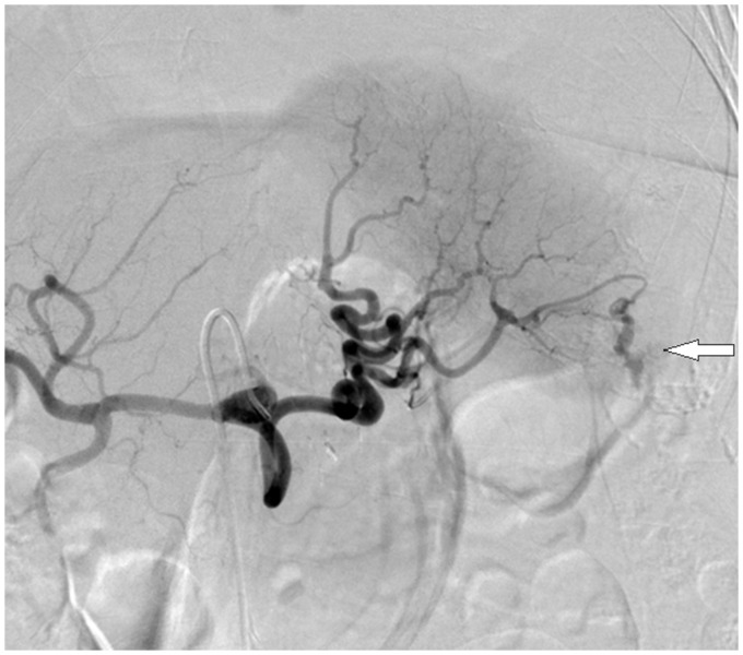
The procedure was performed under conscious sedation within 90 min of arrival at the ED. Briefly, standard right common femoral arterial access was obtained with placement of a 5F vascular sheath (Avanti Sheath Set, Cordis, Bridgewater, NJ, USA). The celiac trunk ostium was stenotic but was successfully cannulated using a Simmons 1 catheter (Merit Medical, South Jordan, UT, USA). Digital subtraction angiography confirmed active contrast extravasation from the inferior splenic pole (Fig. 2). A microcatheter (Renegade STC, Boston Scientific, Natick, MA, USA) was used to selectively cannulate the bleeding lower pole splenic artery branch. Four 2 × 4 mm microcoils (Hilal Embo coil, Cook Medical, Bloomington, IN, USA) were placed and hemostasis achieved (Fig. 3). Subsequent selective DSA images confirmed cessation of extravasation with normal enhancement of the remaining splenic parenchyma (Fig. 4). The patient remained hemodynamically stable but described new right inguinal and lower back pain 24 h following the procedure. Given concern for a possible retroperitoneal hematoma, she underwent a repeat abdominal multidetector CT of the abdomen. This showed no evidence of a retroperitoneal hematoma and a stable perisplenic hematoma with no active contrast extravasation. No blood transfusions were administered before, during or after the embolization procedure. Of note, the patient did not develop symptoms or signs of post-embolization syndrome following the procedure. The patient was discharged on the third postprocedural day with a stable hematocrit of 35% and resolution of symptoms. She remained asymptomatic on outpatient clinic review 3 months later.
Fig. 2.
AP spot film from digital subtraction celiac axis arteriogram. Celiac axis DSA demonstrates conventional arterial anatomy and confirms active contrast extravasation from the inferior pole of the spleen (white arrow). Note is made of normal perfusion of the remaining splenic tissue.
Fig. 3.
AP spot film from digital substraction selective lower pole splenic artery arteriogram. Image demonstrates microcatheter with tip positioned at bifurcation of the lower pole splenic artery (white arrow).Of note contrast extravasation not identified due to transient guide wire induced vasospasm.
Fig. 4.
30 degree RAO digital subtraction selective splenic artery arteriogram. Image acquired following coil deployment in the inferior pole splenic artery branch. No active extravasation identified. Note preservation of flow to the remaining splenic parenchyma and a preserved accessory left colic arterial branch (white arrow).
Discussion
Colonoscopy has become the gold standard for diagnosis of colonic pathology. Researchers estimate the number of colonoscopies performed in the US to be in the region of 14.2 million procedures per year (7), with this number projected to increase with the planned introduction of surveillance and screening programs. While considered a safe and well tolerated procedure, complications can occur. A retrospective review of 16,000 colonoscopies reported intraluminal bleeding (4.8 per 1000) and colonic perforation (0.9 per 1000) to be the two most common complications. Of note, this large series described no cases of splenic injury (1). Other rare complications include pneumothorax, volvulus, appendicitis, and retroperitoneal abscess formation.
Splenic injury at colonoscopy is a rare but potentially life-threatening complication. The cause of splenic injury during colonoscopy is likely multifactorial either related to direct trauma on passage of the scope through the splenic flexure, traction on the splenocolic ligament or fibrous adhesions between the spleen and colon that may have developed following pancreatitis, surgery, or inflammatory bowel disease (8). Splenomegaly, anticoagulant therapy, and administration of external pressure during the colonoscopy all contribute to the risk of splenic injury. The liberal use of intravenous sedation has been suggested to increase the risk of splenic injury as patients cannot report pain associated with stretching of the splenocolic ligament (9). In our case the spleen was normal in size and injury to the lower splenic pole was likely secondary to direct traction on the splenocolic ligament on manipulation of the endoscope, possibly exacerbated by adhesion formation following prior abdominal surgery.
A retrospective review by Kamath et al. gives an incidence rate of 0.001% (4 in 293,000 colonoscopies) (10). A retrospective medicolegal review from Denmark also gave a very low incidence rate for colonoscopy related splenic injury with only eight cases reported over a 14-year period, with an estimated 39,000 colonoscopies performed per year (11). The bulk of the available data on this rare complication comprises case reports and literature reviews. A recent comprehensive literature review identified 93 cases (3). Splenic injury was found to be more common in women (67%) and followed an “uneventful” colonoscopy in the majority of cases (63%). Ninety-four percent of patients presented with abdominal pain. Of the 78 cases in which hemodynamic status was documented, the majority (65%) described hypotension on presentation with an associated tachycardia seen in 42%. Twenty-six percent of patients, as in our case, presented 24 h or more following the procedure. On review of management 74% of patients were treated with splenectomy and a further 4% with mesh splenorrhaphy. Non-operative management was sufficient in 22% with only three cases of SAE described in this review. Ninety-eight percent of available pathology reports following splenectomy described a morphologically normal spleen. In all, five deaths were reported, two following splenectomy and one following SAE. Overall the reported mortality following splenectomy (2 deaths following 72 splenectomy procedures) for colonoscopy-related splenic injury is low, however, morbidity associated with altered immune status and long-term antibiotic prophylaxis has not been evaluated in this patient cohort.
SAE is now an accepted, safe, and effective means of controlling splenic hemorrhage non-operatively (6). Concern for post-splenectomy infection has increased interest in splenic preservation therapies. Proximal embolization of the main splenic artery is appropriate in diffuse splenic hemorrhage, with multiple small bleeding vessels, when rapid embolization of the entire spleen is required or vessel tortuosity prevents more distal catheter position (12). The main disadvantage of main splenic artery occlusion is that it may prevent or at least complicate a repeat procedure in the event of further bleeding. Selective embolization, when possible, allows targeting of specific bleeding vessels while preserving normal arterial supply to the remaining spleen. Although some studies suggest an increased rate of splenic infarction following super selective embolization (13), these small infarcts are rarely of clinical significance. It may be postulated that splenic injury following colonoscopy is most likely to occur to the inferior portion of spleen attached to the splenocolic ligament, and superselective embolization may allow targeted control of the localized area of hemorrhage whilst preserving perfusion of the remaining splenic tissue. No published series, however, has specified the precise anatomic location of the causative splenic laceration.
Although the role of SAE in the successful non-operative management of splenic injury is well accepted, we have identified only seven reported cases in the literature describing this technique in the treatment of splenic laceration following colonoscopy (12,14–17). In all seven cases proximal embolization of the main splenic artery was performed using endovascular coils. In five cases a satisfactory outcome was achieved with control of splenic hemorrhage and prompt patient discharge (14–17). One case required subsequent splenectomy, however, the reason for this was not discussed (18). In a second case the patient was deemed unsuitable for operative management of the splenic laceration despite hemodynamic instability given his poor respiratory function. He underwent SAE with satisfactory hemostasis but died from respiratory complications 6 days following embolization (12).
The under-representation of SAE as an adjunct to non-operative management when compared to splenectomy in this patient population may relate to diagnostic delays and the availability of angiography given many cases were reported before 1990. A more rapid diagnosis in the era of contrast-enhanced CT is noted on more recent case reports.
The presence of active contrast extravasation on multidetector CT is predictive of likely failure of non-operative management in an otherwise stable patient with splenic injury following blunt trauma (19,20). A similar outcome is likely in patients with splenic injury following colonoscopy and guided management in our case.
Left upper quadrant pain following colonoscopy is not uncommon and without performing routine CT after every colonoscopy the true incidence of clinically insignificant splenic injury will never be known. With increasing numbers of colonoscopies being performed and the liberal use of CT in the evaluation of abdominal pain, the number of cases diagnosed is likely to increase. With only five articles relating to splenic injury during colonoscopy described in the radiology literature and only two cases reported in the interventional radiology literature, awareness of this rare complication, and the role of interventional radiology in its management may be low in our community.
In conclusion, splenic injury is an uncommon but potentially life-threatening complication of colonoscopy. When this diagnosis is considered a contrast-enhanced CT should be performed as this will not only yield a diagnosis, but also influence management approach, guide patient selection and assist with procedure planning. In hemodynamically stable patients, super selective embolization is a safe and effective means of controlling localized splenic hemorrhage, increasing probability of successful non-operative management and thus preserving splenic function.
References
- 1.Levin TR, Zhao W, Conell C, et al. Complications of colonoscopy in an integrated health care delivery system. Ann Intern Med 2006; 145: 880–886 [DOI] [PubMed] [Google Scholar]
- 2.Wherry DC, Zehner H. Colonoscopic fiberoptic endoscopic approach to the colon and polypectomy. Med Ann DC 1974; 43: 189–192 [PubMed] [Google Scholar]
- 3.Shankar S, Rowe S. Splenic injury after colonoscopy: Case report and review of literature. Ochsner J 2011; 11: 276–281 [PMC free article] [PubMed] [Google Scholar]
- 4.Michetti CP, Smeltzer E, Fakhry SM. Splenic injury due to colonoscopy: analysis of the world literature, a new case report, and recommendations for management. Am Surg 2010; 76: 1198–1204 [DOI] [PubMed] [Google Scholar]
- 5.van der Vlies CH, van Delden OM, Punt BJ, et al. Literature review of the role of ultrasound, computed tomography, and transcatheter arterial embolization for the treatment of traumatic splenic injuries. Cardiovasc Intervent Radiol 2010; 33: 1079–1087 [DOI] [PMC free article] [PubMed] [Google Scholar]
- 6.Haan JM, Biffl W, Knudson MM, et al. Western Trauma Association Multi-Institutional Trials Committee. Splenic embolization revisited: a multicenter review. J Trauma 2004; 56: 542–547 [DOI] [PubMed] [Google Scholar]
- 7.Seef LC, Richards TB, Shapiro JA, et al. How many endoscopies are performed for colorectal cancer screening? Results from CDS’s survey of endoscopic capacity. Gastroenterology 2004; 127: 1670–1667 [DOI] [PubMed] [Google Scholar]
- 8.Ahmed A, Eller PM, Schiffman FJ. Splenic rupture: an unusual complication of colonoscopy. Am J Gastroenterol 1997; 92: 1201–1204 [PubMed] [Google Scholar]
- 9.Pfefferkorn U, Hamel CT, Viehl CT, et al. Haemorrhagic shock caused by splenic rupture following routine colonoscopy. Int J Colorectal Dis 2007; 22: 559–560 [DOI] [PubMed] [Google Scholar]
- 10.Kamath AS, Iqbal CW, Sarr MG, et al. Colonoscopic splenic injuries: incidence and management. J Gastrointest Surg 2009; 13: 2136–2140 [DOI] [PubMed] [Google Scholar]
- 11.Petersen CR, Adamsen S, Gocht-Jensen P, et al. Splenic injury after colonoscopy. The Patient Insurance Association, Copenhagen, Denmark. Endoscopy 2008; 40: 76–79 [DOI] [PubMed] [Google Scholar]
- 12.de Vries J, Ronnen HR, Oomen AP, et al. Splenic rupture following colonoscopy, a rare complication. Neth J Med 2009; 67: 230–233 [PubMed] [Google Scholar]
- 13.Bessoud B, Denys A, Calmes JM, et al. Nonoperative management of traumatic splenic injuries: is there a role for proximal splenic artery embolization? Am J Roentgenol 2006; 186: 779–785 [DOI] [PubMed] [Google Scholar]
- 14.Stein DF, Myaing M, Guillaume C. Splenic rupture after colonoscopy treated by splenic artery embolization. Gastrointest Endosc 2002; 55: 946–948 [DOI] [PubMed] [Google Scholar]
- 15.Holubar S, Dwivedi A, Eisdorfer J, et al. Splenic rupture: an unusual complication of colonoscopy. Am Surg 2007; 73: 393–396 Erratum: 2007;73:1198 [PubMed] [Google Scholar]
- 16.Parker WT, Edwards MA, Bittner JG, 4th, et al. Splenic hemorrhage: an unexpected complication after colonoscopy. Am Surg 2008; 74: 450–452 [PubMed] [Google Scholar]
- 17.Corcillo A, Aellen S, Zingg T, et al. Endovascular treatment of active splenic bleeding after colonoscopy: a systematic review of the literature. Cardiovasc Intervent Radiol 2013; 36: 1270–1279 [DOI] [PubMed] [Google Scholar]
- 18.Fishback SJ, Pickhardt PJ, Bhalla S, et al. Delayed presentation of splenic rupture following colonoscopy: clinical and CT findings. Emerg Radiol 2011; 18: 539–544 [DOI] [PubMed] [Google Scholar]
- 19.Marmery H, Shanmuganathan K, Alexander MT, et al. Optimization of selection for nonoperative management of blunt splenic injury: comparison of MDCT grading systems. Am J Roentgenol 2007; 189: 1421–1427 [DOI] [PubMed] [Google Scholar]
- 20.Schurr MJ, Fabian TC, Gavant M, et al. Management of blunt splenic trauma: computed tomographic contrast blush predicts failure of nonoperative management. J Trauma 1995; 39: 507–512 discussion 512–513 [DOI] [PubMed] [Google Scholar]


