Abstract
We present a case of a gentleman in his 70s with extensive subcutaneous emphysema. Usually self-limiting, subcutaneous emphysema around the thoracic inlet can rarely lead to airway and cardiovascular compromise by compression of structures in the neck. This patient presented with a large pneumothorax on a background of chronic obstructive pulmonary disease (COPD). This was initially treated with an intrapleural chest drain. However, after removal of this drain, the patient developed subcutaneous emphysema and later signs of tension pneumothorax. Further intrapleural chest drains were required. One of these chest drains produced a broncho-subcutaneous fistula, which contributed to extensive subcutaneous emphysema. He developed symptoms of dysphonia and dysphagia. A subcutaneous drain was inserted for palliation of his symptoms and to improve his quality of life. His symptoms improved significantly after insertion of this subcutaneous drain. There are only a handful of case reports published on interventions to relieve subcutaneous emphysema.
Keywords: Chronic obstructive pulmonary disease, lung injury, pneumothorax, subcutaneous emphysema, subcutaneous tissue
Introduction
This case deals with the management of extensive subcutaneous emphysema in a patient with a broncho-subcutaneous fistula. Subcutaneous emphysema around the thoracic inlet can rarely lead to airway and cardiovascular compromise by compression of the upper airway and jugular vessels.
Case Report
A gentleman in his early 70s with a history of severe chronic obstructive pulmonary disease (COPD) presented to the emergency department with acute onset shortness of breath and associated back pain. Oxygen was given with the aim of maintaining oxygen saturation between 88% and 92% due to his COPD. Arterial blood gas upon admission showed hypoxia with decompensated respiratory acidosis. An urgent chest X-ray showed a large right-sided pneumothorax. A 12Fr intrapleural chest drain was inserted into the right axilla using the Seldinger technique. Clinically, the bubbling underwater seal and the respiratory swing of fluid confirmed chest tube patency. The patient improved rapidly after chest drain insertion.
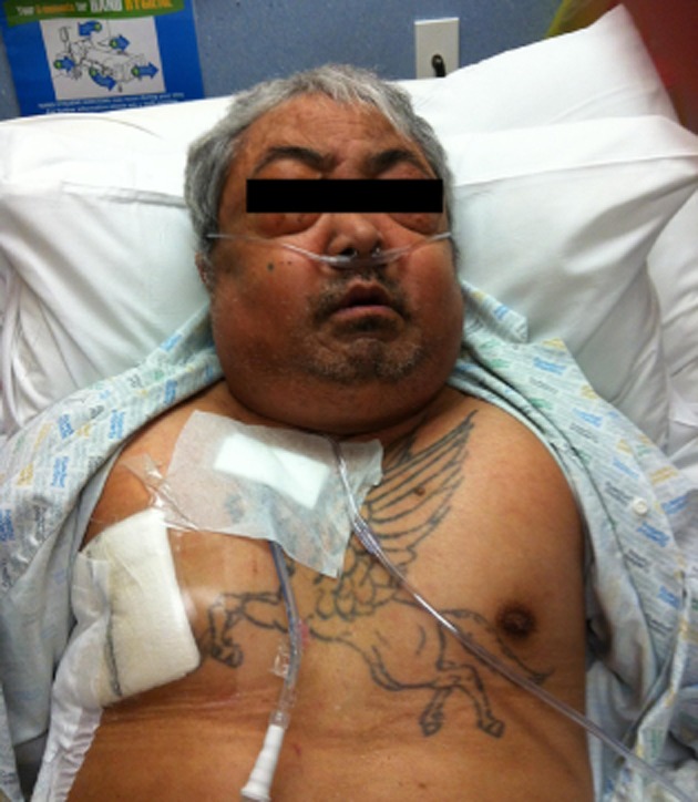
Two days later, the lung was re-inflated on chest X-ray and there had been no air leak seen for 24 h. The chest drain was removed. However, a chest X-ray 4 h after showed extensive subcutaneous emphysema in the chest wall. The subcutaneous emphysema progressed rapidly and the patient was found to have clinical signs of tension pneumothorax. Needle decompression was performed prior to insertion of a large bore 26Fr chest drain in the right axilla. The drain functioned normally after insertion with substantial air leak. Despite this, the subcutaneous emphysema progressed to involve the face, neck, abdomen, and arms (Fig. 1). A chest computed tomography (CT) was requested, which showed that the chest drain traversed the pleural space and entered the lung parenchyma. This intrapulmonary drain was removed and a further two chest drains were inserted into the pleural space, their position was confirmed by chest CT. The skin incision made for the original drain spontaneously sealed over, creating a broncho-subcutaneous fistula.
Figure 1.

Pre-subcutaneous drain insertion. Pleural drains can be seen exiting from right axilla and right apical area. Extensive subcutaneous emphysema was observed.
The surgical emphysema extended to involve the lower limbs over a 2-week period. The patient developed dysphonia and dysphagia, and was unable to see due to palpebral closure. A decision was made to insert a subcutaneous drain with the aim of palliation of symptoms and to improve his quality of life.
We used the approach described by Sherif and Ott [1] as a general guide. After subcutaneous local anesthetic, we made a small (1.5 cm) transverse incision on the left chest wall in the mid-clavicular line, midway between the clavicle and nipple. There was an immediate hiss of air when the subcutaneous plane was reached. Using blunt dissection, we created a 6-cm vertically oriented tract just superficial to the pectoral fascia. We used a 12Fr Seldinger chest drain, and in addition to the available five holes, we created an extra two drainage holes at the side with a scalpel blade and enlarged the apical hole by cutting off the tip. The drain was oriented superiorly in the subcutaneous plane. It was secured with a stay stitch and was placed on low pressure suction of 5 cm H2O. Within the hour, the subcutaneous emphysema had improved to the extent that the patient was able to open his eyes. His dysphonia and dysphagia resolved as he continued to improve over the next 5 days.
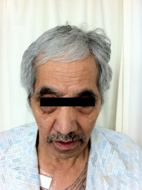
The air leak from both the intrapleural and the subcutaneous chest drains diminished and ceased, indicating closure of the broncho-subcutaneous fistula. The drains were removed sequentially with no recurrence of pneumothorax or subcutaneous emphysema (Fig. 2). Prior to discharge, the patient's mood had improved and he had returned to his baseline level of function.
Figure 2.

Patient prior to discharge, after removal of subcutaneous and pleural drains. Resolution of subcutaneous emphysema.
Discussion
Subcutaneous emphysema occurs when air is trapped under the skin. Both penetrating and blunt trauma to the respiratory and gastrointestinal tract can cause subcutaneous emphysema. Air forced into the interstitial tissues around the pulmonary vasculature travels back toward the hilum, leading to pneumomediastinum, and this eventually tracts into the soft tissue of the neck, face, and chest wall. In our patient, spontaneous pneumothorax was the initial event, exacerbated by an iatrogenic broncho-subcutaneous fistula created by the intrapulmonary drain.
If severe, subcutaneous emphysema can cause compression of the upper airway and jugular venous compression, which can lead to airway and cardiovascular compromise [2]. Clinically, patients can develop dysphonia and dysphagia as air dissects into the tissue planes of the neck. In rare cases, where subcutaneous emphysema causes progressive dyspnea, definitive airway management with tracheostomy may be required.
A variety of techniques to manage subcutaneous emphysema have been described in case reports. Herlan et al. [3] reported success in managing four patients who developed massive spontaneous subcutaneous emphysema with bilateral 3-cm infraclavicular incisions down to pectoralis fascia. Terada and Matsunobe [4] reported using a trochar-type chest tube as a subcutaneous drain. We elected to use the technique described by Sherif and Ott [1] as a guide because this case report gave a clear outline of the technique where they placed a Jackson-Pratt drain in the tract dissected. We used a 12Fr Seldinger drain instead because this was readily available to us. Following this, the drain was placed on gentle suction to ensure both tube patency and resorption of air. A case report by Srinivas et al. [5] describes the use of a modified microcatheter and active compressive massage face downward and arms upward toward the catheter to facilitate drainage.
A small number of different techniques have been described in the literature to manage subcutaneous emphysema. The superiority of each technique is debatable, but we found that the technique of Sherif and Ott [1] was both effective and safe.
Disclosure Statements
No conflict of interest declared.
Appropriate written informed consent was obtained for publication of this case report and accompanying images.
References
- 1.Sherif HM, Ott DA. The use of subcutaneous drains to manage subcutaneous emphysema. Tex. Heart Inst. J. 1999;26:129–131. [PMC free article] [PubMed] [Google Scholar]
- 2.Williams DJ, Jaggar SI, Morgan CJ. Upper airway obstruction as a result of massive subcutaneous emphysema following accidental removal of an inter-costal drain. Br. J. Anaesth. 2005;94:390–392. doi: 10.1093/bja/aei039. [DOI] [PubMed] [Google Scholar]
- 3.Herlan DB, Landreneau RJ, Ferson PF. Massive spontaneous subcutaneous emphysema. Acute management with infra-clavicular “blow holes”. Chest. 1992;102:503–505. doi: 10.1378/chest.102.2.503. [DOI] [PubMed] [Google Scholar]
- 4.Terada Y, Matsunobe S. Palliation of severe subcutaneous emphysema with use of a trocar-type chest tube as a subcutaneous drain. Chest. 1989;31:842–846. doi: 10.1378/chest.103.1.323a. [DOI] [PubMed] [Google Scholar]
- 5.Srinivas R, Singh N, Agarwal R, et al. Management of extensive subcutaneous emphysema and pneumomediastinum by micro-drainage: Time for a re-think? Singapore Med. J. 2008;48:e323–e326. [PubMed] [Google Scholar]


