Summary
In the last few decades, several researches have been conducted in the field of robotic rehabilitation to meet the intensive, repetitive and task-oriented training, with the goal to recover the motor function. Up to now, robotic rehabilitation studies of the upper extremity have generally focused on stroke survivors leaving less explored the field of orthopaedic shoulder rehabilitation. In this review we analyse the present status of robotic technologies, in order to understand which are the current indications and which may be the future perspective for their application in both neurological and orthopaedic shoulder rehabilitation.
Keywords: rehabilitation, robotic, shoulder, upper limb
Introduction
The aim of conventional rehabilitation is to recover the motor function using therapeutic exercises guided by a therapist who moves the patient’s body. An early and repetitive rehabilitation can substantially improve the long-term mobility of the shoulder in both neurological and orthopaedic patients1,2; furthermore, longer and more frequent training sessions have been shown to have beneficial effect in the short term3–5. Traditional rehabilitation techniques rely on well-established standard exercises, carried out by a therapist during in-patient hospital care and continued at home. As the rehabilitation sessions require involvement of a therapist for each patient this entails human and financial resources. In the last decades, in order to meet the intensive, repetitive and task-oriented rehabilitation, numerous and extensive research programs have been conducted in the field of robotic rehabilitation1–4. These systems can provide external assistive support to the human body, helping patients to experience pre-programmed limb movements and to improve related sensory-motor functions through repetitive practices. This may allow the patient to extend their training sessions providing an objective measure of the repeatability that it is hard to achieve with conventional physiotherapy. Up to date, robotic rehabilitation of the upper extremity have focused on stroke survivors studies1,2,5 without significant applications in orthopaedics. Motor disorders of the upper extremities, following orthopaedic or neurological injuries, include joint and muscular stiffness, muscle weakness, spasms, disturbed muscle timing and reduced ability to selectively activate muscles with abnormal synergistic movement patterns of arm and shoulder girdle. In the rehabilitation field, disabilities, residual motor function and efficacy of treatment cannot be quantified reliably as semi-quantitative evaluation scales are the only established methods to assess motor functions and its changes. Robots could allow quantitative measures of physical properties in a wide range of variation with levels of speed, accuracy, power and endurance over time that are unachievable by humans. Anyway, robots lack flexibility and adaptability, code-independent communication, high level information processing, detection and responsiveness to weak and otherwise undetected significant sensory inputs that characterize humans6–9. In the current study we describe the modern robotic systems for shoulder rehabilitation, focusing on the indications and other potential technologies that combined with robots can increase the benefits of rehabilitation to restore shoulder function.
Shoulder biomechanics
In the evolution of neuro-rehabilitation techniques, trunk stability has been considered essential to balance and coordinate the use of the extremities in daily functional activities10 that cannot be ignored even when we address orthopaedic shoulder rehabilitation. Since trunk muscles work together, their strength should be modulated through an appropriate neural control that allows trunk stability and limb movements; on this regards, there is literature evidence that the trunk is part of the prehension system11. Likewise, the role of shoulder and elbow in upper limb recovery is crucial because hand function cannot be obtained without the proximal control of its position in space. Research findings10,11 in motor control area have shown that during grasping and reaching, or sports motions, such as throwing or catching a ball, the trajectory of shoulder, elbow and hand are tightly coupled. This coupling is also task and situation dependent, such as reaching and grasping an object in different places and/or in different orientations. Soma et al.12 showed that we can dinamically distinguish different grips and arm directions from only around-shoulder muscle activities using EMG and acceleration sensors. For this reason, it’s useless to elaborate a complete shoulder and arm rehabilitation program without considering the biomechanics of both trunk and shoulder complex, especially when the aim is to develope new robotic systems for the upper limb.
The shoulder complex consists of the glenohumeral joint and the shoulder girdle that includes the sterno-clavicular, the acromion-clavicular and the scapulothoracic joints. The movements of these three joints shift the centre of gleno-humeral joint (CGH) and make them a closed kinematic chain, in which they cannot move independently. On these basis, the real physical therapy on the shoulder complex is performed by moving the humerus which consequently leads to shoulder girdle movement. Shoulder joint commonly means gleno-humeral joint with three degrees of freedom (DOFs) and this induces us to describe shoulder range of motion (ROM) with a three DOFs ball and socket model13. Vertical and lateral translations4,13,14 are shoulder girdle movements considered dominant and the only ones used to describe the effective shoulder movements. Nevertheless, humeral movements are associated with scapular movements: this is the so-called “scapulo-humeral rhythm” that changes with different planes of humeral elevation and with different angles of shoulder internal or external rotation. Furthermore, we have to consider individual difference in glenohumeral kinematics, so that, in patients with orthopaedic or neurological impairments who are unable to move by themselves the shoulder girdle, robotic assistance may be a valid rehabilitative option. This kind of patients tends to compensate the loss of shoulder motion with trunk movements that have the final effect to reduce the effectiveness of rehabilitation. Hence, the patient’s body should be fixed to limit compensatory movements and increase the use of shoulder girdle.
In order to align joint axes between the robot and the patient, the robot itself must follow the change of the CGH caused by shoulder girdle movement. If the robotic shoulder system is simplified, that means modelled on a three DOFs ball and socket joint, there will be a misalignment between the robot and the patient’s rotation axis due to the change of the CGH. This misalignment causes discomfort to the patient and leads to reduced work space for rehabilitation. Furthermore, whereas the robot moves excessively despite the misalignment, patients might get hurt with a joint glide. The complete human arm model for robotic systems has six DOFs that are as follow: shoulder girdle elevation/depression, shoulder girdle protraction/retraction, shoulder flexion/extension, shoulder abduction/adduction, shoulder internal/external rotation and elbow flexion/extension1,4,8.
Classification of robotic systems for shoulder rehabilitation
Robotic devices appear to be suitable for application under certain conditions and modalities that allow: i) individually adjust the rehabilitative training protocol with due accuracy, ii) obtain replication and congruity with residual motor function and treatment targets, iii) quantitatively assess baseline conditions and monitor changes during training. A robotic system traditionally comprises some major components8, namely:
– a mechanical structure with degrees of freedom consistent with the tasks to be executed;
– joint-controlling actuators, either electric or pneumatic;
– proprioceptive and exteroceptive sensors providing information on the machine functional status and interaction with environment;
– sequences of tasks to be executed as detailed by the system computer in suitable language;
– a computer generating the signals that control the robot joints, processing the signals transmitted by the sensors and instructing the motor controllers;
– man/machine interface receiving information/instructions from users (therapist/patient) and providing online feedback.
Robot can compensate for the patient’s inadequate strength or motor control at speeds individually calibrated on the residual motor functions, while constant feedback provides the patient with subjective perception of improvement. A variety of sensory, motor and cognitive inputs are needed, such as patient’s subjective control of voluntary movements, surface somatosensory inputs, proprioceptive static and dynamic information, pertinent visual information (e.g. virtual reality)15. In this perspective, motor performance is expected to improve in speed and precision of movement thanks to the repetition of calibrated and replicable exercises in intensive training programs8.
Robotic systems for rehabilitation can be classified or analysed from several points of view8,14,16. According to the control strategy, robots can be programmed to assist patient’s motion in different modes: i) passive: the robot moves patient’s arm, ii) active unassisted: the subject executes the exercise and the robot provide no help, iii) active assisted: the subject attempts to move and the robot provides assistance when there are some voluntary but inadequate movements, iv) resistive: the subjects is required to perform an exercise against an antagonist force provided by the robot.
According to their mechanical characteristics, robots can be classified into, at least, three main groups: a) exoskeletons, b) end-effectors (also called “operational type machines” or “manipulators”) c) and cable-driven.
Exoskeletons
Exoskeleton robotized prostheses and devices for rehabilitation are typically designed to match and align their mechanical joints to human limb joints, in order to achieve articular decoupling and a good coverage of the hole arm ROM4,13,14. Connected to each arm segment, they can independently control most of the articular DOFs and this characteristic gives them an important therapeutic advantage over end-effector based robots. Instead, the major drawback is related to the difficulty of faithfully reproducing all the DOFs of the articular complexes and of ensuring their alignment with those of the patients. As discussed previously, the shoulder complex is the most representative examples of these aspects and so, the development of upper limb exoskeletons needs to face important problems. Some systems have been optimized to face and limit singularities drawbacks at the shoulder, in order to exploit as much as possible the human shoulder range of motion. The mechanism design has to cope with two distinct and relevant aspects of upper limb rehabilitation: adaptability and compensation of shoulder displacements to prevent undesired shoulder internal stresses due to joint axes misalignments. Modern exoskeletons should collaborate with the patient in achieving the final goal of the therapy and, consequently, a significant transfer of torques from the human articulations to the mechanical joints (and vice versa) is required13. Exoskeletons usually reproduce shoulder and elbow articulations by a spherical joint centred on the humeral head and a revolute joint aligned to the elbow rotational axis. When human and robot DOFs are not coherent and properly aligned, the robot generates parasitic forces on the patient at the attachment points, that means on the joints themselves. These efforts may not only injure the patient, but also cause pain and long-term damage to healthy joints.
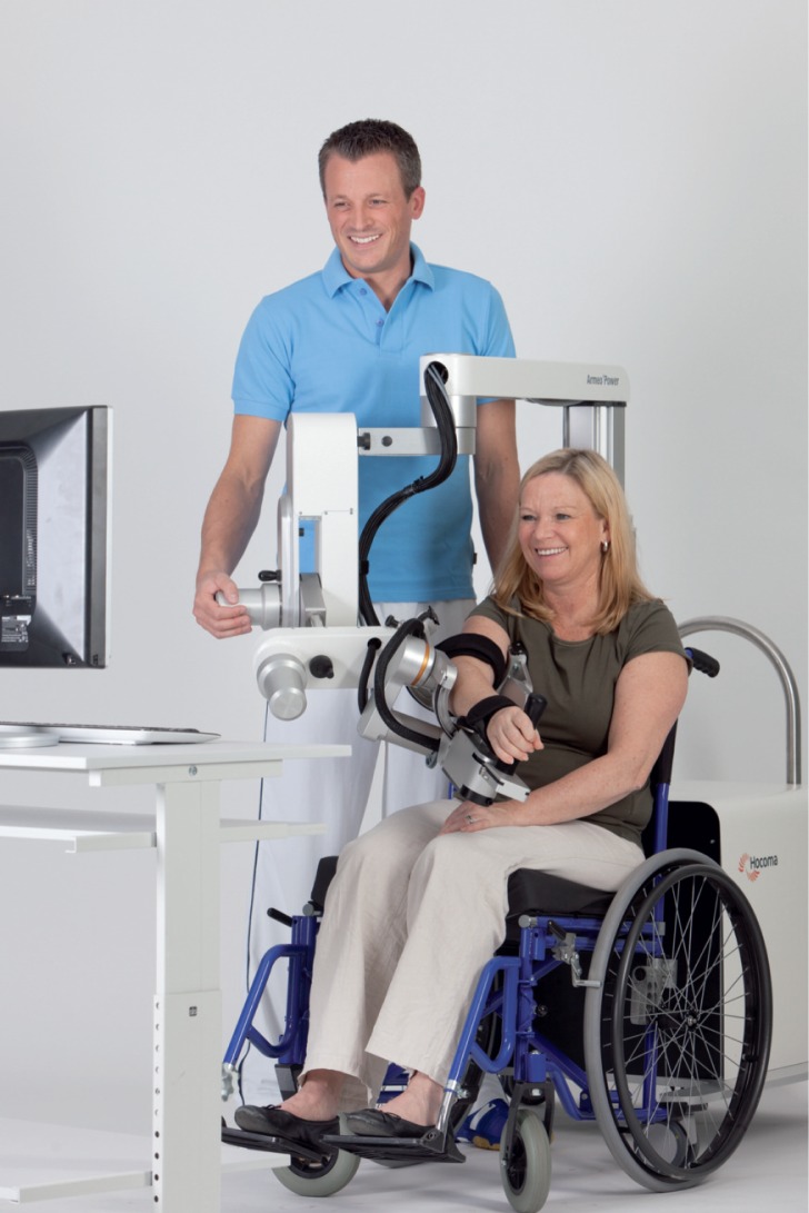
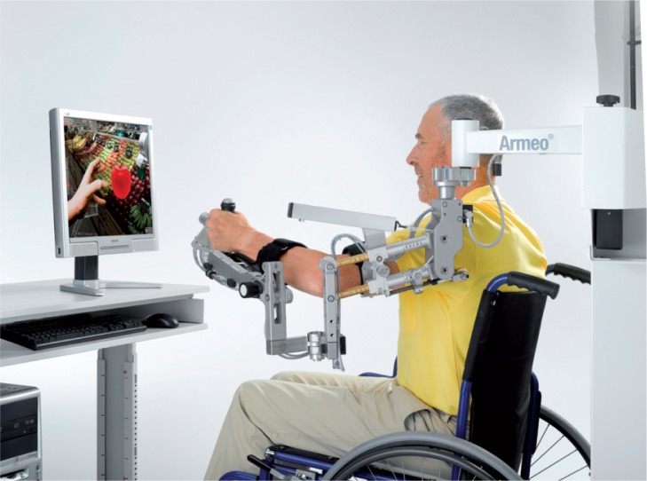
The existing exoskeletons are numerous and differ from each other by the number of DOFs they manage and by the technical solutions implemented to obtain these features4,14. For example, the robots CADEN-74,17 and L-EXOS4,18 don’t possess any of the translational DOFs of the shoulder girdle, but they play on the DOFs of the trunk, which is not rigidly fixed on the proximal end of the exoskeleton to compensate for these missing DOFs. The robots Armin III19,20 and Intelli Arm4 possess an additional vertical translational DOF coupled to arm elevation; the horizontal translational DOF is not taken onto account (Figs. 1,2). The robot MEDARM (Motorized Exoskeleton Device for Advanced Rehabilitation of Motor Function)14 is the most advanced as it uses two DOFs for shoulder girdle elevation/depression and protraction/retraction. However, misalignment occurs because this mechanism assumes the path of CGH to be a circular motion at the sterno-clavicular joint. Another category of exoskeletons3, not much developed, try to overcome the problems related to the alignment of robot and patient joints with a poly-articulate structure whose principle action consists in exerting on both sides of each joint the only efforts required for their mobilization. However, this robots have not yet been tested on patients and it is well known that the amplitudes reach with them are always lower than those normally reached by healthy people.
Figure 1.
Armeo® Power: an exoskeleton based on the ARMin technology (reprint with permission by Hocoma, Swiss Federal Institute of Technology, Zurich - Switzerland).
Even if the most advanced exoskeleton manages all the shoulder DOFs, it still requires, before starting the exercises, to be adapted to the patient size and adjusted to ensure the alignment of the mechanical and biological articulation. The time needed for this operation may not be negligible compared to the duration of a session, and thus detrimental to the interests of robot aided therapy. Exoskeletons are heavy machine that are not easily transportable, have high prices, and the patient is at risk of fractures. In addition, ROM and workspace are insufficient for rehabilitation due to collision of their components with each other3,4,13.
End-effectors
End-effector robots3,21,22 restrict the patient/machine interaction at the end-effector level, that means that they are connected to the patient at a single point, usually the forearm or the hand. They require little or no adjustment to patient’s size and morphology, but, obviously, they do not control all the upper limb DOFs, especially the ones of shoulder joint and shoulder girdle. The MIT-MANUS22 is the most used end-effector designed for clinical neurological application and developed at the Massachussets Institute of Technology (MIT), (Boston, MA, USA): it consists of two DOFs serial robot that may influence or interact with the patient’s arm over a working plane allowing the patient to execute reaching movements only in the horizontal plane. The GENTLE22 is an end-effector connected to the distal end of the arm through a three DOFs spherical joint and a wrist orthosis in order to position the forearm in the 3D space. The MIME (Mirror Image Movement Enhancer)4 consists of a six DOFs industrial robot manipulator connected to the forearm by means of a splint and it is able to position and orient the forearm in a 3D workspace.
Cable-driven
Cable-based or cable-driven parallel manipulators support and manipulate patient’s arm by different wires operating independently by different motors23. Cables are joined with an end-effector and a fixed frame through external connectors. The end-effector can be moved by changing the cable’s lengths, while preventing any cables from becoming slack. The structures are modular and have good inertial behaviour due to the fact that this kind of systems have small moving masses consisting of only cables and end-effector. In addition, they are easy to be transported, have low cost and simple maintenance, which are relevant characteristics for possible commercial use. One important drawback is the physical nature of cables that can only pull and not push. In addition, they compare the human shoulder to a simplified mechanical spherical joint with three DOFs and they have no control on shoulder joint and shoulder girdle.
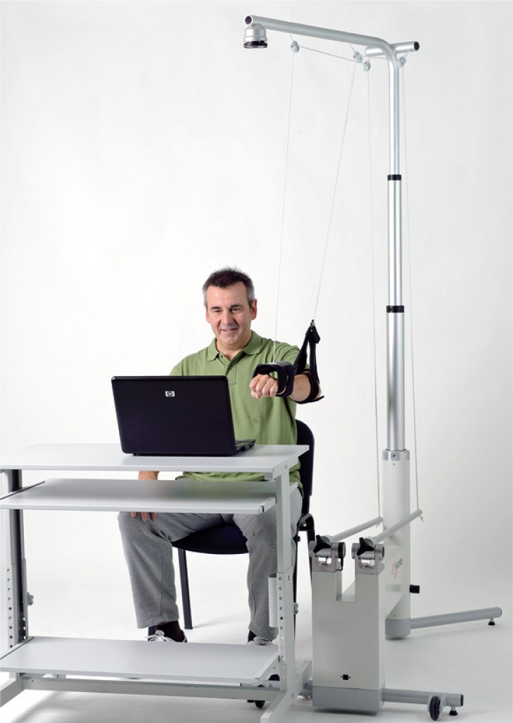
Several cable-based parallel structures have been designed for medical-rehabilitation use, such as23 MACARM (Multi-Axis Cartesian-based Arm Rehabilitation Machine), NeReBot (Neuro-Rehabilitation Robot), and MariBot (Marisa Robot). Their operating principle is simple: once the patient’s forearm is fixed in the splint (or orthosis), the machine can produce stimuli in the upper limb by pulling the cables. Wires can move (or interact with) the patient arm along a pre-planned 3D trajectory and, at the same time, out of path voluntary movements are still permitted, even while robotic assistance is provided. The patient hasn’t the feeling of being restrained by the robot and, at the same time, inertia is reduced to the minimum, requiring no sophisticated controls to create the feeling of low inertia robot (Fig. 3).
Figure 3.
Armeo® Boom: a simplified cable-driven manipulator designed for out-patient clinics and home settings (reprint with permission by Hocoma, Swiss Federal Institute of Technology, Zurich - Switzerland).
Robotic rehabilitation and other technologies
Robotic shoulder mechanical devices can measure speed, direction and strength of residual voluntary activity, can interactively evaluate patients’ movements and assist them in moving the limb through a predetermined trajectory during a given motor task, but with no information on singular muscle activity and no control on scapular compensatory movements.
There is growing interest in combining rehabilitation robots with Functional Electrical Stimulation (FES) to augment the benefit of each approach and extend the impairment range24. Although the muscular activities evoked by FES are different from the natural motor unit recruitment during voluntary muscle contractions, FES could effectively improve muscle strength in rehabilitation training. By accurately stimulating target muscles, FES may also limit the problem of “learned disuse” that chronic patients are gradually accustomed to managing their daily activities without using certain muscles, which has been considered as a significant barrier to maximizing the recovery of motor function and propioception. Currently, FES and rehabilitation robots are still separate systems and have not yet been synchronized at a system level.
Since the beneficial effect of any rehabilitation framework is likely to depend on the presence of proprioception, the robot has not to be simply a machine that imposes passive movements, as industrial robot would do, but a tool that helps the patient to relate force and movement. For this reason, motor rehabilitation is not limited to mechanical or muscular aspects, but is also deeply rooted in motor-cognitive issues, such as motor learning. This is the mission of exploiting the progressive introduction of haptic technologies in the robotic rehabilitation field5,6,9: robots have to provide proper feedbacks to guide the patient in a sensory-motor-type rehabilitative training. Haptics is important because it makes possible the bi-directional interaction between the robot and the patient, and this creates the causal relationship between effort and error that is fundamental for motor learning available to the brain. For example, when patients exercise in a virtual reality (VR) environment, they can monitor their movements and try to mimic the optimal motion patterns that are shown in real time in the virtual scenario. VR can also counterbalance adaptation, prevent boredom and therefore sustain attention by enhancing environmental diversity and promoting the subject’s interest6.
Another interesting technology that could be associated to robotics is the one introduced by Rodriguez et al.25 who have developed a Brain-Robot Interface for rehabilitation that artificially supports the sensory motor feedback loop. This tool permits the synchronization of the subject’s intention, or attempt, with the actual movement of the robot that guides the impaired limb. The relevant electrodes cover parts of the pre-motor, primary motor and somatosensory cortex. This system leads to simultaneous monitoring of positions/velocities of joint and neural signals and could permit future researches and rehabilitation strategies based on the correlations between real movement performance and neural content.
Indications for robotic rehabilitation
The objective of every training process is a relatively long lasting change in the quality of a movement. Each rehabilitative exercise must be intensive and specific in order to have an effective treatment; in addition, treatment itself must be repetitive, functional and motivating, so as to bring about an increase in performance, as well as learning, acquisition and generalization. In order to meet the intensive, repetitive and task-oriented training, robotic technologies can be useful tools to help patients and therapists to complete arm movement and stretch both muscles and soft tissues, thus preventing stiffness and contracture. Moreover, helping a weakened patient to complete a movement through a normal ROM introduces novel sensory-motor integration that otherwise would not be experienced26.
The success of functional joint movements depends on the kinaesthetic sense of the person, which is related with the propioception sense of the musculoskeletal structures of the joints. Since motor impairment is frequently associated with degraded proprioception and somatosensory functions, it is necessary to diagnose and then improve the loss of them. The repeated active exercises have a positive influence not only on motor deficits, but also on defective proprioception. Thus, robot assisted rehabilitation systems can be used not only to provide repetitive exercises, but also to improve proprioception6,10.
The status of motor function and the effect of any therapeutic intervention are generally measured by physiotherapists, using clinical assessment scales that probe specific aspects of subject’s motor behaviour. Although they could be standardised and validated, are prone to human errors that make them less reliable. The measurement obtained is always subjective and depends on the ability of the clinician. Robot devices can have the potential to measure displacements, velocities, forces and quantify other derived parameters. These measures could have the benefit of being objective, reproducible and capturing different aspects of motor improvement. Therefore, they could be successfully employed both for training and evaluation purposes. This topic is of great importance because the purpose is to provide researchers and clinicians with a standardised and reliable tool to evaluate patient’s outcomes with a set of objective, quantitative and highly repeatable measurements.
As stated in the introduction, up to now, most of the researches in the robotic fields regarding the upper extremities have focused on neurological patients suffering from paralysis or paresis due to stroke and traumatic brain injury, therefore we think that it should be interesting to consider additional fields of application such as peripheral nervous system injuries, degenerative pathologies of the central nervous system and muscular dystrophies. In order to the application of robotics in shoulder rehabilitation the major drawback is related to the joint axes misalignments between patient and robot, source of parasitic forces that can induce pain or damage in the healthy joints. In spite of these defects, it is reasonable to consider the robotic rehabilitation treating usefully and safely shoulder instability, stiffness (eg. adhesive capsulitis27), arthroplasty28, rotator cuff tears29 or other tendon ruptures30 (Tab. 1).
Table 1.
Potential applications of robotics in shoulder rehabilitation.
| Shoulder Pathologies | |
|---|---|
| Orthopaedic | Neurological |
| Instability | Paralysis/paresis |
| Conservative | Post-stroke |
| Postoperative | Post-traumatic brain injuries |
|
| |
| Stiff shoulder | Peripheral nervous system injuries |
| Idiopatic | |
| Postoperative | |
| Post-traumatic | |
|
| |
| Arthroplasty | Degenerative diseases of the central nervous system |
|
| |
| Rotator cuff tears | Muscular dystrophies |
| Other tendon or muscle ruptures Pectoralis major Deltoid | |
Discussion
This paper analyses the present status of robotic technologies in the field of shoulder rehabilitation. Up to now, robotics and virtual reality, have proven to be applicable in the area of neuro-rehabilitation but not in the orthopaedic one. Their use in the treatment of the paretic upper limb appears promising, especially in post-stroke patients with upper limb impairment where results have been positive in terms of motor recovery, but poor in functional outcomes16. As previously described, all robotic systems developed nowadays have still too many drawbacks that limit the safe and thorough approach needed in orthopaedics. If the goal is to improve the ability to make functional movements, it seems better to have patients who perform functional movements, that means using a large number of DOFs of the upper limb. This requires the development of more sophisticated, but completely safe, multiple DOFs robotic therapy devices and, at the same time, the objective measurement of motor performance is important to identify the most beneficial rehabilitation approaches. Despite the increasing use of robotic systems in clinical and research settings, it is still questioned which of the wide variety of available robotic outcome measures are relevant to assess arm movement. The current level of upper limb robotic technology should be considered as an advanced therapeutic tool under the direction of the rehabilitation team, composed by physiatrists and physiotherapists. As such, the robot can handles relatively simple treatments, characterized by repetitive and labour-intensive nature. Clinical decisions should be managed by the physiatrist and, if appropriate, should be planned and executed on the robot. Of course, both physiatrists and physiotherapists need to be adequately trained in the use of the robots for orthopaedic rehabilitation. Research findings on the efficacy and the advantages of robotic supported rehabilitation compared with conventional treatments remain poor. Moreover, a comprehensive scientific rationale and pathophysiological understanding of the mechanisms underlying recovery remain to be devised and discovered. The applicability of novel technologies depends on the efficacy and cost-benefit ratio as much as it requires scientific background, competence and communication to be shared by professionals and scientists from different fields. Further investigations on large samples of patients are required in order to: i) define the relationship between disability and residual function, ii) provide shared criteria of evaluation of both disability and outcome, iii) set new protocols of rehabilitation and identify the future role and use of robotics in the rehabilitation field31.
Last but foremost, upper limb motor function is essential in human daily living activities, for reaching and grasping, as well as for exploring and manipulating objects that allows the relational and emotional life. It is well-known that both arm and hand movements are under a more complex neural control than the leg and foot movements8, this is the main reason why the level of robotic development for the upper extremities is far from the one reached in the gait field. Which are the differences when comparing the motor impairment and the expectations of neurological and orthopaedic patients? Is there the same utility in investing resources for robotic development between these two rehabilitation fields? Which are the objectives when working with neurological or orthopaedic patients? There are many possible answers, but only a single and unquestionable shared consideration: the nervous system governs human function. Neurophysiology of both normal and pathological movement has the same importance when approaching neurological and orthopaedic rehabilitation. Only integration of knowledge among these two disciplines will produce the best results in the development of any kind of new technologies.
Figure 2.
Armeo® Spring: an ergonomic arm exoskeleton with integrated springs (reprint with permission by Hocoma, Swiss Federal Institute of Technology, Zurich -Switzerland).
References
- 1.Bishop L, Stein J. Three upper limb Robot devices after stroke rehabilitation: a review and clinical perspective. Neuro Rehabil. 2013;33:3–11. doi: 10.3233/NRE-130922. [DOI] [PubMed] [Google Scholar]
- 2.Waldner A, Hesse S, Tomelleri C. Transfer of scientific concept on clinical practice: recent robot assisted training studies. Funct Neurol. 2009;24:173–77. [PubMed] [Google Scholar]
- 3.Dehez B, Sapin J, Shoulde RO. An alignment-free two-DOF rehabilitation robot for the shoulder complex. IEEE International Conference on Rehabilitation Robotics (ICORR); 2011. pp. 1–6. [DOI] [PubMed] [Google Scholar]
- 4.Koo D, Chang PH, Sohn MK, Shin J. Shoulder mechanism design of an exoskeleton robot for stroke patient rehabilitation. IEEE International Conference on Rehabilitation Robotics (ICORR); 2011. pp. 1–6. [DOI] [PubMed] [Google Scholar]
- 5.Timmermans AA, Seelen HA, Willmann RD, Kingma H. Technology-assisted training of arm-hand skills in stroke: concepts on reacquisition of motor control and therapist guidelines for rehabilitation technology design. J Neuroeng Rehabil. 2009;6:1–18. doi: 10.1186/1743-0003-6-1. [DOI] [PMC free article] [PubMed] [Google Scholar]
- 6.Ozkul F, Barkana DE, Demirbas SB, Inal S. Evaluation of elbow joint proprioception with RehabRoby: a pilot study. Acta Orthop Traumatol Turc. 2012;46:332–338. doi: 10.3944/aott.2012.2702. [DOI] [PubMed] [Google Scholar]
- 7.Nunes WM, Rodrigues LAO, Oliveira LP, Ribeiro JF, Carvalho JCM, Goncalves RS. Cable-Based Parallel Manipulator for Rehabilitation of Shoulder and Elbow Movements. IEEE International Conference on Rehabilitation Robotics (ICORR); 2011. pp. 1–6. [DOI] [PubMed] [Google Scholar]
- 8.Pignolo L. Robotics in neuro-rehabilitation. J Rehabil Med. 2009;41:955–960. doi: 10.2340/16501977-0434. [DOI] [PubMed] [Google Scholar]
- 9.Mehrholz J, Platz T, Kugler J, Pohl M. Electromechanical and robot-assisted arm training for improving arm function and activities of daily living after stroke. Cochrane Database of Systematic Reviews. 2008;4:CD006876. doi: 10.1002/14651858.CD006876.pub2. [DOI] [PubMed] [Google Scholar]
- 10.Bartolo M, Don R, Ranavolo A, Serrao M, Sandrini G. Kinematic and neurophysiological models: future application in neurorehabilitation. J Rehabil Med. 2009;41:986–987. doi: 10.2340/16501977-0413. [DOI] [PubMed] [Google Scholar]
- 11.Cholewicki J, Panjabi M, Khachatryan A. Stabilizing function of trunk flexor-extensor muscles around a neutral spine posture. Spine. 1997;22:2207–2221. doi: 10.1097/00007632-199710010-00003. [DOI] [PubMed] [Google Scholar]
- 12.Soma H, Horiuchi Y, Gonzales J, Yu W. Preliminary results of online classification of upper limb motions from around-shoulder muscle activities. IEEE International Conference on Rehabilitation Robotics (ICORR); 2011. pp. 1–6. [DOI] [PubMed] [Google Scholar]
- 13.Malosio M, Pedrocchi N, Vicentini F, Tosacchi LM. Analysis of elbow joint misalignment in upper limb exoskeleton. IEEE International Conference on Rehabilitation Robotics (ICORR); 2011. pp. 1–6. [DOI] [PubMed] [Google Scholar]
- 14.Lo HS, Xie SQ. Exoskeleton Robots for upper limb rehabilitation: state of the art and future prospects. Med Eng Phys. 2012;34:261–268. doi: 10.1016/j.medengphy.2011.10.004. [DOI] [PubMed] [Google Scholar]
- 15.Ambike S, Schmiedeler JP. The leading joint hypothesis for spatial reaching arm motions. Exp Brain Res. 2013;224:591–603. doi: 10.1007/s00221-012-3335-x. [DOI] [PubMed] [Google Scholar]
- 16.Masiero S, Cararo E, Ferraro C, Gallina P, Rossi A, Rosati G. Upper limb rehabilitation robotics after stroke: a perspective from the University of Padua, Italy. J Rehabil Med. 2009;41:981–985. doi: 10.2340/16501977-0404. [DOI] [PubMed] [Google Scholar]
- 17.Perry JC, Rosen J, Burns S. Upper limb powered exoskeleton design. Trans Mechatronics. 2007;12:408–417. [Google Scholar]
- 18.Frisoli A, Bergamasco M, Carboncini M, Rossi B. Robotic assisted rehabilitation in virtual reality with the L-EXOS. Stud Health Technol Inform. 2009;145:40–54. [PubMed] [Google Scholar]
- 19.Nef T, Guidali M, Riener R. ARMin III – arm therapy exoskeleton with an ergonomic shoulder actuation. Appl Bionics Biomech. 2009;6:127–142. [Google Scholar]
- 20.Nef T, Riener R. Comfort two shoulder actuation mechanisms for arm therapy exoskeletons: a comparative study in healthy subjects. Med Biol Eng Comput. 2013;51:781–789. doi: 10.1007/s11517-013-1047-4. [DOI] [PubMed] [Google Scholar]
- 21.Ball SJ, Brown IE, Scott SH. MEDARM: a rehabilitation robot with 5DOF at the shoulder complex. IEEE Int Conf Adv Int Mech.; 2007. [Google Scholar]
- 22.Krebs HI, Ferraro M, Buerger SP, Newbery MJ, Makiyama A, Sandmann M, Lynch D, Volpe BT, Hogna N. Rehabilitation robotics: pilot trial of a spatial extension for MIT-Manus. J Neuro-engineering Rehabil. 2004;1:5. doi: 10.1186/1743-0003-1-5. [DOI] [PMC free article] [PubMed] [Google Scholar]
- 23.Rosati G, Gallina P, Masiero S. Wire-based robots for upper limb rehabilitation. Int J Ass Rob Mech. 2006;7:3–10. [Google Scholar]
- 24.Brunetti F, Garay A, Moreno JC, Pons JL. Enhancing Functional Electrical Stimulation (FES) for emerging rehabilitation robotics in the framework of HYPER project. IEEE International Conference on Rehabilitation Robotics (ICORR); 2011. pp. 1–6. [DOI] [PubMed] [Google Scholar]
- 25.Gomez-Rodriguez M, Grosse-Wentrup M, Hill J, Gharabaghi A, Scholkopf B, Peters J. Towards Brain Robot Interface in stroke rehabilitation. IEEE International Conference on Rehabilitation Robotics (ICORR); 2011. pp. 1–6. [DOI] [PubMed] [Google Scholar]
- 26.Schmidt RA, Lee TD. Motor control and learning: a behavioural emphasis. 4th ed. Champaign, IL: Human Kinetics Publishers; 2005. [Google Scholar]
- 27.D’Orsi GM, Giai Via A, Frizziero A, Oliva F. Treatment of adhesive capsulitis: a review. Muscles Ligaments Tendons J. 2012;2:70–78. [PMC free article] [PubMed] [Google Scholar]
- 28.Merolla G. Shoulder replacement in advanced glenohumeral osteoarthritis: current concepts review. OA Orthopaedics. 2013;1:7. [Google Scholar]
- 29.Giai Via A, De Cupis M, Spoliti M, Oliva F. Clinical and biological aspects of rotator cuff tears. Muscles Ligaments Tendons J. 2013;3(3):70–79. doi: 10.11138/mltj/2013.3.2.070. [DOI] [PMC free article] [PubMed] [Google Scholar]
- 30.Merolla G, Paladini P, Campi F, Porcellini G. Pectoralis major tendon rupture. Surgical procedures review. Muscles Ligaments Tendons J. 2012;2:96–103. [PMC free article] [PubMed] [Google Scholar]
- 31.Padulo J, Oliva F, Frizziero A, Maffulli N. Muscles, Ligaments and Tendons Journal. Basic principles and recommendations in clinical and field science research. MLTJ. 2013;4:250–252. [PMC free article] [PubMed] [Google Scholar]


