Abstract
The present study was undertaken to investigate the possible involvement of cattle in the epidemiology of peste des petits ruminants (PPR) as subclinical carriers. Cattle were exposed experimentally to PPR virus (PPRV) infection or placed in contact with PPR infected goats. Clinical samples including heparinized/EDTA blood, plasma, peripheral blood monocyte cells (PBMCs) and clotted blood (for serum) were collected periodically from 21 days post infection (dpi) to 397 dpi (21, 45, 50, 57, 65, 95, 111, 119, 148, 190, 203 and 397 dpi) and tested for PPRV antigen, nucleic acid and antibody. Exposed cattle seroconverted and maintained PPRV specific haemagglutinin antibodies and detectable PPRV antigen/nucleic acid in blood, plasma and PBMCs from 21 to 397 dpi. PPRV was recovered from blood and PBMC collected from experimental animals at 21 dpi, initially in B95a cells and then adapted to Vero cells. The study indicated that PPRV can infect cattle subclinically and PPRV antigen/nucleic acid persist in cattle for at least 397 days.
Keywords: Cattle, Experimental studies, PPR, Subclinical infection
Peste des petits ruminants virus (PPRV), the causative agent of PPR, is a member of the Morbillivirus genus of the Paramyxoviridae family. The seroprevalence of PPR in cattle [2], buffaloes [4, 9] and wildlife (Dorcas gazelle, Gemsbok) [7] has been reported and the infectious virus has been described from gazelles, buffaloes and camels [1, 9, 10]. The subclinical form of PPR in cattle may indicate novel aspects on the epidemiology and pathogenesis of PPRV with implications on the mechanism of adaptation of the virus in a new host niche. In this study, the ability of PPRV to cause subclinical infection in cattle exposed to the virus either directly or by exposure to infected goats was demonstrated. PPRV was isolated after 21 days post infection (dpi) from infected cattle PBMCs and the detection of PPRV antigen/nucleic acid in blood, plasma, PBMCs and antibody in serum samples from 21 to 397 dpi.
Calves (Nos. 182, 185 and 190) were kept in direct contact with four goats that had active PPRV experimental infection with virulent goat-adapted PPRV as described earlier [11]. Two calves (Nos. 92 and 95) were inoculated with 5 ml of 10 % PPRV infected goat spleenic suspension, subcutaneously as described earlier [11]. Nasal, oral and rectal swabs were collected from the cattle up to 14 dpi. The heparinised/EDTA blood and clotted blood were collected for PBMC and serum preparation, respectively from all the cattle after 21dpi periodically up to 397 dpi (21, 45, 50, 57, 65, 95, 111, 119, 148, 190, 203 and 397 dpi).
Serum samples were monitored for the presence of PPRV-specific antibodies by PPR competitive-ELISA (c-ELISA) [12], while, whole blood, PBMCs, plasma and swabs were subjected to sandwich ELISA (s-ELISA) [13] and RT-PCR assays [3, 6, 8] for the detection of PPRV antigen and nucleic acid, respectively. Indirect fluorescent antibody tests (IFAT) for antigen demonstration were carried out using PBMCs from s-ELISA positive animals.
Vero cells (CCL-18, ATCC) and B95a (EBV-transformed marmoset B) cell line, were propagated in growth media (EMEM and RPMI-1640, respectively) containing 10 % fetal bovine serum (FBS) and maintained with 2 % FBS. The whole blood and PBMCs were used for isolation of virus in both cell lines. Primary isolation of virus from cattle PBMCs was carried out in B95a and Vero cells using PBMCs that were positive by both s-ELISA and indirect FAT. Briefly, isolated PBMCs −1 × 10² cells (by co-cultivation) and blood −500 µl (by adsorption) with either 1 × 10⁶ B95a or Vero cells in a 25 cm2 tissue culture flask, incubated in 5 % CO2 and observed for morphological changes for 48–72 h with a change of media every alternate day. B95a cell harvests showing evidence of morbillivirus specific cytopathic effect (CPE), such as large syncytia, fusion and cording of cells at the third passage after isolation were freeze-thawed twice and further used to infect Vero cell. The virus (PPRV/Cattle/Muk/07) was passaged four-times in Vero cells and isolated. The cell culture adapted virus at the fifth passage after isolation from cattle was confirmed by s-ELISA, RT-PCR assays and sequencing (GenBank Acc. Nos. EF641263 and EF641264).
PPRV was detected in cattle following experimental infection and it remains to be seen as to whether the virus is in the process of adaptation to a new host or whether a subclinical infection is resulting due to the relative non-permissiveness of the host to the virus. Most of the clinical samples from animals tested positive for PPRV antigen in s-ELISA, except a blood sample from a cattle exposed indirectly and four PBMC samples each from cattle exposed directly and indirectly. PPRV exposed cattle revealed the presence of virus antigen in blood samples (blood/plasma/PBMC) (Table 1). At 397 dpi the s-ELISA was suddenly giving strongly positive in all the cattle. This is a very interesting observation as it indicates that the virus may have adapted in some way in the system, so the multiplication of the virus load in the blood may be high, which needs to be further studied.
Table 1.
Results of sandwich ELISA (s-ELISA) and RT-PCR assays employed to detect PPRV antigen/nucleic acid in blood samples collected after twenty-first days post infection (dpi) to 397 dpi from experimentally infected cattle
| Animal no | Mode of exposure to PPRV | Assays employed on different samples | Days post infection (dpi) | |||||||||||
|---|---|---|---|---|---|---|---|---|---|---|---|---|---|---|
| 21 | 45 | 50 | 57 | 65 | 95 | 111 | 119 | 148 | 190 | 203 | 397 | |||
| 92 | S/C inoculation | s-ELISAa | ++ | ++ | +++ | + + | ++ | + | – | – | – | – | ++ | +++ |
| RT-PCRb | + | + | + | + | + | + | – | – | – | + | + | + | ||
| 95 | s-ELISAa | ++ | ++ | +++ | +++ | +++ | ++ | + | ++ | + + | + | ++ | ++ | |
| RT-PCRb | + | + | + | + | + | + | + | + | + | + | + | + | ||
| 190 | Direct contact with infected goats | s-ELISAa | + | + | + | + | ++ | + | ++ | + | + | + | ++ | +++ |
| RT-PCRb | + | + | + | + | + | + | + | + | + | + | + | + | ||
| 185 | s-ELISAa | ++ | ++ | ++ | ++ | + | + | + | + | + + | + | ++ | +++ | |
| RT-PCRb | + | + | + | + | + | + | + | + | + | + | + | + | ||
| 182 | s-ELISAa | – | + | + | +++ | + | + | + | ++ | ++ | ++ | +++ | +++ | |
| RT-PCR | + | + | + | + | + | + | + | + | + | + | + | + | ||
aSandwich ELISA with reactivity in terms of OD496 value against each samples ranges mentioned in the parenthesis +++, strongly positive (0.76–1.4); ++, positive (0.186–0.67);+, Weakly positive (0.115 to 0.184); −, negative (0.089 to 0.096)
bN gene-based RT-PCR assay for PPRV nucleic acid detection
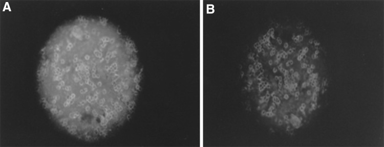
The PBMCs from an animal, (#182, highly reactive in s-ELISA) after 21 days post infection showed immunofluorescence in FAT (Fig. 1). To verify the presence of PPRV in cattle exposed to infected goats, PPRV N, M and F gene-specific RT-PCR assays were performed and the results indicate that some of the blood, plasma and PBMC samples were positive for either of the RT-PCR assays. The samples were scored more positive with N gene-based PCR than that of the M gene- and F gene-based RT-PCR assays, as reported earlier. Earlier, natural transmission of PPRV from sheep and goats to cattle (in-contact transmission) has been demonstrated [2] without any clinical signs but the cattle were seroconverted and protected against rinderpest virus challenge [5].
Fig. 1.

Detection of PPRV specific antigen from PBMCs collected from infected cattle (animal # 182) using PPRV anti-N monoclonal antibody (MAb) (a) and anti-H MAb (b) in IFAT (magnification at ×10). N protein staining showing characteristic diffused fluorescent foci but that was localizing on the cell surface in case of H MAb staining
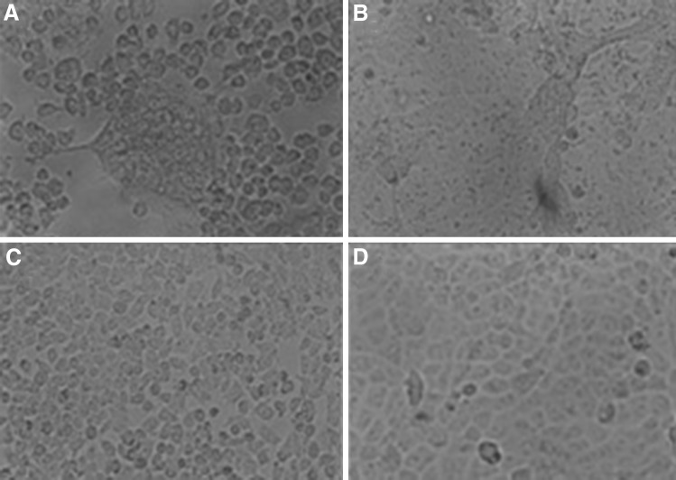
Virus was isolated from blood and PBMC samples collected from animals after 21 dpi exposed directly or indirectly. The characteristic CPE in B95a cells were syncytia and presence of large fusion masses. Initial isolation attempts in Vero cells were unsuccessful. However, B95a adapted virus, at passage three, produced CPE in Vero cells (Fig. 2). PPRV isolates either in B95a or Vero cells were detected by PPRV different gene-specific RT-PCR assays and serological assays. Infection was productive after four blind passages only in B95a cells. However, harvest from infected B95a cells induced productive infection in Vero cells on sixth passage. Virus isolation was successfully recovered from three animals (95, 182 and 190), where as inconclusive results obtained from two cattle (92 and 185). The titre observed for the isolated adapted virus in the Vero cells was 104.5 TCID50/ml. Further attempt was not made for the isolation of virus from infected cattle at different intervals, as the known virus was inoculated into the unnatural host to know the persistence of virus especially to detect virus antigen/nucleic acid in the blood samples.
Fig. 2.
Micrograph showing characteristic cytopathic effect of PPRV in B95a cells (a) and Vero cells (b) recovered from infected cattle with uninfected B95a (c) and Vero cells (d)
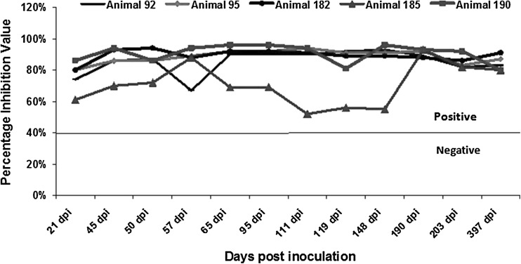
There was an agreement between antigen detection and virus isolation from PBMC of different animals. The results demonstrate the presence of virus in subclinically infected cattle that were exposed to PPRV. However, oral, nasal and rectal swabs that were screened up to 14 dpi were negative for PPRV antigen/nucleic acid detection in s-ELISA/RT-PCR assays. This indicates that the cattle pose no risk in transmitting the disease or secreting the virus in the natural secretion of the animals. The infected cattle seroconverted by 21 dpi and remained seroconverted until the end of the study (397 dpi). The animals maintained a high titer of PPRV-specific haemagglutinin inhibition antibodies in terms of PI values ranging from 55 to 96 % for a period from 21 to 397 dpi (Fig. 3) with detectable PPRV antigen in blood (Table 1).
Fig. 3.
Graph showing the PPR competitive ELISA results of screening of serum samples for PPRV antibody with PI values. Line shows negative/positive cut-off
The most significant finding of this study is that PPRV antigen and nucleic acid were detected for up to 397 dpi in cattle indicating that the virus is likely to be persisting in cattle for this time period. It is also interesting that virus was isolated from the cattle at 21 dpi. Further, it is clear from the experiment that (i) PPRV can infect cattle subclinically although their exact tropism is unknown. Data seems to indicate that PPRV persists in cattle for long periods (ii) PPRV was not detected in the secretions and excretions tested suggesting that they pose no or low risk in transmission of the virus to other animals (iii) PPRV can be isolated from PBMC of subclinically infected cattle. Further studies are needed to investigate whether the target species i.e. goat and sheep maintain PPRV for long periods subclinically and where the virus persists in subclinically infected cattle.
Acknowledgments
The authors wish to thank the Director, Indian Veterinary Research Institute (IVRI), for providing all the facilities and staff of Rinderpest and Allied Disease Laboratory, Division of Virology, IVRI, Mukteswar for their assistance.
Footnotes
A. Sen, P. Saravanan and V. Balamurugan have equal contribution.
References
- 1.Abu-Elzein EME, Housawi FMT, Bashareek Y, Gameel AA, Al-Afaleq AI, Anderson E. Severe PPR infection in gazelles kept under semi-free range conditions. J Vet Med B. 2004;51:68–71. doi: 10.1111/j.1439-0450.2004.00731.x. [DOI] [PubMed] [Google Scholar]
- 2.Anderson Mc Kay. The detection of antibodies against peste des petits ruminants virus in cattle, sheep and goats and the possible implications to rinderpest control programmes. Epidemiol Infect. 1994;112(1):225–231. doi: 10.1017/S0950268800057599. [DOI] [PMC free article] [PubMed] [Google Scholar]
- 3.Balamurugan V, Sen A, Saravanan P, Singh RP, Singh RK, Rasool TJ, Bandyopadhyay SK. One-step multiplex RT-PCR for detection of PPR virus in clinical samples. Vet Res Commun. 2006;30:655–666. doi: 10.1007/s11259-006-3331-3. [DOI] [PubMed] [Google Scholar]
- 4.Balamurugan V, Krishnamoorthy P, Veeregowda BM, Sen A, Rajak KK, Bhanuprakash V, Gajendragad MR, Prabhudas K. Seroprevalence of peste des petits ruminants in cattle and buffaloes from Southern Peninsular India. Trop Anim Health Prod. 2012;44(2):301–306. doi: 10.1007/s11250-011-0020-1. [DOI] [PubMed] [Google Scholar]
- 5.Dardiri AH, De Boer CJ, Hamdy FM. Response of American goats and cattle to peste des petits ruminants. In proceedings of the 19th annual meeting of the American association of veterinary laboratory Diagnosticians, 1976; pp. 337–344.
- 6.Forsyth MA, Barrett T. Detection and differentiation of rinderpest and peste-des-petits-ruminants viruses in diagnostic and experimental samples by polymerase chain reaction using P and F gene-specific primers. Virus Res. 1995;39:151–163. doi: 10.1016/0168-1702(95)00076-3. [DOI] [PubMed] [Google Scholar]
- 7.Furley W, Taylor P, Obi TU. An outbreak of peste des petits ruminants in a zoological collection. Vet Rec. 1987;121:443–447. doi: 10.1136/vr.121.19.443. [DOI] [PubMed] [Google Scholar]
- 8.George A, Dhar P, Sreenivasa BP, Singh RP, Bandyopadhyay SK. The M and N genes-based simplex and multiplex PCRs are better than the F or H gene-based simplex PCR for peste-des-petits-ruminants virus. Acta Virol. 2006;50:217–222. [PubMed] [Google Scholar]
- 9.Govindarajan R, Koteeswaran A, Venugopalan AT, Shyam G, Shaguna S, Shaila MS, Ramachandran FS. Isolation of pestes des petits ruminants virus from an outbreak in Indian buffalo (Bubalus bubalis) Vet Rec. 1997;141:573–574. doi: 10.1136/vr.141.22.573. [DOI] [PubMed] [Google Scholar]
- 10.Khalafalla AI, Saeed IK, Ali YH, Abdurrahman MB, Kwiatek O, Libeau G, Obeida AA, Abbas Z. An outbreak of peste des petits ruminants (PPR) in camels in the Sudan. Acta Trop. 2010;116(2):161–165. doi: 10.1016/j.actatropica.2010.08.002. [DOI] [PubMed] [Google Scholar]
- 11.Saravanan P, Sen A, Balamurugan V, Rajak KK, Bhanuprakash V, Palaniswami KS, Nachimuthu K, Thangavelu A, Dhinakarraj G, Hegde R, Singh RK. Comparative efficacy of peste des petits ruminants (PPR) vaccines. Biologicals. 2010;38(4):479–485. doi: 10.1016/j.biologicals.2010.02.003. [DOI] [PubMed] [Google Scholar]
- 12.Singh RP, Sreenivasa BP, Dhar P, Shah LC, Babdyopadhyay SK. Development of monoclonal antibody based competitive-ELISA for detection and titration of antibodies to peste des petits ruminants virus. Vet Microbiol. 2004;98:3–15. doi: 10.1016/j.vetmic.2003.07.007. [DOI] [PubMed] [Google Scholar]
- 13.Singh RP, Sreenivasa BP, Dhar P, Bandyopadhyay SK. A sandwich-ELISA for the diagnosis of peste des petits ruminants (PPR) infection in small ruminants using anti-nucleocapsid protein monoclonal antibody. Arch Virol. 2004;149:2155–2170. doi: 10.1007/s00705-004-0366-z. [DOI] [PubMed] [Google Scholar]


