Abstract
A simple, rapid and sensitive diagnostic assay for Foot-and-mouth disease (FMD) is required for deployment in the field. In this study, development of Reverse Transcription-Loop Mediated Isothermal Amplification (RT-LAMP) assay based on the 3D polymerase gene for specific and rapid detection FMD virus (FMDV) was carried out. The assay was optimised with viral RNA extracted from serotype O, A and Asia 1 FMDV vaccine strains, which resulted a reliable amplification at 65 °C for 60 min. The amplified RT-LAMP products were identified by agarose gel electrophoresis with ethidium bromide staining or observation by naked eye for the presence of turbidity and colour change following the addition of hydroxyl naphthol blue (HNB). The specificity of the assay was demonstrated by the absence of amplification of genome extracted from other viruses or cellular origin. With respect to analytical sensitivity the developed RT-LAMP assay was found more sensitive than routinely used multiplex PCR (mPCR). Further, the assay was evaluated with RNA extracted from cell cultured isolates (n = 50), tongue epithelial samples (n = 150) and semen samples from infected bulls (n = 13). In conclusion, RT-LAMP with HNB dye was shown to be simple, specific and sensitive assay for rapid diagnosis of FMDV infection. Further, the assay has the potential for field deployment and use for rapid FMDV surveillance in India.
Electronic supplementary material
The online version of this article (doi:10.1007/s13337-014-0211-2) contains supplementary material, which is available to authorized users.
Keywords: Foot and mouth disease, FMD virus, Diagnosis, Loop mediated isothermal amplification assay, mPCR
Introduction
Foot-and-mouth disease (FMD) is a highly contagious and economically important disease of both cloven-hoofed and wild animals [1]. The causative agent, foot-and-mouth disease virus (FMDV), a positive strand RNA virus, belongs to the genus Aphthovirus within the family Picornaviridae [3]. FMDV exists as seven serologically and immunologically distinct serotypes (O, A, C, Asia 1, SAT-1, -2, -3) [19], which are further sub-divided into distinct topotypes/genotypes based on phylogenetic analysis [10, 11]. FMD is endemic in India caused by serotypes O, A and Asia 1 [20].
During FMD outbreak, diagnosis is primarily dependent on the early recognition of clinical signs of the disease by the farmer and rapid reporting to the relevant authorities for confirmation. Typical cases of FMD are characterised by the formation of vesicles and epithelial erosions in the tongue, hard and soft palate, coronary band, and feet. However, FMD may not be distinguished clinically from other vesicular and look alike diseases. Therefore, confirmatory diagnosis is made by testing clinical samples in the reference laboratory for the presence of FMD virus and/or viral antigen. Molecular tool such as multiplex PCR [5] detects viral genome, and can be used on samples which fail to grow on tissue culture. Multiplex PCR (mPCR) has been shown to be an efficient and sensitive tool for FMDV detection at the Central laboratory, Project Directorate on Foot-and-mouth Disease (PDFMD), Mukteswar, India. However, during the course of large scale outbreaks in an FMD endemic country like India, the mPCR method is slow and cumbersome to perform.
The loop-mediated isothermal amplification (LAMP) method offers an attractive alternative to PCR based methods and it has been shown to be highly accurate for the detection of RNA viruses [8, 13, 22, 23]. The assay is being increasingly used for rapid detection and typing of emerging viruses, such as West Nile, severe acute respiratory syndrome, dengue, Japanese encephalitis, Chikungunya, and so forth [12, 15]. LAMP is based on the principle of strand displacement reaction that forms stem-loop structure products utilizing the Bst DNA polymerase enzyme and a set of 4 specially designed primers [16] and the reaction time is further reduced when two additional loop primers are added to a mixture [15]. LAMP amplifies target nucleic acid under isothermal conditions, usually between 60 and 65 °C [16]. Therefore, only simple equipment, such as a heating block or water bath, is required, in place of thermal cyclers as in PCR. In addition, RNA can directly be used as starting material as in PCR. The large amounts of gene products from LAMP reaction and the presence of by-product (e.g., magnesium pyrophosphate) make it possible to give diagnosis based on visual judgment of the turbidity or colour change of the reaction mixture with the addition of SYBR Green [12, 14, 16] or pre added hydroxyl naphthol blue [6]. For the first time in India this study describes the development and preliminary validation of a one-step, single tube RT-LAMP assay with HNB dye, targeting the FMDV 3D gene, for rapid detection of three serotypes of FMDV circulating in India.
Materials and methods
Virus isolates
Virus isolates were either cell culture adapted virus stocks or in the form of infected epithelial suspension collected from vesicular lesions. FMDV vaccine strains, O/IND R2/75, A/IND 40/00, and Asia 1/IND 63/72 in the form of infected cell culture supernatants were initially used as reference strains for the development of the RT-LAMP assay. For evaluation of the RT-LAMP assay, cell culture adapted viruses (n = 50) belonging to the serotypes O, A and Asia 1 and tongue epithelial samples (n = 150) were used.
Oligonucleotide primers
A conserved region of 3D RNA polymerase encoding gene of the FMDV genome was selected as a suitable target for designing the primers. The nucleotide sequences corresponding to the 3D polymerase coding region of various topotypes/lineages of FMDV serotypes O, A and Asia 1 were obtained from the sequence database of PDFMD, Mukteswar. For the RT-LAMP assay, six primers comprising two inner primers [forward inner primer (LMP-3D F2) and backward inner primer (LMP-3D R2)], two outer primers (LMP-3D F1 and LMP-3D R1) and two loop primers (LMP-3D F3 and LMP-3D R3), were designed using the Primer Explorer version 4 software. The amplified region lies between nucleotide 1183 and 1382 of 3D polymerase with an amplicon size of 199 bp (Supplementary fig. 1). The details of designed primers are mentioned in Supplementary table 1.
RNA extraction
Total RNA was extracted from 140 µl of original tongue epithelial suspension or from cell culture supernatant using QIAamp viral RNA mini kit (QIAGEN, Hilden, Germany) as per the manufacturer’s instruction. However, while extracting RNA from epithelial samples, an additional step of homogenization using QIA shredder columns (Qiagen, Germany) was included.
Optimization of the RT-LAMP assay conditions
Initially, the effect various concentrations of MgSO4 (2–14 mM), betaine (0–1.2 M), dNTPs (0.2–2 mM), the amplification temperature (60–67 °C), the reaction time (5–75 min) and the ratio of outer and inner primers (1:1–1:12) were identified to optimize the LAMP assay in order to amplify the nucleic acids extracted from FMDV vaccine strains of O, A and Asia 1 serotypes. Once optimized, the reaction was performed in a total 25 µl reaction volume, using 12.5 µl of reaction mixture buffer (2×) [(40 mM Tris–HCl (pH 8.8), 20 mM KCl, 12 mM MgSO4, 20 mM (NH4)2SO4, 0.2 % Triton, 2 M betaine (New England Biolabs) and 1.6 mM each of four dNTPs (fermentas)], 1 μl of primer mix at a ratio of 5 pmol of external primers: 50 pmol internal primers, 1 μl (8 units) Bst DNA polymerase (New England Biolabs); 0.2 μl (10 U/μl) of AMV reverse transcriptase (New England Biolabs); 7.3 μl of nuclease-free water; and 2 μl of RNA along with 1 μl of hydroxyl naphthol blue (3 mM). The reaction mixture was incubated at 65 °C for 60 min in a water bath. The reaction was terminated by heating at 80 °C for 10 min. BHK-21 cellular RNA and distilled water were used as negative control, while FMD viral RNA from cell culture suspension served as positive control in LAMP reactions.
Detection of RT-LAMP product
RT-LAMP products were detected either by agarose gel electrophoresis or observation by naked eye. RT-LAMP products were separated electrophoretically in 2 % agarose gel and visualized under U.V. light after staining with ethidium bromide. To make the assay simple and rapid two naked eye visualization methods were used for specific detection of LAMP products. Those are presence of turbidity in the reaction tube due to formation of magnesium pyrophosphate for positive LAMP reaction and the colour change (violet to sky blue in positive reactivity) due to the addition of hydroxyl naphthol blue (HNB; Sigma-Aldrich, St. Louis).
Multiplexed polymerase chain reaction (mPCR)
Multiplex PCR reaction was carried out in a 10 µl of reaction mix using Hot Start Taq DNA Polymerase as described [5]. The PCR includes universal FMD virus specific NK-61 primer (5′ GACATGTCCTCCTGCATCTG, negative-sense) and three serotype specific forward primers namely DHP13, DHP15 and DHP9 against O, A and Asia1, respectively. The basic PCR reaction mix contained l µl of RT product, l µl of 10X PCR buffer, 0.6 µl of 25 mM MgCl2, 0.4 µl of 10 mM dNTP mix, 4 pmols each of virus specific and serotype-specific primers, 0.25U of HotstarTaq DNA polymerase, 3.6 µl of RNase-free water. The thermal profile followed was one cycle of denaturation (95 °C for 15 min); 30 cycles of denaturation (95 °C for 30 s), annealing (60 °C for 30 s) and extension (72 °C for 30 s) followed by one cycle of final extension (72 °C for 7 min). The generated PCR products were visualized by running 2 % agarose gel and ethidium bromide staining. Serotypes were differentiated based on amplicon size i.e. 249, 376, 537 bp specific for serotype O, A and Asia1 respectively.
Sensitivity and specificity of RT-LAMP assay
Analytical sensitivity of RT-LAMP assay was evaluated and compared with mPCR using RNA extracted from 10-fold dilution series (from 10−1 to 10−12) of O, A and Asia 1 virus culture. The initial virus titers for O, A and Asia 1 FMDV were 6.33, 5.00 and 5.57 TCID50/50 µl respectively. This RT-LAMP products were visualized by changed in colour due to pre addition of HNB dye. The specificity of RT-LAMP assay was evaluated by cross-reactivity test with nucleic acid extracted from other viruses such as Blue Tongue Virus (BTV), Orf. Viral RNA from vaccine strains O/IND R2/75, A/IND 40/00, and Asia 1/IND 63/72 was used as positive control and RNA extracted from healthy goat and buffalo tongue epithelium, mock infected BHK-21 cell was used as negative control in each test. The specificity of RT-LAMP products were further confirmed by restriction enzyme digestion by Bam HI (Fermentas, USA) restriction enzyme and DNA sequencing. Briefly restriction enzyme digestion was performed in 20 µl reaction mixture containing 1× Tango buffer (Fermentas), 5 µl of RT-LAMP product, 20 IU of Bam HI (Fermentas) and incubated at 37 °C for 4 h and visualised in agarose gel.
Evaluation of RT-LAMP assay using field samples
To determine the performance of RT-LAMP assay on a variety of FMDV isolate from the field, clinical samples (n = 263) consisting of tongue epithelia (n = 150), semen samples (n = 13) and cell culture adopted virus (n = 50), were tested using both mPCR and RT-LAMP assay. These samples collected from different parts of India representing a broad coverage of inter- and intra-typic genetic variation of FMDV prevailing in India. Out of 150 tongue epithelial samples, n = 50 samples were both Ag-ELISA and mPCR positive, n = 50 samples were Ag-ELISA negative but mPCR positive, and n = 50 samples were both Ag-ELISA and mPCR negative.
Results
Optimization of assay conditions
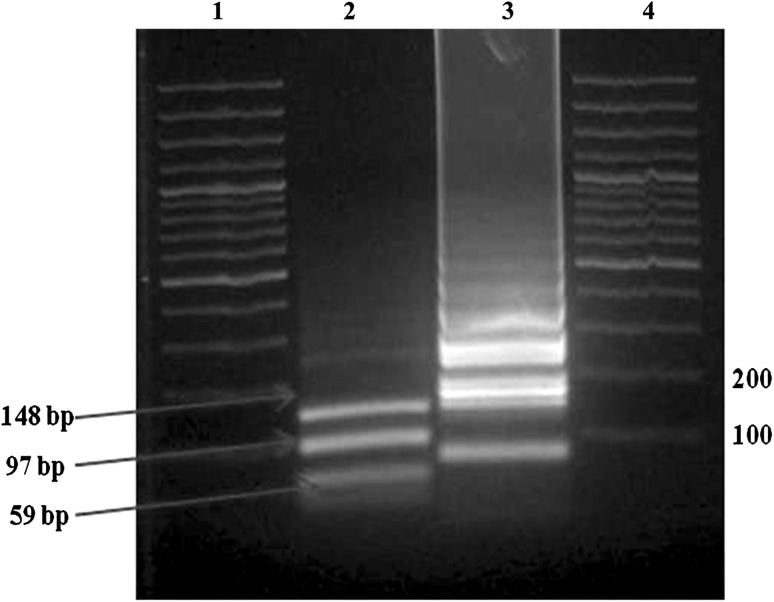
RT-LAMP assay was initially optimized using viral RNA from the FMDV vaccine strains belonging to the serotypes O, A and Asia 1 at different annealing temperature, reaction time, various concentration of Mg+2 ion, dNTPs and betaine. Taken together, the optimum temperature for RT-LAMP reaction to produce bright and distinct ladder-like pattern on the agarose gel was obtained at 65 °C (Supplementary fig. 2). A reaction time of 60–65 min was found optimum for FMDV genome detection. A minimum 6 mM Mg2+ ion concentration was needed to give a positive reaction product, but no additional change was observed by increasing the Mg2+ ion concentration up to 12 mM. It was observed that as betaine concentration was increased, the amounts of RT-LAMP reaction products were also increased; however, at the concentration of 1 M betaine the maximum amplification was detected. The optimal ratio of primers (inner-outer) was found to be 10:1 with the concentration of 50 and 5 pmol, respectively. After restriction digestion of RT-LAMP product with Bam HI enzyme, the specific ladder like pattern was disappeared and three fragments with approximate size of 148, 97 and 59 bp were obtained (Fig. 1).
Fig. 1.
Agarose gel electrophoresis and restriction analysis of RT-LAMP products. Lane 1 and 4 100-bp plus DNA ladder (Fermentas), lane 2 RT-LAMP product of FMDV digested with BamHI, lane 2 RT-LAMP products of FMDV
Sensitivity and specificity of RT-LAMP assay
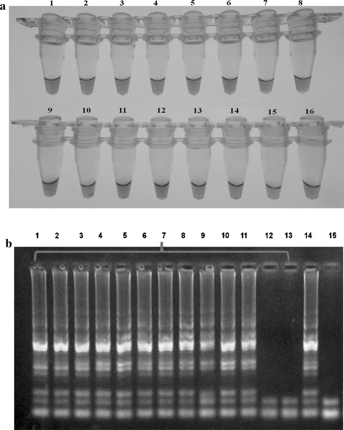
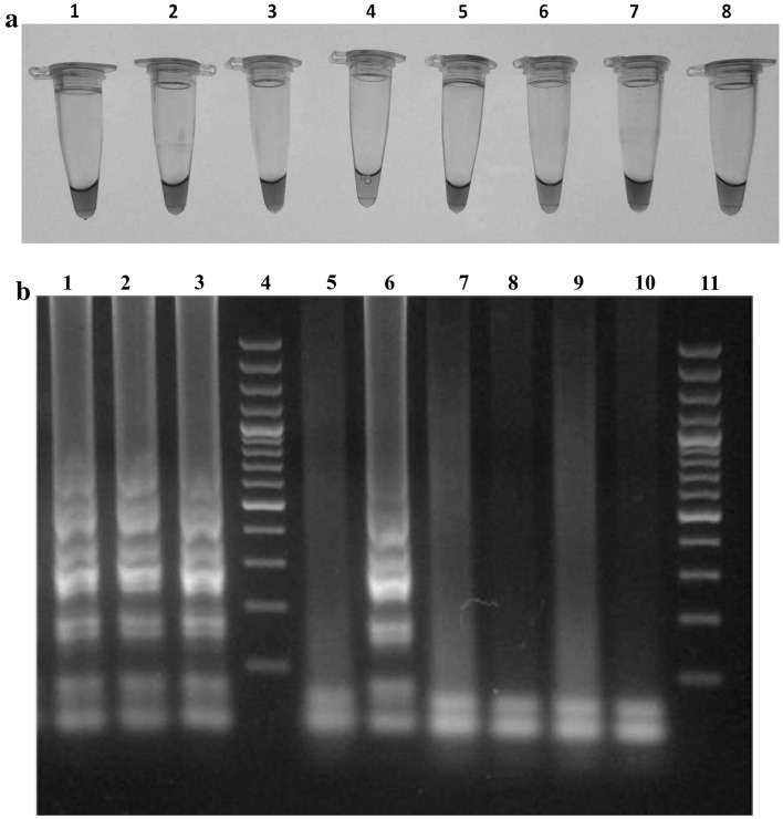
The sensitivities of the RT-LAMP and mPCR assays were analysed using viral RNA extracted from 10-fold dilution series of virus culture. The detection limit of RT-LAMP assay was 4.2 × 10⁻⁴, 2 × 10⁻⁶ and 1.1 × 10⁻⁴ TCID50/ml per reaction mixture for FMDV serotypes O, A and Asia1 respectively. While detection limit of mPCR was 158, 1.5 and 15 TCID50/ml for serotype of O, A and Asia1of FMDV, respectively. High level of analytical sensitivity of RT LAMP was observed compared to mPCR with a detection limit of 0.01 fg/µl (Fig. 2a, b) as compared to 1,000 fg/µl in mPCR. Therefore the analytical sensitivity of RT-LAMP assay was found higher than that of the conventional gel based mPCR assay. Further, it was also found that the currently developed RT-LAMP assay did not cross react with BTV, CSFV and yielded a negative reaction with RNA isolated from healthy goat and buffalo tongue epithelium (Fig. 3a, b). The specificity of RT-LAMP reaction was also tested by visual turbidity method, RE digestion and nucleotide sequencing (data not shown).
Fig. 2.
Comparative sensitivity of LAMP assay; serial 10-fold dilutions of control RNA (100 ng) was tested using LAMP and mPCR a Sensitivity of RT-LAMP monitored by visual observation and detected up to 0.01 fg/µl of RNA (tube 11), tube 14- positive control and tubes 15 & 16- negative control, b Agarose gel electrophoresis of RT-LAMP products of above. Lane (ladder like pattern of LAMP products may be seen) 14 positive control, lane 15 negative control
Fig. 3.
Specificity of LAMP assay a Tubes 1–3: Color change from violet to sky blue indicates LAMP positivity for serotype O, A and Asia1 FMDV RNA, respectively. While no color change was observed with RNA isolated from Blue Tongue Virus (BTV), Classical Swine Fever Virus (CSFV), and healthy tongue epithelium of bovine, respectively (Tubes 6,7, 8). Tube 4: negative control and tube 5: positive control. b Agarose gel electrophoresis of RT-LAMP products of the above LAMP products. Lanes 1–3 FMDV serotypes O, A and Asia1, respectively. Lane 4 and 11 100-bp plus DNA ladder (Fermentas). Lane 5 negative control, lane 6 positive control, lane 7, 8, 9 and 10 BTV, CSFV, healthy tongue epithelium of goat and sheep, respectively
Evaluation of RT-LAMP assay with field samples
A detailed evaluation of the assay was performed using RNA extracted from clinical samples obtained from suspected cases of FMD along with cell culture FMDV isolates. During this evaluation, the result of RT-LAMP assay was compared with that of the mPCR assay. All the cell culture FMDV isolates are found positive by both mPCR and RT-LAMP assays. Out of 150 suspected tongue epithelium samples 139 samples were found positive for FMDV by RT-LAMP assay, where as only 105 samples were found positive for FMDV by mPCR assay (Table 1). Similarly more number of semen samples was found positive for FMDV by RT-LAMP assay (12/13) as compared to mPCR assay (6/13). Therefore, both the analytical and diagnostic sensitivity of RT-LAMP assay were detected to be higher than the conventional mPCR currently being used at PD-FMD.
Table 1.
Diagnostic sensitivity of different tests against clinical samples
| Samples type | Total number of samples | mPCR positive (%) | RT-LAMP positive (%) |
|---|---|---|---|
| Tongue epithelium | 150 | 105 (70) | 139 (92) |
| Semen | 13 | 6 (46) | 12 (92) |
| Total | 183 | 111 | 141 |
Discussion
An essential component of FMD control strategy includes the deployment of rapid, on-site diagnostic assays to confirm the initial cases of clinical infection. In India, this is of paramount importance, as most of the time, FMD susceptible species remains in close proximity to each other. Therefore, speed of diagnosis is essential in maximizing the efficiency of control measures which are implemented to stop the spread of infection. At present, detection of FMDV is carried out by a combination of Ag-ELISA, RT-PCR followed by virus cultivation in cell culture. Though these methods are reliable and accurate, they require skilled operator, complex laboratory set-up and several hours to perform. Further transportation of clinical samples from the suspected outbreak sites to the diagnostic laboratory in suitable cold-chain condition can be major constraint for diagnosis and in making subsequent decision for FMD control. Therefore, there is an urgent need of on-site diagnostic assay that would allow rapid confirmation of clinical FMD diagnosis in India. Here we described the development and evaluation of a RT-LAMP assay for rapid diagnosis of FMD. The assay may be used as a supportive method for taking rapid measurements at the site of a suspected FMD outbreak before detailed diagnosis of FMD in the laboratory.
Owing to the high degree of sequence conservation of 3D polymerase gene across different serotypes of FMDV, in the current study a RT-LAMP assay targeting this gene was developed and evaluated for detection of FMDV field isolates from India. The results demonstrated that 3D gene based RT-LAMP assay detected only FMDV (O, A and Asia I) RNA from clinical samples but not the nucleic acid of any other analyzed viral or cellular origin. This high specificity is due to the use of six primers that recognise eight distinct regions on the target sequence. Pilot experiments carried out to optimise various factors that affect the performance of the RT-LAMP assay had shown that Mg2+ concentration and ratio of primers (10:1:5 in this study) are important as described earlier [9]. Further it was observed that increasing betaine concentration increased the RT-LAMP reaction product and the concentration at 1 M gave the maximum amplification product. Two mechanisms have been suggested for the action of betaine: (i) Betaine makes DNA templates accessible for DNA polymerase, and (ii) it is able to destabilise GC- rich DNA sequence [2, 7, 17, 18]. One of the attractive features of the RT-LAMP assay is the ability for the user to monitor the success of amplification by naked eye visualization of a white precipitate. Further, the positive reaction could easily be identifiable with same intensity as that of the ethidium bromide stained gel electrophoresis by addition of HNB. This facilitates the rapid screening of clinical samples without the use of hazardous ethidium bromide and simultaneously prevents aerosol volatilization of RT-LAMP products and its subsequent cross-over contamination. Therefore the currently developed RT-LAMP assay is a simple diagnostic tool in which reaction being carried out in a single microfuge tube by mixing optimised components of the assay and incubating the mixture at 65 °C for 60 min. Additionally, no thermal cycler instrument is required, as no heat denaturation step is used with the template RNA.
With respect to the conventional mPCR, RT-LAMP has the advantage of reaction simplicity and detection sensitivity. In this study, a comparative evaluation of analytical sensitivity between mPCR and RT-LAMP assay had shown that RT-LAMP assay is more sensitive then routinely used mPCR. As the detection limit of RT-LAMP assay was 4.2 × 10⁻⁴, 2 × 10⁻⁶ and 1.1 × 10⁻⁴ TCID50/ml for FMDV serotypes O, A and Asia1 respectively, the assay can be used for diagnosis of clinical samples with very low FMD viral titre. Due to the unavailability of samples, the RT-LAMP assay has not been analysed with nucleic acid from viruses causing other vesicular diseases (SVD, VSV). However, when the assay had analysed with nucleic acid from viruses causing look alike diseases such as BTV and ORF, it was found negative. Further, the developed RT-LAMP assay found negative with RNA extracted from BRV (assay has been conducted at PIADC, USA; Dr L.L. Rodriguez personal communication). Therefore, as mentioned above the RT-LAMP assay is specific for detection FMDV in addition to having high sensitivity.
During the current study a comparative evaluation of diagnostic sensitivity was carried out between conventional mPCR and RT-LAMP assay with field samples (tongue epithelium), cell cultured isolates and semen samples collected from FMD infected bulls. With respect to the tongue epithelial samples (n = 50) that were found negative by both typing ELISA and mPCR, 36 out of 50 samples were found positive in RT-LAMP assay with a sensitivity of 72 %. This is due to higher analytical sensitivity of RT-LAMP assay compared to that of mPCR. Negativity in mPCR assay may be due the various PCR inhibitors that are carried over during the RNA isolation procedure. If this is true, unlike PCR, RT-LAMP assay is not inhibited by various contaminates and can be performed after relatively crude sample extraction procedures. Currently used nucleic acid extraction procedure is too elaborate for filed application and simpler procedures are needed. In this case, RT-LAMP assay can be used for amplification of FMDV RNA directly on filter paper bound samples, as described for other diseases [21]. Additionally, 12 out of 13 semen samples were found positive by RT-LAMP assay. Consequently RT-LAMP assay can be used for rapid screening of cryopreserved semen prior to artificial insemination.
During the comparative analysis between mPCR and RT-LAMP assay it was found that clinical samples (n = 6) that are found positive by mPCR and detected as negative in RT-LAMP assay. The failure of RT-LAMP assay to detect certain clinical samples may probably due to the sequence change of some isolates at the primer binding sites [4]. Therefore, further development will be aimed to identify these mismatches and introduction of redundancy at RT-LAMP primers. Alternatively, a cocktail of primers incorporating all the possible mismatches can be developed and used in future.
In conclusion, the present study demonstrates that the RT-LAMP assay was developed to detect the conserved region of FMDV 3D polymerase gene. It is a simple, rapid, sensitive and reliable diagnostic tool to be used as an ‘on-site’ diagnostic test in the field and by developing countries. Another important practical advantage of the RT-LAMP technique is that it is suitable for use in resource-poor settings and few molecular and technical skills are required for execution of the assay procedure.
Electronic supplementary material
Acknowledgments
We are thankful to Indian Council of Agricultural Research, New Delhi, for providing financial support and necessary facilities to carry out this work. We are also thankful Dr. Manmohan M. Parida, Division of Virology, Defence Research and Development Establishment, Gwalior 474002, Madhya Pradesh, for his kind support. Technical assistance of Shri N.S. Singh, Mr. B. Das and Mr. L.K. Pandey are highly acknowledged.
References
- 1.Alexandersen S, Zhang Z, Donaldson AI, Garland AJ. The pathogenesis and diagnosis of foot-and-mouth disease. Comp Pathol. 2003;129:1–36. doi: 10.1016/S0021-9975(03)00041-0. [DOI] [PubMed] [Google Scholar]
- 2.Baskaran N, Kandpal RP, Bhargava AK, Glynn MW, Bale A, Weissman SM. Uniform amplification of a mixture of deoxyribonucleic acids with varying GC content. Genome Res. 1996;6:633–638. doi: 10.1101/gr.6.7.633. [DOI] [PubMed] [Google Scholar]
- 3.Belsham GJ. Distinctive features of foot-and-mouth disease virus, a member of the picornavirus family; aspects of virus protein synthesis, protein processing and structure. Prog Biophys Mol Biol. 1993;60:241–260. doi: 10.1016/0079-6107(93)90016-D. [DOI] [PMC free article] [PubMed] [Google Scholar]
- 4.Dukes JP, King DP, Alexandersen S. Novel reverse transcription loop-mediated isothermal amplification for rapid detection of foot-and mouth disease virus. Arch Virol. 2006;151:06–1093. doi: 10.1007/s00705-005-0708-5. [DOI] [PubMed] [Google Scholar]
- 5.Giridharan P, Hemadri D, Tosh C, Sanyal A, Bandyopadhyay SK. Development and evaluation of a multiplex PCR for differentiation of foot-and-mouth disease virus strains native to India. J Virol Methods. 2005;126:1–11. doi: 10.1016/j.jviromet.2005.01.015. [DOI] [PubMed] [Google Scholar]
- 6.Goto M, Honda E, Ogura A, Nomoto A, Hanaki K. Colorimetric detection of loop mediated isothermal amplification reaction by using hydroxynaphthol blue. Biotechniques. 2009;46:167–172. doi: 10.2144/000113072. [DOI] [PubMed] [Google Scholar]
- 7.Hatem S, Mansour El-M. An inexpensive and rapid diagnostic method of Koi Herpesvirus (KHV) infection by loop-mediated isothermal amplification. Virol J. 2005;2:83. doi: 10.1186/1743-422X-2-83. [DOI] [PMC free article] [PubMed] [Google Scholar]
- 8.Hong TC, Mai QL, Cuong DV, Parida M, Minekawa H, Notomi T, Hasebe F, Morita K. Development and evaluation of a novel loop-mediated isothermal amplification method for rapid detection of severe acute respiratory syndrome coronavirus. J Clin Microbiol. 2004;42:1956–1961. doi: 10.1128/JCM.42.5.1956-1961.2004. [DOI] [PMC free article] [PubMed] [Google Scholar]
- 9.Ihira M, Yoshikawa T, Enomoto Y, Akimoto S, Ohashi M, Suga S, Nishimura N, Ozaki T, Nishiyama Y, Notomi T, Ohta Y, Asano Y. Rapid diagnosis of human herpesvirus 6 infection by a novel DNA amplification method, loop-mediated isothermal amplification. J Clin Microbiol. 2004;42:140–145. doi: 10.1128/JCM.42.1.140-145.2004. [DOI] [PMC free article] [PubMed] [Google Scholar]
- 10.Knowles NJ, Samuel AR. Molecular epidemiology of foot-and-mouth disease virus. Virus Res. 2003;91:65–80. doi: 10.1016/S0168-1702(02)00260-5. [DOI] [PubMed] [Google Scholar]
- 11.Mohapatra JK, Subramaniam S, Pandey LK, Pawar SS, De A, Das B, Sanyal A, Pattnaik B. Phylogenetic structure of serotype A foot-and-mouth disease virus: global diversity and the Indian perspective. J Gen Virol. 2011;92:873–879. doi: 10.1099/vir.0.028555-0. [DOI] [PubMed] [Google Scholar]
- 12.Mori Y, Nagamine K, Tomita N, Notomi T. Detection of loop-mediated isothermal amplification reaction by turbidity derived from magnesium pyrophosphate formation. Biochem Biophys Res Comm. 2001;289:150–154. doi: 10.1006/bbrc.2001.5921. [DOI] [PubMed] [Google Scholar]
- 13.Mori N, Motegi Y, Shimamura Y, Ezaki T, Natsumeda T, Yonekawa T, Ota Y, Notomi T, Nakayama T. Development of a new method for diagnosis of rubella virus infection by reverse transcription-loop-mediated isothermal amplification. J Clin Microbiol. 2006;44:3268–3273. doi: 10.1128/JCM.00803-06. [DOI] [PMC free article] [PubMed] [Google Scholar]
- 14.Nagamine K, Watanabe K, Ohtsuka K, Hase T, Notomi T. Loopmediated isothermal amplification reaction using a nondenatured template. Clin Chem. 2001;47:1742–1743. [PubMed] [Google Scholar]
- 15.Nagamine K, Hase T, Notomi T. Accelerated reaction by loopmediated isothermal amplification using loop primers. Mol Cell Probes. 2002;16:223–229. doi: 10.1006/mcpr.2002.0415. [DOI] [PubMed] [Google Scholar]
- 16.Notomi T, Okayama H, Masubuchi H, Yonekawa T, Watanabe K, Amino N, Hase T. Loop-mediated isothermal amplification of DNA. Nuc Acids Res. 2000;28:E63. doi: 10.1093/nar/28.12.e63. [DOI] [PMC free article] [PubMed] [Google Scholar]
- 17.Rajendrakumar CS, Suryanarayana T, Reddy AR. DNA helix destabilization by proline and betaine: possible role in the salinity tolerance process. FEBS Lett. 1997;410:201–205. doi: 10.1016/S0014-5793(97)00588-7. [DOI] [PubMed] [Google Scholar]
- 18.Rees WA, Yager TD, Korte J, Von Hippel PH. Betaine can eliminate the base pair composition dependence of DNA melting. Biochemistry. 1993;32:137–144. doi: 10.1021/bi00052a019. [DOI] [PubMed] [Google Scholar]
- 19.Robson KJ, Harris TJ, Brown F. An assessment by competition hybridization of the sequence homology between the RNAs of the seven serotypes of FMDV. J Gen Virol. 1977;37:271–276. doi: 10.1099/0022-1317-37-2-271. [DOI] [PubMed] [Google Scholar]
- 20.Subramaniam S, Pattnaik B, Sanyal A, Mohapatra JK, Pawar SS, Sharma GK, Das B, Dash BB. Status of Foot-and-mouth Disease in India. Transboundary and emerging diseases. Transbound Emerg Dis. 2013;60(3):197–203. doi: 10.1111/j.1865-1682.2012.01332.x. [DOI] [PubMed] [Google Scholar]
- 21.Workenesh A, Rob S, Tsehaynesh M, Wendelien DZ, Yohannes M, Jaap G, William AP, Michel PB, Georgios P. Use of Dried Spots of Whole Blood, Plasma and Mother’s Milk Collected on Filter Paper for Measurement of Human Immunodeficiency Virus Type 1 Burden. J Clin Microbiol. 2007;45(3):891. doi: 10.1128/JCM.01919-06. [DOI] [PMC free article] [PubMed] [Google Scholar]
- 22.Yashpal SM, Kuldeep S, Naveen K, Sathish BS, Vinita R, Ritu R, Rajeev R, Balasubramanian G, Parida MM. Rapid detection of human rotavirus using NSP4 gene specific reverse transcription loop-mediated isothermal amplification assay. Indian J Virol. 2013;24(2):265–271. doi: 10.1007/s13337-013-0147-y. [DOI] [PMC free article] [PubMed] [Google Scholar]
- 23.Yoda T, Suzuki Y, Yamazaki K, Sakon N, Kanki M, Aoyama I, Tsukamoto T. Evaluation and application of reverse transcription loop-mediated isothermal amplification for detection of noroviruses. J Med virol. 2007;79:326–334. doi: 10.1002/jmv.20802. [DOI] [PMC free article] [PubMed] [Google Scholar]
Associated Data
This section collects any data citations, data availability statements, or supplementary materials included in this article.


