Abstract
Focal spot size is one of the crucial factors that affect the image quality of any x-ray imaging system. It is, therefore, important to measure the focal spot size accurately. In the past, pinhole and slit measurements of x-ray focal spots were obtained using direct exposure film. At present, digital detectors are replacing film in medical imaging so that, although focal spot measurements can be made quickly with such detectors, one must be careful to account for the generally poorer spatial resolution of the detector and the limited usable magnification. For this study, the focal spots of a diagnostic x-ray tube were measured with a 10-μm pinhole using a 194-μm pixel flat panel detector (FPD). The two-dimensional MTF, measured with the Noise Response (NR) Method was used for the correction for the detector blurring. The resulting focal spot sizes based on the FWTM (Full Width at Tenth Maxima) were compared with those obtained with a very high resolution detector with 8-μm pixels. This study demonstrates the possible effect of detector blurring on the focal spot size measurements with digital detectors with poor resolution and the improvement obtained by deconvolution. Additionally, using the NR method for measuring the two-dimensional MTF, any non-isotropies in detector resolution can be accurately corrected for, enabling routine measurement of non-isotropic x-ray focal spots. This work presents a simple, accurate and quick quality assurance procedure for measurements of both digital detector properties and x-ray focal spot size and distribution in modern x-ray imaging systems.
Keywords: focal spot measurement, Noise Response method, pinhole camera, flat panel detector, Focal Spot
1. INTRODUCTION
The total imaging performance [1, 2] of any x-ray imaging system depends significantly on the x-ray focal spot. The finite size of the focal spot introduces geometrical unsharpness [2, 3] that degrades system resolution [3]. The effect of the finite size focal spot and geometrical un-sharpness on total system performance can be evaluated using generalized linear system metrics such as the GMTF and the GDQE [1, 2, 4–7] and, hence, accurate measurement of the focal spot is essential. For focal spot measurements, a pinhole camera [8, 9] or a slit camera [10] is generally used. In the past, focal spot size measurements were carried out using high resolution analog film detectors with special arrangements to allow for large magnifications. Currently, digital detectors are replacing analog film systems and the availability of film processors is limited in the clinic. Focal spot measurements can be carried out using digital detectors with a pinhole or slit camera [11]; however, where digital detectors are an integral part of a C-arm angiographic gantry, the limited magnification available combined with the reduced digital detector spatial resolution may easily result in inaccurate measurements. Digital detector blur may be removed by deconvolution with the detector MTF; however, MTF measurement techniques such as the slanted slit [12] or the slanted edge [13] methods, only give a one dimensional MTF. Use of a one dimensional MTF would not be able to remove the detector blur except in the dimension measured unless the MTF were radially symmetric. Our group recently developed a new method to measure the full two dimensional detector MTF. This new method known as the Noise Response (NR) method [14] uses the 2D noise power spectrum of the detector which is a function of the two dimensional MTF. Once we have obtained the 2D detector MTF, it can be used to deconvolve the 2D pinhole image of the focal spot to accurately obtain the focal spot size independent of detector blurring.
2. METHOD AND MATERIALS
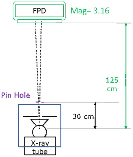
A standard flat panel detector (FPD) with 600 micron thick CsI(Tl) and 1024X1024 194-micron pixels (Varian PaxScan 2020+, Palo Alto, CA) mounted on a C-arm gantry (Infinix, Toshiba Medical Systems Corporation) was used for this study. A standard 10 micron pinhole (Fluke Biomedical, Model-01-633) was used to image the focal spot. The experimental set-up is shown in Figure 1. The pinhole was put on the x-ray tube beam exit port at a distance of 30 cm from the focal spot position as shown in Figure 1. Pinhole images were taken at the maximum magnification of 3.16. This magnification was achieved with the FPD at 125 cm from the focal spot. The small focal l spot with a nominal size of 0.30 mm was imaged for this study. These pinhole images of the small focal spot included detector blurring, and the detector MTF were determined to facilitate deconvolution. For the FPD MTF measurement, single-frame flat field images for different exposure values were acquired. After offset and flat field correction, the images were used to determine the 2D Noise Power Spectrum with the standard Fourier Transform Method [15]. The 2D presampled detector MTF was calculated from the quantum noise components of the NPS using the NR method [14].
Figure 1.
Experimental set-up for focal spot measurement
The effect of detector blurring by the FPD was eliminated by effectively deconvolving the pinhole images with the detector PSF. The actual deconvolution was performed in frequency space by taking the Fourier transform of the measured focal spot PSF and dividing by the NR derived FPD’s 2D MTF and finally transforming the result back to obtain the corrected focal spot distribution.
The pinhole images were taken for the small focal spot and each was in turn corrected for FPD blur. Once corrected, the full width at tenth maximum of the one-dimensional profiles of the pinhole images were obtained to estimate the focal spot width parallel to the anode cathode direction and perpendicular to the anode-cathode direction. For the comparison, 1D profiles in both directions were obtained for the pinhole images with detector blur, i.e. without the correction. These FWTMs were scaled for the magnification and the focal spot widths in both the directions were recorded.
In addition, pinhole images of the focal spot were obtained with a high resolution electron multiplying CCD (EMCCD) detector, with 8 μm pixel size and 100 μm thick CsI(Tl) phosphor to provide a direct measurement for comparison with the results obtained with the deconvolved FPD results.
3. RESULTS
The surface plot of measured two dimensional MTF for the FPD is shown in Figure 2. In Figure 2, the y-axis represents the anode-cathode direction and the x-axis represents the direction perpendicular to the anode-cathode axis. Figure 2 shows that the MTF for the FPD is not radially symmetric.
Figure 2.
Plot of the 2 D MTF for the FPD demonstrating anisotropy. y-axis is in the anode-cathode direction and x-axis is perpendicular to the anode-cathode direction.
For a comparison of focal spot width, the 1D profile of the small focal spot in the anode –cathode direction is shown in Figure 3 obtained for a magnification value of 3.16 and scaled to the focal spot plane. A similar comparison for the perpendicular direction for the small focal spot is shown in Figure 4. A summary of the FWTM measurements for both directions appears in Table 1. For the small focal spot, we found that the focal spot length was 0.61 mm when measured in the anode-cathode direction without removing the detector blur. After deconvolving with the detector MTF, the focal spot length was measured to be 0.57 mm. We measured 0.64 mm and 0.61 mm with and without detector blur respectively for the perpendicular direction. For comparison, we also show the results of using the high resolution detector to obtain accurate focal spot measurements directly in Figures 3 and 4. We measured 0.57 mm and 0.61 mm for the small focal spot in parallel and perpendicular directions, respectively. The measurements of the focal spot dimension in both the directions with the FPD after deconvolution also show a close agreement with the measurements taken with the high resolution detector. The differences between the line profiles for the deconvolved FPD and high resolution detector near the center is because of the presence of finite detector blur present in the high resolution detector image.
Figure 3.
Comparison of 1 D profiles of the small focal spot in a direction parallel to the A–C axis with and without detector blur obtained with a Mag = 3.16 and scaled to a magnification of 1.0.
Figure 4.
Comparison of 1 D profiles of the small focal spot in a direction perpendicular to the A–C axis with and without detector blur obtained for a Mag = 3.16 and scaled to a magnification of 1.
Table 1.
Tenth value width of focal spot profiles measured parallel and perpendicular to the anode-cathode axis
| Direction | FPD with Detector Blur | FPD without Detector Blur | High Resolution Detector |
|---|---|---|---|
| Parallel | 0.61 mm | 0.57 mm | 0.57 mm |
| Perpendicular | 0.64 mm | 0.61 mm | 0.61 mm |
4. CONCLUSION
In this study, we show that focal spot measurement with a gantry-mounted digital x-ray imager can be done easily and accurately even if large magnification is not available to reduce the effect of detector blurring. Use of deconvolution with the detector MTF enables accurate focal spot size without the need for a high-resolution detector. In this study, we showed our results for the measurement of the small focal spot because the effect of detector blurring is relatively more important than for a larger focal spot size. The significance of the detector deconvolution will be more apparent for even smaller focal spot sizes. Use of the Noise Response method also enabled us to measure the 2D detector MTF and to get insight into the non-isotropy of the MTF that could not be easily visualized with any other standard measurement method. This determination of the 2D detector MTF allowed 2D correction of the measured x-ray focal spot. These techniques using the 2D detector MTF could lead to more complete and improved quality evaluation procedures for both x-ray tube and detector.
Acknowledgments
This work was partially supported by NIH Grant R01EB002873.
References
- 1.Kyprianou IS, Rudin S, Bednarek DR, Hoffmann KR. Study of generalized MTF and DQE for a new micro- angiographic system. Proc SPIE. 2004;5368:349–360. doi: 10.1117/12.533512. [DOI] [PMC free article] [PubMed] [Google Scholar]
- 2.Kyprianou I, Rudin S, Bednarek DR, Hoffmann KR. Generalizing the MTF and DQE to include x-ray scatter and focal spot unsharpness: Application to a new micro-angoigraphic system. Med Phys. 2005;32:613–626. doi: 10.1118/1.1844151. [DOI] [PubMed] [Google Scholar]
- 3.Muntz EP. Analysis of the significance of scattered radiation in reduced dose mammography including magnification effects scatter suppression and focal spot and detector blurring. Med Phys. 1979;6:110–117. doi: 10.1118/1.594540. [DOI] [PubMed] [Google Scholar]
- 4.Jain A, Kuhls-Gilcrist A, Bednarek DR, Rudin S. Generalized two-dimensional (2D) linear system analysis metrics (GMTF, GDQE) for digital radiography systems including the effect of focal spot, magnification, scatter, and detector characteristics. Proc of SPIE. 2010:7622–19. doi: 10.1117/12.845293. [DOI] [PMC free article] [PubMed] [Google Scholar]
- 5.Jain A, Bednarek DR, Rudin S. Effect of Focal Spot Sizes and Magnification On the Total System Performance for a High Resolution Detector System Using Generalized Linear System Metrics (GMTF, GDQE) MO-FF-A4–1 AAPM. 2010 [Google Scholar]
- 6.Yadava G, Rudin S, Kuhls-Gilcrist AT, Bednarek KRHDR. Generalized Objective Performance Assesment of a new high- sensitivity Micro angiographic fluoroscopic (HSMAF) Imaging system. Proc SPIE. 2008;6913:29. doi: 10.1117/12.769808. [DOI] [PMC free article] [PubMed] [Google Scholar]
- 7.Yadava G, Kyprianou IS, Rudin S, Bednarek DR, Hoffmann KR. Generalized Performance Evaluation of X-Ray Image Intensifier Compared with a Micro angiographic System. Physics of Medical Imaging; Proc. SPIE; 2005. pp. 419–429. [DOI] [PMC free article] [PubMed] [Google Scholar]
- 8.Robinson A, Grimshaw GM. Measurement of the focal spot size of diagnostic X-ray tubes- a comparison of pinhole and resolution methods. British Journal of Radiology. 1975;48:572–580. doi: 10.1259/0007-1285-48-571-572. [DOI] [PubMed] [Google Scholar]
- 9.Law J. Measurement of focal spot size in mammography X-ray tubes. British journal of Radiology. 1993;66:44–50. doi: 10.1259/0007-1285-66-781-44. [DOI] [PubMed] [Google Scholar]
- 10.Everson JD, Gray JE. Focal-Spot Measurement: Comparison of Slit, Pinhole and Star Resolution Pattern Techniques. Radiology. 1987;1651:261–264. doi: 10.1148/radiology.165.1.3628780. [DOI] [PubMed] [Google Scholar]
- 11.Rong XJ, Krugh KT, Shepard SJ, Geiser WR. Measurement of focal spot size with slit camera using computed radiography and flat panel based digital detectors. Med Phys. 2003;30(7):1968–1975. doi: 10.1118/1.1579583. [DOI] [PubMed] [Google Scholar]
- 12.Fujita H, Tsai DY, Itoh T, Doi K, Morishita J, Ueda K, Ohtsuka A. A simple method for determining the modulation transfer function in digital radiography. IEEE Trans Med Imaging. 1992;11(1):34–39. doi: 10.1109/42.126908. [DOI] [PubMed] [Google Scholar]
- 13.Samei E, Flynn MJ, Reimann DA. A method for measuring the presampled MTF of digital radiographic systems using an edge test device. Med Phys. 1998;25(1):102–113. doi: 10.1118/1.598165. [DOI] [PubMed] [Google Scholar]
- 14.Kuhls-Gilcrist A, Jain A, Bednarek D, Hoffmann K, Rudin S. Accurate MTF measurement in digital radiography using noise response. Med Phys. 2010;37:724–735. doi: 10.1118/1.3284376. [DOI] [PMC free article] [PubMed] [Google Scholar]
- 15.Dobbins JT, 3rd, Ergun DL, Rutz L, Hinshaw DA, Blume H, Clark DC. DQE(f) of four generations of computed radiography acquisition devices. Med Phys. 1995;22(10):1581–1593. doi: 10.1118/1.597627. [DOI] [PubMed] [Google Scholar]


