

Abstract
INTRODUCTION
Inflatable penile prostheses (IPP) have been a successful method of treating men with erectile dysfunction since the early 1970s. IPP are comprised of two intracorporal cylinders, a scrotal pump and a fluid reservoir.
PRESENTATION OF CASE
We present a case of a retained reservoir in a sixty eight year old gentlemen presenting with a cystic abdominal mass and bothersome LUTS, 15 years after the removal of the penile components of a three-piece penile prosthesis. Percutaneous drainage of the cyst was performed, with four litres of purulent fluid evacuated. A midline laparotomy was required to remove the reservoir and drain the collection completely.
DISCUSSION
Inflammatory reaction and subsequent erosion of an IPP reservoir is an infrequent but severe complication of IPP insertion, replacement or infection. Infection remains the primary indication for penile prosthesis removal and in this setting removal of the reservoir is routine. A thorough literature search has identified that in the non-infective setting, the routine removal of the original reservoir is not standard practice during three-component IPP replacement. In patients with a history of IPP presenting with new LUTS, reservoir erosion should be considered in the differential diagnosis and investigation with cystoscopy and computed tomography included early in the investigatory armament of the urologist.
CONCLUSION
It is our belief that a defunctionalized reservoir serves no purpose; rather it can only cause trouble in the future. Consequently, at our institution we do not leave defunctionalized reservoirs in situ.
Keywords: Erectile dysfunction, Inflatable penile prosthesis, Lower urinary tract symptoms, Prosthesis reservoir
1. Introduction
Inflatable penile prostheses (IPP) have been a successful method of treating men with erectile dysfunction since the early 1970s. IPP are comprised of two intracorporal cylinders, a scrotal pump and a fluid reservoir. IPP reservoirs come in a variety of sizes and configurations. These reservoirs may be placed in the space of Retzius, in a pre-peritoneal or retroperitoneal location, or in layers of the abdominal wall. Complications relating to IPP include patient dissatisfaction, mechanical failure, infection, herniation and occasionally migration. In the non-infective setting, the routine removal of the original reservoir is not standard practice during three-component IPP replacement.
We present a case of a retained reservoir presenting as a cystic abdominal mass and bothersome LUTS, 15 years after the removal of the penile components of a three-piece penile prosthesis.
2. Presentation of case
A sixty eight year old gentlemen who had undergone removal of a three-piece penile prosthesis and placement of a malleable prosthesis 15 years previously presented with symptoms of urgency, urinary frequency, and a feeling of incomplete bladder emptying. His past medical history included erecticle dysfunction secondary to diabetes related vascular disease, end stage renal failure requiring haemodialysis thrice weekly and ischaemic heart disease.
With respect to his penile prosthesis the patient reported a mechanical failure shortly after the initial IPP was inserted in 1998. This prompted the replacement of the three piece device with a two piece malleable prosthesis. At the time of the initial IPP removal the reservoir was left in situ. The initial IPP procedure was performed in a different institution and attempts made to track the make and model of the device were unsuccessful.
Whilst awaiting investigations for his lower urinary tract symptoms he developed an episode of acute urinary retention. A Foley catheter was inserted, however required the guidance of a flexible cystoscope. Cystoscopy demonstrated a normal bladder mucosa, with an indentation on the anterior surface of the bladder wall. This was followed up with a computed tomography (CT) which demonstrated a 10 cm × 10 cm cyst, presumed to be of dermoid origin. The patient went on to have a transurethral resection of the prostate as it was thought his urinary retention was secondary to bladder outflow obstruction. However his urinary symptoms persisted.
A follow up scan seven months later demonstrated an increase in the size of the cyst to 13 cm × 12 cm, which appeared to be abutting and exerting a mass effect upon the anterior aspect of the bladder wall. The scan also suggested that the cyst contained a foreign body likely to represent a retained penile prosthesis reservoir (Fig. 1). At this point a percutaneous aspiration of the cyst was offered to the patient however he was not keen for any intervention as his symptoms were manageable. The patients urinary tract symptoms continued to deteriorate over the course of nine months and on examination a large anterior abdominal mass was palpable. A further CT scan showed the cyst surrounding the retained reservoir had enlarged substantially, measuring 21 cm in maximum axial diameter (Fig. 2).
Fig. 1.
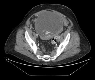
Computed tomography showing the retained penile prosthesis reservoir within the cystic mass.
Fig. 2.

A computed tomography scan demonstrating the cyst at its maximum axial diameter (21 cm) caused by the retained penile prosthesis reservoir.
At this point the patient agreed to surgical intervention, as the symptoms were unbearable. An initial attempt to remove the reservoir was made percutaneously. Under ultrasound and fluoroscopic guidance the cyst was punctured in the lower midline with an 18G percutaneous access needle through which a guidewire (Sensor, Boston Scientific) was advanced into the cyst. Serial fascial dilators (Cook) followed by balloon dilatation (Nephromax, Boston Scientific) allowed the insertion of a 30F Amplatz sheath to create a controlled access into the cyst. Four litres of purulent fluid were evacuated and the cyst thoroughly washed out through a 24Ch nephroscope (Wolff) to obtain a clear view of the retained reservoir. The size of the reservoir did not allow removal through the Amplatz sheath, therefore a limited midline laparotomy was performed using the sheath as guide to enter the cyst. The reservoir was removed intact. After further washout a 24Ch Robinson drain was left in the cavity and the abdomen closed in layers.
The microbiology analysis identified pus cells however there was no organism visualized on gram stain and no growth when cultured. The patient was reviewed in the outpatient department six weeks post operatively. The drain was removed in the clinic. His urinary tract symptoms which were the initial presenting complaint had resolved completely. He remains symptom free six months post operatively.
3. Discussion
Inflammatory reaction to the foreign body and subsequent erosion of an IPP reservoir is an infrequent but severe complication of IPP insertion, replacement or infection. There have been three mechanisms proposed whereby an IPP reservoir may injure the bladder: gradual erosion into the bladder; inadvertent surgical placement into the bladder; and laceration of the bladder when an IPP reservoir is re-inflated during a revision procedure. The concern regarding leaving an empty reservoir during implant removal cases remains controversial.
We present the first case whereby a retained reservoir has led to the formation of a large reactive cyst causing pressure effect on the bladder.
A number of previous publications have described de-functionalized IPP reservoir complications. In 1999, Munoz1 described a patient who had a three component IPP placed, but 2 years later had the cylinders and pump removed and replaced with a two-component IPP. Two years thereafter, with no signs of urinary tract or peri-prosthetic infection, he developed intravesical erosion of the de-functionalized IPP reservoir, requiring laparotomy and reservoir removal.
In 2002, Jones et al. reported four cases of intravesical erosion of retained, de-functionalized IPP reservoirs in patients who only had their cylinders and pump removed. These patients presented 3–15 years (mean 7 years) postoperatively, and required either endoscopic or open-reservoir removal.2
There have been reports of retained reservoirs migrating and eroding into sigmoid colon,3 ileal conduit,4 neobladders,5 seminal vesicles6 and ureters.7 Levine and Hoeh describe six cases of reservoir complications including reservoir herniation, direct inguinal herniation, bladder laceration, ectopic reservoir placement, iliac vein compression and a vascular laceration.8
Infection remains the primary indication for penile prosthesis removal and in this setting removal of the reservoir is routine. However the need to remove the reservoir in the setting of prosthesis malfunction or patient dissatisfaction may be questioned, for it has been thought to be a relative innocuous entity. In 2004, Rajpurkar et al. published a series of 98 IPP revision surgeries in 85 patients. It was their practice to insert new cylinders, pump and reservoir, but to leave the original reservoir in situ. With a mean of 50 months of follow up, they reported one infection, and no complications related to the originally retained reservoir. They concluded that routine removal of the original reservoir was not required during three-component IPP replacement.9 Those in favour of this approach suggest that pursuing a retained reservoir may be difficult due to extensive scar and may require extra time as well as a secondary incision to extract.
Erosion of retained prosthetic materials can have disastrous consequences that can be avoided if complete explantation is carried out immediately. Kava and Burdick-Will recently described their case series of retained foreign bodies complicated by infections necessitating removal. Hardware retained includes tubing connectors, rear tip extenders and reservoirs. They devised an implant-specific checklist to ensure removal of all device-related foreign bodies when explanting an infected penile prosthesis.10 We advocate the use of such a checklist during removal of penile prosthesis in the non infected setting. In particular the use of pre-operative imaging is indispensible in confirming the presence and location of the retained foreign bodies.
In patients with a history of IPP presenting with new LUTS, inflammatory reactions of the reservoir should be considered in the differential diagnosis and investigation with cystoscopy and computed tomography included early in the investigatory armament of the urologist.
4. Conclusion
In conclusion based on our case report and the literature described it is our belief that a defunctionalized reservoir serves no purpose; rather it can only cause trouble in the future. Consequently, at our institution we do not leave defunctionalized reservoirs in situ.
Conflict of interest
The authors of this manuscript have no conflicts of interest to disclose as described by International Journal of Surgical Case Reports. The results presented in this paper have not been published previously in whole or part.
Funding
No funding was obtained for the purposes of this study.
Ethical approval
Written informed consent was obtained from the patient for publication of this case report and accompanying images. A copy of the written consent is available for review by the Editor-in-Chief of this journal on request.
Author contributions
Hamid Abboudi, Marco Bolgeri, Andrew Chetwood – writing; Rajesh Nair – writing and data analysis; Andy Symes, Philip Thomas – review and writing.
Key learning points.
-
•
Patients presenting with LUTS require a detailed history including previous urological surgery.
-
•
Always consider removing all three parts of an inflatable penile prosthesis where the device has failed or become infected.
Contributor Information
Hamid Abboudi, Email: hamid.abboudi@doctors.org.uk.
Marco Bolgeri, Email: marcobolg@hotmail.com.
Rajesh Nair, Email: drrajnair@hotmail.com.
Andrew Chetwood, Email: andrewchetwood@doctors.org.uk.
Andrew Symes, Email: andy.symes@bsuh.nhs.uk.
Philip Thomas, Email: philip.thomas@bsuh.nhs.uk.
References
- 1.Munoz J.J., Ellsworth P.I. The retained penile prosthesis reservoir: a risk. Urology. 2000;55(6):949. doi: 10.1016/s0090-4295(99)00601-9. [DOI] [PubMed] [Google Scholar]
- 2.Jones L., Ryan R., Ghobriel A., Wilson S. The retained reservoir in inflatable penile prosthesis explantation. J Urol suppl. 2002;167(150) [Google Scholar]
- 3.Leach G.E., Shapiro C.E., Hadley R., Raz S. Erosion of inflatable penile prosthesis reservoir into bladder and bowel. J Urol. 1984;131(6):1177–1178. doi: 10.1016/s0022-5347(17)50862-9. [DOI] [PubMed] [Google Scholar]
- 4.Godiwalla S.Y., Beres J., Jacobs S.C. Erosion of an inflatable penile prosthesis reservoir into an ileal conduit. J Urol. 1987;137(2):297–298. doi: 10.1016/s0022-5347(17)43986-3. [DOI] [PubMed] [Google Scholar]
- 5.Tran C.N., Boncher N., Montague D.K., Angermeier K.W. Erosion of Inflatable Penile Prosthesis Reservoir into Neobladder. J Sex Msed. 2013 doi: 10.1111/jsm.12239. [DOI] [PubMed] [Google Scholar]
- 6.Agrawal V., Rickards D., Ralph D.J. Ejaculatory pain as a result of inflatable penile prosthesis reservoir compressing a seminal vesicle. Urology. 2006;68(4):888. doi: 10.1016/j.urology.2006.05.002. [DOI] [PubMed] [Google Scholar]
- 7.Jiann B.P., Ou C.W., Lin J.T., Huang J.K. Compression of ureter caused by a retained reservoir of penile prosthesis. Int J Impot Res. 2006;18(3):316–317. doi: 10.1038/sj.ijir.3901402. [DOI] [PubMed] [Google Scholar]
- 8.Levine L.A., Hoeh M.P. Review of penile prosthetic reservoir: complications and presentation of a modified reservoir placement technique. J Sex Med. 2012;9(11):2759–2769. doi: 10.1111/j.1743-6109.2012.02807.x. [DOI] [PubMed] [Google Scholar]
- 9.Rajpurkar A., Bianco F.F. Jr, Al-Omar O., Terlecki R., Dhabuwala C. Fate of the retained reservoir after replacement of 3-piece penile prosthesis. J Urol. 2004;172(2):664–666. doi: 10.1097/01.ju.0000131454.51640.a3. [DOI] [PubMed] [Google Scholar]
- 10.Kava B.R., Burdick-Will J. Complications associated with retained foreign bodies from infected penile implants: proposal for the use of an implant-specific checklist at the time of device removal. J Sex Med. 2013;10(6):1659–1666. doi: 10.1111/jsm.12145. [DOI] [PubMed] [Google Scholar]