Abstract
Cardiac rhabdomyoma, the primary cardiac tumor most often diagnosed in children, is frequently present in patients with tuberous sclerosis. Most pediatric patients with rhabdomyoma are asymptomatic; however, various electrocardiographic abnormalities can be detected, such as Wolff-Parkinson-White syndrome, ectopic atrial tachycardia, and atrioventricular node dysfunction. We describe the case of a 10-month-old infant girl who had tuberous sclerosis and multiple cardiac rhabdomyomas. Her electrocardiographic presentation was notable for dome-shaped T waves and no ST segment in some leads. To our knowledge, this electrocardiographic finding has not been described in patients with tuberous sclerosis and cardiac masses.
Keywords: Arrhythmias, cardiac/etiology; electrocardiography; heart neoplasms/pathology; infant; rhabdomyoma/ultrasonography; tuberous sclerosis/pathology
Tuberous sclerosis is an autosomal dominant disorder characterized by the development of hamartomas in multiple organ systems, including the skin, brain, heart, lungs, kidney, and liver. The disease has an estimated prevalence of 1 in 6,000 individuals.1 The most frequent cardiac manifestation of the disease is rhabdomyoma, which is thought to occur in at least 60% of children with tuberous sclerosis. Although these tumors are often clinically silent, they can cause hemodynamic compromise and rhythm disturbances, including ectopic atrial tachycardia, atrioventricular node dysfunction, and ventricular preexcitation. We report the case of an infant who was diagnosed with tuberous sclerosis and cardiac rhabdomyomas, and we discuss our noteworthy electrocardiographic (ECG) findings.
Case Report
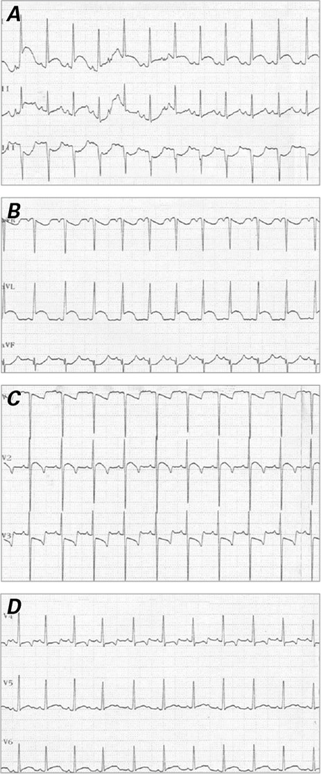
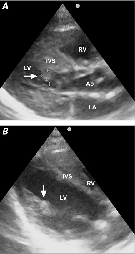
In February 2012, a 10-month-old girl with a diagnosis of epilepsy and tuberous sclerosis was referred by our hospital's pediatric neurology department for cardiac examination. The girl had been delivered vaginally at 39 weeks' gestation, with no complications. Except for an earlier epileptic seizure, no cardiovascular or other systemic symptoms were present. Examination revealed normal growth, multiple hypopigmented skin lesions, a regular cardiac rhythm with no murmur, and normal vital signs (pulse rate, 120 beats/min; respiratory rate, 32 breaths/min; and blood pressure, 70/30 mmHg). Results of laboratory tests, including a complete blood count, serum electrolytes, thyroid function, and troponin I, were all normal. No tumors were detected in the patient's brain or visceral organs, and a chest radiograph showed no cardiomegaly. A 12-lead ECG revealed sinus rhythm with dome-shaped T waves and no ST segment in leads I, III, aVL, and aVF (Fig. 1). The corrected QT interval was 360 ms. Echocardiograms showed multiple rhabdomyomas in the interventricular septum, the posterior wall of the left ventricle (LV), the papillary muscle of the mitral valve, and the moderator band of the right ventricle (RV) (Fig. 2). No obstruction of the intracavitary inflow or outflow tract of either ventricle was apparent. Echocardiograms revealed normal systolic and diastolic function, no hypokinetic or akinetic regions, and normal sizes and locations of the proximal segments of the right and left main coronary arteries. A 24-hour Holter ECG yielded the same findings as did the initial ECG. In June 2012, follow-up examinations in the departments of pediatric cardiology and pediatric neurology revealed no change.
Fig. 1.

Twelve-lead electrocardiograms. Just after the QRS complex, dome-shaped T waves are seen in A) leads I and III and B) leads aVL and aVF, but not in C) leads V1 through V3 or D) V4 through V6.
Fig. 2.

Echocardiograms show rhabdomyomas attached to the A) interventricular septum (arrow) and B) papillary muscle of the mitral valve (arrow).
Ao = aorta; IVS = interventricular septum; LA = left atrium; LV = left ventricle; RV = right ventricle
Discussion
Tuberous sclerosis is a neurocutaneous syndrome that affects the brain, heart, skin, and other organs. Józwiak and colleagues2 evaluated 154 patients who had tuberous sclerosis and found that 74 had cardiac rhabdomyomas (48%). The tumors were most often in the RV (35%), interventricular septum (33%), LV (22%), left atrium (5%), and right atrium (5%); 61% were clinically silent. The main clinical manifestations of the tumors were arrhythmias (23%), murmurs (14.9%), and heart failure (5.4%). Only one of the 154 patients had LV outflow tract obstruction. Our patient had multiple rhabdomyomas in the RV, LV, and interventricular septum; however, we detected no cardiac dysfunction.
Various rhythm abnormalities can occur in tuberous sclerosis. Shiono and colleagues3 reported that 12 of their 21 patients presented with one or more ECG abnormalities. Ventricular tachycardia, atrioventricular block, and preexcitation syndrome are most often reported.4–6 Intramural rhabdomyomas are thought to interrupt the conduction pathways, lead to ectopic electrical foci or an accessory electrical circuit, and produce preexcitation.2 Rhabdomyomas naturally regress over time: half of patients experience partial resolution and 18% have complete resolution.2 Because spontaneous regression is observed, tumor resection is indicated only in patients whose arrhythmias are refractory to medical management.
Dome-shaped T waves and an absent ST segment can suggest hypothyroidism.7 In our patient, the absence of relevant physical findings, the normal QRS voltages, and the normal results of thyroid function tests helped us to rule out hypothyroidism. In addition, her normal cardiac function and serum troponin I level suggested a very low probability of cardiac ischemia.
The dome-shaped T waves were not present in all the ECG leads. This might be due to mechanical compression of the intracardiac masses in the electrical pathway of the heart. However, we found no relationships between the locations of the tumors and the ECG results.
In patients with tuberous sclerosis, ECG and echocardiography should be performed routinely even when the results of physical examination are normal. In addition, it is prudent to monitor these patients closely. Despite no clinically important findings in our patient, we continued to perform serial examinations. To our knowledge, the ECG finding of dome-shaped T waves and absent ST segment has not been described previously in patients with tuberous sclerosis and cardiac masses.
Footnotes
From: Department of Pediatric Cardiology, Konya Training and Research Hospital, 42060 Konya, Turkey
References
- 1.Ahlsen G, Gillberg IC, Lindblom R, Gillberg C. Tuberous sclerosis in Western Sweden. A population study of cases with early childhood onset. Arch Neurol. 1994;51(1):76–81. doi: 10.1001/archneur.1994.00540130110018. [DOI] [PubMed] [Google Scholar]
- 2.Jozwiak S, Kotulska K, Kasprzyk-Obara J, Domanska-Pakiela D, Tomyn-Drabik M, Roberts P, Kwiatkowski D. Clinical and genotype studies of cardiac tumors in 154 patients with tuberous sclerosis complex. Pediatrics. 2006;118(4):e1146–51. doi: 10.1542/peds.2006-0504. [DOI] [PubMed] [Google Scholar]
- 3.Shiono J, Horigome H, Yasui S, Miyamoto T, Takahashi- I, gari M, Iwasaki N, Matsui A. Electrocardiographic changes in patients with cardiac rhabdomyomas associated with tuberous sclerosis. Cardiol Young. 2003;13(3):258–63. [PubMed] [Google Scholar]
- 4.Akalin F, Baysoy G, Ozturk B, Yalcin Y, Ekici G, Yilmaz Y. A case of tuberous sclerosis presenting with dysrhythmia in the first day of life. Turk J Pediatr. 2004;46(1):79–81. [PubMed] [Google Scholar]
- 5.Hirakubo Y, Ichihashi K, Shiraishi H, Momoi MY. Ventricular tachycardia in a neonate with prenatally diagnosed cardiac tumors: a case with tuberous sclerosis. Pediatr Cardiol. 2005;26(5):655–7. doi: 10.1007/s00246-004-0714-5. [DOI] [PubMed] [Google Scholar]
- 6.Venugopalan P, Babu JS, Al-Bulushi A. Right atrial rhabdomyoma acting as the substrate for Wolff-Parkinson-White syndrome in a 3-month-old infant. Acta Cardiol. 2005;60(5):543–5. doi: 10.2143/AC.60.5.2004977. [DOI] [PubMed] [Google Scholar]
- 7.Ertugrul A. A new electrocardiographic observation in infants and children with hypothyroidism. Pediatrics. 1966;37(4):669–72. [PubMed] [Google Scholar]


