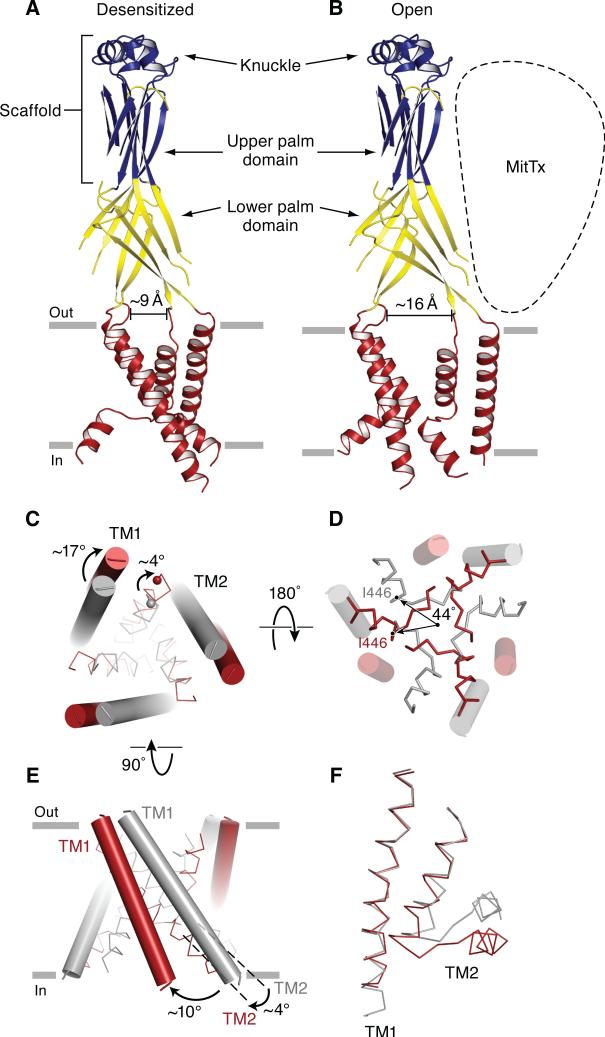
Figure 4.
Gating movements in the extracellular and transmembrane domains deduced from comparison of the desensitized state and MitTx-bound structures. Cartoon representation of the desensitized state (A) and the Δ13-MitTx state (B) where the structurally conserved scaffold is blue, the malleable lower palm domain is yellow, and the transmembrane domains are red. Measurements use the Cα atoms Ala 424 on adjacent subunits as landmarks. (C) View of the transmembrane domains from the extracellular side illustrates the iris-like rotation of TM1 and TM2a. TM1 and TM2a are in cylinders and ribbon representation, respectively. In panels C-F the desensitized and Δ13-MitTx structures are gray and red, respectively. (D) View of the transmembrane domains from the intracellular side shows that TM2a rotates by ~44°, in the plane of the membrane. (E) View of the transmembrane domains perpendicular to the bilayer illustrating tilts of ~10° and ~4° by TM1 and TM2, respectively. (F) Close-up view of the transmembrane helices of one subunit. The TM1 domain of the Δ13-MitTx structure is superimposed onto the TM1 domain of the desensitized state structures using Cα positions of residues Cys 50 to Phe 70. Both TM1 and TM2 are shown in ribbon representations. See also Figure S3.

