

Abstract
The sliding filament model of muscle contraction, put forward by Hugh Huxley and Jean Hanson in 1954, is 60 years old in 2014. Formulation of the model and subsequent proof was driven by the pioneering work of Hugh Huxley (1924–2013). We celebrate Huxley’s integrative approach to the study of muscle contraction; how he persevered throughout his career, to the end of his life at 89 years, to understand at the molecular level how muscle contracts and develops force. Here we show how his life and work, with its focus on a single scientific problem, had impact far beyond the field of muscle contraction to the benefit of multiple fields of cellular and structural biology. Huxley introduced the use of x-ray diffraction to study the contraction in living striated muscle, taking advantage of the paracrystalline lattice that would ultimately allow understanding contraction in terms of single molecules. Progress required design of instrumentation with ever-increasing spatial and temporal resolution, providing the impetus for the development of synchrotron facilities used for most protein crystallography and muscle studies today. From the time of his early work, Huxley combined electron microscopy and biochemistry to understand and interpret the changes in x-ray patterns. He developed improved electron-microscopy techniques, thin sections and negative staining, that enabled answering major questions relating to the structure and organization of thick and thin filaments in muscle and the interaction of myosin with actin and its regulation. Huxley established that the ATPase domain of myosin forms the crossbridges of thick filaments that bind actin, and introduced the idea that myosin makes discrete steps on actin. These concepts form the underpinning of cellular motility, in particular the study of how myosin, kinesin, and dynein motors move on their actin and tubulin tracks, making Huxley a founder of the field of cellular motility.
Introduction
The year 2014 is the 60th birthday of the sliding filament model of muscle contraction, first put forward in 1954 by Hugh Huxley and Jean Hanson. The formulation of the model, and its subsequent proof, was driven in a large part by the pioneering work of Hugh Huxley (1924–2013). In this memorial review, we celebrate his integrative approach to learning how muscles contract. We illustrate how his ideas and approach to science led to seminal new concepts and developments in technology that continue to have wide-ranging impact on biophysics, cellular biology, and structural biology. Although these methods have gone on to enable biophysicists and cell biologists to study numerous processes at the molecular level in living cells and tissues, we recognize that Huxley’s focus and passion were always to understand the contractile process at the molecular level in living muscle in real time. Several fine tributes to Hugh Huxley have been published elsewhere (1–5). Our aim here is to give a flavor of how Huxley thought about and did science, informed by his ingenuity and inventiveness, his consummate hands-on approach, focus and drive. It is, therefore, not intended as a comprehensive, critical review of the field, or even of just the work of Huxley and his colleagues. We apologize in advance to those whose work we short-shrifted in this process.
Why muscle?
Skeletal muscle was likely the best-studied tissue when Huxley began his graduate work with John Kendrew in 1949 at the newly-formed Medical Research Council (MRC) Unit for Work on the Molecular Structure of Biological Systems at the Cavendish Lab in Cambridge, England. For an historical review of early research on muscle contraction, see Needham (6) and Szent-Gyorgyi (7). For a century, the striations of the sarcomere (Fig. 1) had been known from light microscopy studies to change during contraction. Actin and myosin were understood to be separate proteins that form filaments and interact to produce ATP-dependent contraction. Cambridge was for decades a center of muscle research with luminaries such as Kenneth Bailey with S. V. Perry, A. V. Hill, and Joseph and Dorothy Needham studying the biochemistry, energetics, and mechanics of skeletal muscle. Although theories of muscle contraction abounded, there was no consensus. Huxley’s reviews and memoirs (8–10) provide personal accounts of his early career, and how he chose to work on muscle contraction within this intellectual milieu.
Figure 1.

(A) Sarcomere pattern of striated muscle. Longitudinal section of frog muscle and diagram of corresponding overlapping thick, myosin-containing filaments and thin, actin-containing filaments (from Huxley (9), with permission). (B) First published scheme showing sliding filaments, related to changes in the sarcomere pattern at different lengths (from Hanson and Huxley (29), with permission).
X-Ray Diffraction: A Tool to Study Physiology
Huxley has recounted how, after an early (and somewhat painful, it seems) brush with macromolecular crystallography, he preferred to work on a more biological system than that of individual proteins (8,9). Then as now, x-ray diffraction remains the only technique to detect nanometer-scale structural changes in real physiological time in living muscle. However, this was far from obvious when Huxley started his work. The Astbury group in Leeds, England pioneered x-ray fiber diffraction of muscle proteins (11), showing that these proteins had the diffraction features expected from α-helices but that these features did not change after stretch of the muscle or after contraction. The first small angle x-ray diffraction studies of muscle from the F. O. Schmitt lab at MIT (12) and early electron microscopy (EM) images showed filaments (13,14), indicating some degree of order (as recounted in Huxley (8)). Based on these meager clues, Huxley realized he would need an exceptionally bright source of x-rays coupled to a specialized instrument to resolve the ∼40-nm basic repeats in muscle in hydrated specimens (9,10).
True to a pattern that would characterize the rest of his career, whenever Huxley needed a new instrument or technique, he looked around for possible solutions and then applied his ingenuity to adapt them to his use. In this case, he designed and built a compact small-angle x-ray camera with a fine-focus x-ray tube developed by Ehrenberg in Bernal’s lab (15) and a high-voltage supply made of ex-war-surplus parts that could deliver relatively high-intensity x-rays through very narrow slits to live frog muscle to resolve the weak x-ray reflections. With this then-innovative setup, Huxley was able to make some profound observations and some prescient predictions (16–18), reviewed in Huxley (10). He was able to resolve multiple reflections on the equator in x-ray patterns from resting muscle indexing on the ∼40-nm periodicity, corresponding to the inter-thick filament spacing in the sarcomere (Fig. 2 A). He also obtained x-ray patterns from muscle in rigor (Fig. 2 B) where he noticed that although the interfilament spacings were the same, the relative intensities of the first two reflections were reversed compared to resting living muscle. Long before crossbridges were seen in the EM, Huxley postulated that the filaments at the hexagonal lattice position were made of myosin and those at the trigonal position were made of actin, and that the changes in equatorial intensities were due to the formation of cross-links between them. At that time, there was no other direct evidence for this arrangement. Axial x-ray patterns showed periodicities that he measured to be orders of 41.5 nm (now known to be 43 nm) that did not change with stretch. Perhaps in compensation for his lucky guess about the equatorial pattern, he mistakenly ascribed these periodicities to actin (reviewed in Huxley (10)), later correctly assigned to myosin by Worthington (19) and Elliott and Worthington (20). These x-ray observations set the stage and prepared Huxley’s mind for sliding filaments, as would be revealed in the light and electron microscopy studies that followed.
Figure 2.

Representative x-ray diffraction patterns from muscle collected at various stages of Huxley’s career. (A and B) Equatorial patterns from resting and rigor muscle taken with his first x-ray camera and reported in his Ph.D. thesis (86). Note the reversal of relative intensities of the 1,0 and 1,1 equatorial reflections when going from relaxed to rigor (adapted from Huxley (9), with permission). (C) Resting pattern from frog muscle taken with a rotating anode generator and a Huxley-Holmes mirror monochromator camera (adapted from Huxley and Brown (47), with permission). (D) Integrated intensity of the 14.5-nm myosin-based meridional reflection during a quick release experiment collected at DESY (adapted from Huxley et al. (55) with permission). (E) Comparison of the ∼2.7-nm reflection from actin (A) and the ∼2.8-nm reflection from myosin (M) under isometric contracting conditions (C) as compared to relaxed (R) conditions. The inward movement of the reflections reflects the stretch of the filaments under contracting conditions (adapted from Huxley et al. (74) with permission). (F) The fine structure in the 14.5-nm meridional reflection (M3) in a diffraction pattern from contracting frog muscle due to the interference of myosin heads on either side of the M-line in a thick filament (H. E. Huxley and T. C. Irving, unpublished).
Sliding Filaments: Electron Microscopy, Thinner and Thinner Sections
Upon completing his Ph.D. in 1952, aware that EM had the potential to reveal muscle structures in a way he could relate to his x-ray studies, Huxley moved to the Massachusetts Institute of Technology in Cambridge, Massachusetts, to learn EM with the biophysics pioneer, F. O. Schmitt. It happened that Jean Hanson, who had been studying the changes in the sarcomere pattern of myofibrils during contraction using light microscopy at King’s College, London, England (21), soon joined the lab, also to learn EM. True to form, from the start Huxley was constantly improving the existing EM methods for application to muscle. The x-ray studies told him that the thick-to-thick filament center-to-center distance was ∼40 nm. Isolating single layers of thick filaments would require sections much thinner than this. He partnered with A. J. Hodge and D. Spiro in developing a microtome that could make thinner sections that would allow visualization of the double array of filaments predicted by his x-ray studies (22,23). And there they were. Hanson and Huxley wrote, “thin filaments of actin extend from the Z-line through the I-band and through one half of the A-band” (24), noting that there were two sets of filaments in the A-band. When the myosin was selectively extracted in the presence of ATP, the A-band disappeared, leaving thin filaments. These too disappeared when the actin was selectively extracted.
It was not until the landmark 1954 paper (25) that Huxley and Hanson proposed the sliding filament model, after quantifying parameters of glycerinated myofibrils during stretch and shortening, and relating the results to their EM studies. They noted that as the I-band shortens, the H-zone disappears whereas there is no change in the A-band at different sarcomere lengths (Fig. 1 B). A. F. Huxley (no relation) and Niedergerke (26) saw the same band pattern changes at the light microscope level in muscle fibers using an interference microscope. The classic length-tension relationship that related function to the sliding filament model was years away (27), although it was known from A. V. Hill’s studies (and even earlier in work by Schwann) that during isometric contraction, tension is maximal at the rest length of muscle (reviewed in Needham (6)). Reflecting the caution of the investigators that was also typical of the times, the models of the sliding filaments were not published until 1957 (Fig. 1 B) (28), after additional quantitative measurements using interference microscopy, biochemistry, and electron microscopy (28–30).
Skepticism: Exacting Experiments and Even Thinner Sections Support the Sliding Filament Model
Huxley returned to Cambridge, England in 1954, and at the end of 1955 took a position at University College, London to continue his EM studies. In 1962, he became a founding member of the MRC Laboratory of Molecular Biology (LMB) in Cambridge, England. The micrographs published in the 1953–1954 papers showed filaments, but to see individual filaments would require even thinner sections as well as precise orientation of the section in relation to the myofibril axis. The idea of contraction based on sliding filaments was still avant-garde, because at the time the prevailing view was that the protein filaments themselves contract (7). His remarkable images, such as those in Fig. 3, A and B (30), clearly showed the overlap between thick and thin filaments, the crossbridges between the filaments, the relationship between the size of the H-zone and sarcomere length, and the constant distance between the Z-line and the edge of the H-zone. He recognized that the bridges between the thick and thin filaments were sites of actin-myosin interaction, and introduced the idea that they attach and detach with a minimum step distance of 5.4 nm—the size of an actin monomer—during shortening, well within the range of later experimental determinations of the crossbridge working stroke (31). Huxley suggested the crossbridges were part of myosin, although they “do not reveal the basic mechanism involved in contraction” (30).
Figure 3.

(A) Thin section of skeletal muscle showing interdigitating thick and thin filaments, from a circa-1957 image. In this image, and others like it, Huxley noted that the thin filaments end at the H-zone, the crossbridges between the thick and thin filaments, the thickening of the thick filaments at the M-band, the dimensions of the thick and thin filaments, and the interfilament spacing (from a photo in Huxley’s collection). (B) Cross-section through the overlap region of skeletal muscle showing the double hexagonal array of thick and thin filaments. One of the fundamental insights driving all Huxley’s work was that the paracrystalline lattice of filaments offered an ensemble approach to understand the behavior of single molecules (from a circa-1957 photo in Huxley’s collection). (C) Negatively-stained native thick filaments showing crossbridges except for bare central region and tapered ends (adapted from Huxley (37), with permission). (D) Negatively-stained synthetic myosin filaments showing bare central region free of crossbridges, leading to postulation of the bipolar nature of the thick filaments (adapted from Huxley (37), with permission). (E) Actin filaments decorated with myosin S1 (showing the polar arrowheads) with the same polarity along the entire filament (from Huxley’s collection, date unknown).
Looking back, it is difficult to appreciate the skepticism that greeted the sliding filament model, and that the model was not widely accepted until the mid-to-late 1960s. Sliding mechanisms as a mode of mechanochemical transduction are now the paradigm for dynein-based ciliary and flagellar motility, and motility of filamentous and nonfilamentous myosins and kinesins in a wide variety of cytoskeletal motile processes. Beyond the technical advances made in the course of their work, Hanson and Huxley were among the first to use electron microscopy for a physiological experiment accompanied by rigorous quantitative analyses at a time when EM was predominantly used to describe the ultrastructure of cells and tissues. We note in passing that Huxley was also the first to show that the T-tubules of skeletal muscle are invaginations of the plasma membrane, continuous with the extracellular space (32), a critical aspect of the excitation-contraction coupling mechanism.
Hanson and Huxley (24), followed up by other groups (33,34), noted fine S-filaments that run from Z-line to Z-line, but they did not incorporate a third filament system into the sliding filament model or their thinking, engendering years of controversy (reviewed in dos Remedios and Gilmour (35)). We now know the elastic protein, first called connectin, now titin, forms a third set of filaments in the sarcomere that have multiple, critical roles in the mechanics and intracellular regulation of muscle even if they do not appear to have a direct role in the central actin-myosin force generating mechanism (36).
Electron Microscopy to Study Structure and Function of Isolated Proteins
In the 1950s, the structures of actin and myosin molecules or filaments and origin of the crossbridges in thin sections were unknown. By comparing negatively-stained EM images of myosin filaments assembled from purified myosin with thick filaments isolated from muscle (37), Huxley saw for the first time the bipolar nature of myosin filaments with crossbridges extending from both ends, leaving a bare zone in the middle (Fig. 3, C and D). The structure of native thin filaments was similar to that Hanson and Lowy had seen in filaments made from purified actin (38). By 1963, the proteolytic fragments of myosin, HMM and LMM, were well known (39). One can imagine Huxley’s excitement at seeing arrowheads on actin filaments when he added HMM (Fig. 3 E). The polarity inferred in the sliding filament model was realized in the bipolar structure of myosin combined with the opposite polarity of actin filaments extending from Z-lines. In the 1963 paper, Huxley proposed that myosin formed the crossbridges and that sliding of thick and thin filaments past each other depended on crossbridges interacting with actin in an ATP-dependent manner (37). In years to come, myosin HMM, and later myosin S1 decoration of actin, became diagnostic tools to identify filaments as actin and to define their polarity in cells. The terms “barbed” and “pointed” have become generic.
The classic images in Huxley’s 1963 article (37) again depended on improved methodology. Huxley and Zubay (40) modified an earlier negative-staining method (41) by using uranyl acetate as the stain, and perforated grids where “one gently breathed on the [drying collodion] so that the surface appeared slightly cloudy” (40) to create holes in the collodion film. On the EM grids, after dissolving the collodion and staining, the specimen over the holes was thin, embedded only in a layer of stain, allowing visualization of structural details.
Further development of EM methods to study macromolecular structure remained a focus within the Structural Studies Division at the MRC-LMB, where Huxley served as joint head from 1975 to 1987 (and Deputy Director of the LMB from 1978 to 1987) until he moved to Brandeis University in Waltham, Massachusetts. DeRosier and Klug (42) developed a method for three-dimensional reconstruction of EM images. The first application in muscle research was to actin filaments. Because the actin filament is helical, the image of a single filament contains all the structural views, without needing to tilt the specimen (43). Although low-resolution by present standards, the structures that Moore et al. (44) obtained of pure actin and native thin filaments (that contain tropomyosin-troponin), with and without myosin-S1, were astounding (Fig. 4, A and B). The structures revealed the tilted and slewed appearance of the myosin head that became the basis of future studies and modeling of x-ray diffraction data. In addition, they modeled the position of tropomyosin along the helical actin filament, reinforcing Hanson and Lowy’s proposal that tropomyosin lies in the grooves of the actin helix (38), setting the stage for the steric blocking model (see below). The Structural Studies Division at the LMB continued to be a frontier for EM methodology and structure determination, a focus that continues to this day. Electron microscopy is now a mainstream technique for structure determination of macromolecular assemblies at near-atomic resolution.
Figure 4.

Three-dimensional reconstruction of thin filaments. (A) End-on view of a model of actin-myosin S1. The S1 heads are tilted and skewed relative to the actin filament axis (from a circa-1969 photo in Huxley’s collection). (B) Superposition of a reconstruction of a native thin filament (actin with tropomyosin-troponin, red) with actin-myosin S1 (blue) to illustrate the extra material (tropomyosin) in the thin filament (adapted from Moore et al. (44), with permission). (C) A composite end-on view of actin-tropomyosin-myosin S1 based on EM and x-ray data where tropomyosin (TM) is shown in the active state (solid contours) and relaxed state (dotted contours), where it was postulated that it could block cross-bridge attachment (steric-blocking model) (adapted from Huxley (64), with permission).
First X-Ray Diffraction Measurements from Contracting Muscle
After 1963, Huxley’s major focus returned to x-ray diffraction. This was a period of competition between Huxley and colleagues (including Ken Holmes, John Haselgrove, Bill Longley, and Wilf Brown) at Cambridge and the King’s College, London group (that included Roy Worthington, Gerald Elliott, Jack Lowy, and Barry Millman). This rivalry was a kind of technological arms-race whereby considerable ingenuity in both groups was devoted to extracting maximum performance out of their x-ray generators to collect diffraction data from contracting muscle. The Cambridge group held a technological edge primarily in the development of the Huxley-Holmes mirror-monochromator diffraction camera, and rotating anode x-ray generators of ever-increasing power (see Fig. 3 C). Huxley gives a lively account of these developments (45). This competition culminated in two monumental papers by Elliott et al. (46) and Huxley and Brown (47), which compared resting, isometrically contracting, and rigor muscle. In addition to the rich harvest of structural details for the myofilaments resulting from these studies, one of the key conclusions was that the major axial periodicities from the thick and thin filaments are constant, within experimental error, between rest and isometric contractions, confirming the sliding filament theory of contraction. In either contraction or rigor, the majority of the structural changes could be attributed primarily to changes in the thick filament, presumably due to movement of myosin crossbridges, not to large-scale changes in the thin filament. This work ushered in a time of synthesis in the muscle field culminating in Huxley’s 1969 paper in Science (48) proposing the well-known swinging crossbridge mechanism for muscle contraction. We note that Huxley put forward a cycling crossbridge model in a 1958 Scientific American article (49), showing he had been thinking in those terms for a long time.
Although there was a widespread sense that the so-called problem of muscle was solved at the time of the 1972 Cold Spring Harbor Symposium on muscle contraction, it was no doubt frustrating for Huxley that the work he and others had done did not provide direct evidence that individual myosin crossbridges actually move axially during contraction in synchrony with the force-producing events as suggested by the swinging cross-bridge model. For such evidence, he had to wait for the next major technological advance. And it was a while in coming.
Development of Synchrotron Radiation as a Tool for Structural Biology
The now widespread availability of synchrotron radiation for structural biology is one of the most outstanding success stories in science. How the use of synchrotron radiation began has been recounted in the literature (45,50,51). In the mid-1960s, Ken Holmes realized that electron synchrotrons, large machines initially developed for high-energy physics experiments, emitted hard x-rays that might be useful for structural biology. In 1971, Rosenbaum, Holmes and Witz (52), using the Deutsches Elektronen-Synchrotron (DESY), in Hamburg, Germany, published an x-ray pattern from insect flight muscle, the first diffraction pattern taken from anything using synchrotron radiation, using a beam that was then ∼100-fold brighter than that available from the best rotating anodes. In 1969, Huxley chaired a committee of the planned European Laboratory of Molecular Biology (EMBL) that proposed an organization to foster technology development that was beyond the scope of any one university or laboratory. The future promise of synchrotron radiation for structure determination was quickly appreciated, and the opportunity presented by the synchrotron radiation facilities at Hamburg, Germany helped tip the balance toward creation of the EMBL in 1974. The EMBL, headquartered in Heidelberg, Germany, now has outstations in Hamburg and Grenoble, France that are critical components of the world’s scientific infrastructure.
Whereas development of beamline technology continued for some years on DESY, the first production x-ray beamlines (X11 and X13) had to wait until the construction of the DORIS storage ring and the EMBL outstation at DESY in 1975. The flux delivered by these beamlines, along with fast electronic detectors, allowed ∼1000× faster data acquisition than with the best rotating anodes of the time (53). The goal of the experiments was to try to detect evidence that myosin crossbridges moved axially during rapid mechanical transients, designed to synchronize crossbridges, of the type pioneered by Huxley and Simmons (54). Huxley was then able, for the first time, to obtain x-ray diffraction evidence (Fig. 3 D) that there were structural changes in the myosin heads, as reported by changes in the intensity of the myosin meridional x-ray reflection at 14.5 nm. The changes did indeed appear to be synchronous with the force-producing events (55–57), as predicted by Reedy et al. (58), based on studies comparing relaxed and rigor glycerinated insect flight muscle. The X11 and X13 beamlines at the DESY storage ring at Hamburg went on to have a long and productive life with a significant impact on many fields of structural biology.
Regulation of Contraction: Steric Blocking Model
As the sliding filament mechanism became accepted, attention turned to how muscle contraction is regulated, where EM and x-ray studies from Huxley’s group were again critical. By the 1960s, it was established that Ca2+ is the regulatory ion, and tropomyosin-troponin on the actin filament is the target (59–61). The pieces of the puzzle were in place to propose the steric blocking model. The focus became the second-order actin layer line attributed to tropomyosin, consistent with its position along the actin filament (62). Haselgrove (63), Huxley (64), Lowy and Vibert (65), Vibert et al. (66), and Parry and Squire (67) reported changes in the actin layer lines during contraction in x-ray studies. The patterns were modeled and interpreted to show an azimuthal shift in the position of tropomyosin away from the outside of the filament, where myosin binds toward a more central position upon activation, or in rigor when myosin binds to actin (Fig. 4 C). The change does not depend on myosin, because it takes place when muscles are stretched to lengths where the thick and thin filaments do not overlap (63,64,66,68).
Subsequent improvements in sensitivity and time resolution at DESY (see above), enabled Huxley working with Kress et al. (69) to show that the changes in the second actin layer line occur after activation but before the movement of the myosin crossbridges toward the actin filaments. The results pointed to the change in the position of tropomyosin on the actin filament as the first observable structural step in contraction, in response to Ca2+ binding to troponin C on the thin filament—a prerequisite for myosin binding and full force development. Definitive proof for the structural changes in the thin filament upon Ca2+ binding came later (70). It is now accepted that in vertebrate skeletal muscle, both Ca2+ binding to troponin and myosin binding to actin are required for full activation (71,72); however, the details of this process remain unresolved.
Stretchy Myofilaments
When Huxley retired from the LMB, he moved to Brandeis University in Waltham, Massachusetts where he was Professor of Biology (1987–1997), Professor Emeritus (1997–2013), and Director of the Rosenstiel Basic Medical Sciences Research Center at Brandeis from 1988–1994. While continuing with EM studies (73), his main interest was to use the more powerful beam and the then-new imaging-plate detector technology at the Cornell High Energy Synchrotron Source in Ithaca, New York. To his surprise, the experiments revealed unanticipated small spacing changes, ∼0.2–0.3% in the myofilament lengths, in the higher-order meridional reflections at ∼2.7 nm (actin) and ∼2.8 nm (myosin) (Fig. 2 E). These studies (74) were paralleled by those conducted by K. Wakabayashi et al. (75) in Japan, and the two papers appeared back-to-back in Biophysical Journal in 1994. These changes in filament length were near the limit of detectability, circa. 0.3%, but nonetheless required radical reformulation of the main crossbridge theories (e.g., A.F. Huxley and Simmons (54)), which assumed that all the compliance was in the crossbridges and none in the myofilament backbones (see A.F. Huxley and Tideswell (76)).
Seeing Crossbridges Move: Late X-Ray Diffraction Experiments
Huxley was a major motivator, through his collaboration with Tom Irving, behind the development in the late 1990s of the BioCAT x-ray beamline 18ID at the Advanced Photon Source (APS), Argonne National Laboratory in Lemont, Illinois, which continues to this day as a facility for diffraction studies of noncrystalline biological materials, including muscle. Huxley was well aware that despite years of research including in vitro motility assays and crystallographic studies of myosin S1 in various nucleotide states, direct evidence for axial motions of crossbridges in intact muscle as envisaged in the Huxley 1969 swinging crossbridge model (48) remained elusive. A possible way to achieve such evidence emerged when it was realized that the fine structure in the ∼14.5-nm meridional reflection (Fig. 2 F), first reported by Bordas et al. (77), arose from interference between the diffracted rays from the arrays of crossbridges in the two halves of each thick filament, as first reported by Linari et al. (78). The relative intensities of the interference peaks would be sensitive to small axial motions of crossbridges (78). A productive period followed where the Lombardi group in Florence worked on interference studies on single muscle fibers, e.g., Piazzesi et al. (31), Linari et al. (77), and Reconditi et al. (79,80), mainly at the European Synchrotron Radiation Facility in Grenoble, France, but also at the APS, in friendly competition with Huxley on whole muscle at the APS with Massimo Reconditi working with both groups at different times (Fig. 5). Huxley’s data allowed him to determine crossbridge angles during synchronized isometric contraction after a quick release or during isotonic shortening, and how these angles change with adjustments in load and estimates of the degree of dispersion of crossbridge angles about these averages (81,82). Although this interpretation is not without its critics (83), this led Huxley to feel satisfied that he had finally achieved his goal of seeing axial motions in crossbridges in contracting muscle (9).
Figure 5.
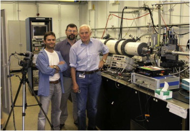
Huxley’s team at the Advanced Photon Source, Argonne National Laboratory, July 2004. (Left to right) Massimo Reconditi, Tom Irving, and Hugh Huxley. Huxley, ever the hands-on experimentalist, preferred to do as much as possible himself, from dissecting the muscles and setting up the electronics, to running the experiments themselves—showing remarkable stamina for synchrotron x-ray experimentation long after the retirement ages of most scientists (photo from T. C. Irving).
Huxley’s Legacy
The sliding filament mechanism was a paradigm shift in our view of muscle contraction. However, proving the idea of sliding filaments to the skeptics was just the beginning. A satisfactory explanation of the mechanism behind how muscle contracts and develops force, and how it is regulated at the molecular level, required advances in 1), both x-ray diffraction and electron microscopy, 2), x-ray crystallography beyond what is described herein, 3), development of in vitro motility and single molecule assays, and 4), application of a lot more contractile protein biochemistry. In the end, it took another 50 years until Huxley convinced himself that he had proven the swinging crossbridge mechanism of muscle contraction. Ever a perfectionist, Huxley would only publish when he was “good and ready,” leading to a small number of primary publications by modern standards. But his influence has been disproportionately far-reaching.
We have seen how Huxley’s single-minded devotion to one important problem—the molecular mechanism of muscle contraction—led to an impressive number of technical advances that he either produced himself or motivated in others, which profoundly influenced multiple fields in cell biology, including cellular motility. Although he was keenly interested in cellular motility (84,85), he left the area to others at the LMB, such as Linda Amos, John Kendrick-Jones, Murray Stewart, and Alan Weeds, and the many postdoctoral fellows and visiting scientists who worked there. Honored as a founder of the field of cellular motility in its broadest sense (3), the American Society for Cell Biology recognized Huxley with the E. B. Wilson Award, the highest honor of the Society, in 1983. As the field moves forward, the goal of deciphering mechanisms of muscle contraction and cellular motility at the single molecule level and even the atomic level are within reach. More realistic molecular dynamics simulations and other improved modeling approaches offer the best hope to relate the x-ray patterns of living muscles to molecular structures. The ability to visualize single molecules in living cells coupled with methods to transition between superresolution light microscopy of living cells and cryo-EM may fulfill a dream Huxley surely had, that we carry on.
Acknowledgments
We thank Maggie McNeely, archivist at Brandeis University, for access to Hugh Huxley’s papers. We also thank the reviewers and numerous other colleagues who provided helpful suggestions and information to us as we tried to sort out details and their essence, reflecting their admiration and respect for Hugh Huxley.
Use of the Advanced Photon Source, an Office of Science User Facility operated for the U.S. Department of Energy Office of Science by Argonne National Laboratory, was supported by the U.S. Department of Energy under Contract No. DE-AC02-06CH11357. This project was supported by grant No. 9-P41-GM103622 from the National Institute of General Medical Sciences, National Institutes of Health (to T.C.I.), and National Institutes of Health RO1 grant No. GM-093065 (to S.E.H.-D.).
References
- 1.Holmes K. Hugh Esmor Huxley (1924–2013) J. Muscle Res. Cell Motil. 2013;34:311–315. doi: 10.1007/s10974-013-9365-6. [DOI] [PMC free article] [PubMed] [Google Scholar]
- 2.Holmes K.C. Hugh Esmor Huxley (1924–2013) Proc. Natl. Acad. Sci. USA. 2013;110:18344–18345. doi: 10.1073/pnas.1318966110. [DOI] [PMC free article] [PubMed] [Google Scholar]
- 3.Pollard T.D., Goldman Y.E. Remembrance of Hugh E. Huxley, a founder of our field. Cytoskeleton (Hoboken) 2013;70:471–475. doi: 10.1002/cm.21141. [DOI] [PubMed] [Google Scholar]
- 4.Spudich J. Memories of Hugh E. Huxley (1924–2013) Mol. Biol. Cell. 2013;24:2769–2771. doi: 10.1091/mbc.E13-08-0454. [DOI] [PMC free article] [PubMed] [Google Scholar]
- 5.Weeds A. Hugh Huxley (1924–2013) Nature. 2013;500:530. doi: 10.1038/500530a. [DOI] [PubMed] [Google Scholar]
- 6.Needham D.M. Cambridge University Press; Cambridge, UK: 1971. Machina Carnis: The Biochemistry of Muscular Contraction in its Historical Development. [Google Scholar]
- 7.Szent-Gyorgyi A. Academic Press; New York: 1951. Chemistry of Muscular Contraction. [Google Scholar]
- 8.Huxley H.E. A personal view of muscle and motility mechanisms. Annu. Rev. Physiol. 1996;58:1–19. doi: 10.1146/annurev.ph.58.030196.000245. [DOI] [PubMed] [Google Scholar]
- 9.Huxley H.E. Fifty years of muscle and the sliding filament hypothesis. Eur. J. Biochem. 2004;271:1403–1415. doi: 10.1111/j.1432-1033.2004.04044.x. [DOI] [PubMed] [Google Scholar]
- 10.Huxley H.E. Memories of early work on muscle contraction and regulation in the 1950’s and 1960’s. Biochem. Biophys. Res. Commun. 2008;369:34–42. doi: 10.1016/j.bbrc.2007.11.130. [DOI] [PubMed] [Google Scholar]
- 11.Astbury W.T. Croonian lecture. On the structure of biological fibres and the problem of muscle. Proc. R. Soc. Lond. B. Biol. Sci. 1947;134:303–328. doi: 10.1098/rspb.1947.0016. [DOI] [PubMed] [Google Scholar]
- 12.Bear R.S. Small-angle x-ray diffraction studies on muscle. J. Am. Chem. Soc. 1945;67:1625–1626. [Google Scholar]
- 13.Hall C.E., Jakus M.A., Schmitt F.O. An investigation of cross striations and myosin filaments in muscle. Biol. Bull. 1946;90:32–50. [PubMed] [Google Scholar]
- 14.Draper M.H., Hodge A.J. Studies on muscle with the electron microscope. 1. The ultrastructure of toad muscle. Aust. J. Exp. Biol. Med. Sci. 1949;27:465–503. [Google Scholar]
- 15.Ehrenberg W., Spear W.E. X-ray micro-radiography of biological specimens. Nature. 1951;168:513–514. doi: 10.1038/168513b0. [DOI] [PubMed] [Google Scholar]
- 16.Huxley H.E. Low-angle diffraction studies on muscle. Discuss. Faraday Soc. 1951;11:148–149. [Google Scholar]
- 17.Huxley H.E. University of Cambridge; Cambridge, UK: 1952. Investigations in Biological Structures by X-Ray Methods. The Structure of Muscle. [Google Scholar]
- 18.Huxley H.E. X-ray analysis and the problem of muscle. Proc. R. Soc. Lond. B Biol. Sci. 1953;141:59–62. doi: 10.1098/rspb.1953.0017. [DOI] [PubMed] [Google Scholar]
- 19.Worthington C.R. Large axial spacings in striated muscle. J. Mol. Biol. 1959;1:398–401. [Google Scholar]
- 20.Elliott G.F., Worthington C.R. Low-angle x-ray diffraction patterns of smooth and striated muscles. J. Physiol. (Lond) 1959;149:32–33. [Google Scholar]
- 21.Hanson J. Changes in the cross-striation of myofibrils during contraction induced by adenosine triphosphate. Nature. 1952;169:530–533. doi: 10.1038/169530a0. [DOI] [PubMed] [Google Scholar]
- 22.Hodge A.J., Huxley H.E., Spiro D. Electron microscope studies on ultrathin sections of muscle. J. Exp. Med. 1954;99:201–206. doi: 10.1084/jem.99.2.201. [DOI] [PMC free article] [PubMed] [Google Scholar]
- 23.Hodge A.J., Huxley H.E., Spiro D. A simple new microtome for ultrathin sectioning. J. Histochem. Cytochem. 1954;2:54–61. doi: 10.1177/2.1.54. [DOI] [PubMed] [Google Scholar]
- 24.Hanson J., Huxley H.E. Structural basis of the cross-striations in muscle. Nature. 1953;172:530–532. doi: 10.1038/172530b0. [DOI] [PubMed] [Google Scholar]
- 25.Huxley H., Hanson J. Changes in the cross-striations of muscle during contraction and stretch and their structural interpretation. Nature. 1954;173:973–976. doi: 10.1038/173973a0. [DOI] [PubMed] [Google Scholar]
- 26.Huxley A.F., Niedergerke R. Structural changes in muscle during contraction; interference microscopy of living muscle fibers. Nature. 1954;173:971–973. doi: 10.1038/173971a0. [DOI] [PubMed] [Google Scholar]
- 27.Gordon A.M., Huxley A.F., Julian F.J. The variation in isometric tension with sarcomere length in vertebrate muscle fibers. J. Physiol. 1966;184:170–192. doi: 10.1113/jphysiol.1966.sp007909. [DOI] [PMC free article] [PubMed] [Google Scholar]
- 28.Huxley H.E., Hanson J. Quantitative studies on the structure of cross-striated myofibrils. I. Investigations by interference microscopy. Biochim. Biophys. Acta. 1957;23:229–249. doi: 10.1016/0006-3002(57)90325-6. [DOI] [PubMed] [Google Scholar]
- 29.Hanson J., Huxley H.E. Quantitative studies on the structure of cross-striated myofibrils. II. Investigations by biochemical techniques. Biochim. Biophys. Acta. 1957;23:250–260. doi: 10.1016/0006-3002(57)90326-8. [DOI] [PubMed] [Google Scholar]
- 30.Huxley H.E. The double array of filaments in cross-striated muscle. J. Biophys. Biochem. Cytol. 1957;3:631–648. doi: 10.1083/jcb.3.5.631. [DOI] [PMC free article] [PubMed] [Google Scholar]
- 31.Piazzesi G., Reconditi M., Lombardi V. Skeletal muscle performance determined by modulation of number of myosin motors rather than motor force or stroke size. Cell. 2007;131:784–795. doi: 10.1016/j.cell.2007.09.045. [DOI] [PubMed] [Google Scholar]
- 32.Huxley H.E. Evidence for continuity between the central elements of the triads and extracellular space in frog sartorius muscle. Nature. 1964;202:1067–1071. doi: 10.1038/2021067b0. [DOI] [PubMed] [Google Scholar]
- 33.Sjostrand F.S. The connections between A- and I-band filaments in striated frog muscle. J. Ultrastruct. Res. 1962;7:225–246. doi: 10.1016/s0022-5320(62)90020-5. [DOI] [PubMed] [Google Scholar]
- 34.Carlsen F., Fuchs F., Knappeis G.G. Contractility and ultrastructure in glycerol-extracted muscle fibers. II. Ultrastructure in resting and shortened fibers. J. Cell Biol. 1965;27:35–46. doi: 10.1083/jcb.27.1.35. [DOI] [PMC free article] [PubMed] [Google Scholar]
- 35.dos Remedios C.G., Gilmour D. Is there a third type of filament in striated muscles? J. Biochem. 1978;84:235–238. doi: 10.1093/oxfordjournals.jbchem.a132113. [DOI] [PubMed] [Google Scholar]
- 36.Linke W.A., Hamdani N. Gigantic business: titin properties and function through thick and thin. Circ. Res. 2014;114:1052–1068. doi: 10.1161/CIRCRESAHA.114.301286. [DOI] [PubMed] [Google Scholar]
- 37.Huxley H.E. Electron microscope studies on the structure of natural and synthetic protein filaments from striated muscle. J. Mol. Biol. 1963;7:281–308. doi: 10.1016/s0022-2836(63)80008-x. [DOI] [PubMed] [Google Scholar]
- 38.Hanson J., Lowy J. The structure of actin filaments and the origin of the axial periodicity in the I-substance of vertebrate striated muscle. Proc. R. Soc. Lond. B Biol. Sci. 1964;160:449–460. doi: 10.1098/rspb.1964.0055. [DOI] [PubMed] [Google Scholar]
- 39.Szent-Gyorgyi A.G. Meromyosins, the subunits of myosin. Arch. Biochem. Biophys. 1953;42:305–320. doi: 10.1016/0003-9861(53)90360-9. [DOI] [PubMed] [Google Scholar]
- 40.Huxley H.E., Zubay G. Preferential staining of nucleic acid-containing structures for electron microscopy. J. Biophys. Biochem. Cytol. 1961;11:273–296. doi: 10.1083/jcb.11.2.273. [DOI] [PMC free article] [PubMed] [Google Scholar]
- 41.Brenner S., Horne R.W. A negative staining method for high resolution electron microscopy of viruses. Biochim. Biophys. Acta. 1959;34:103–110. doi: 10.1016/0006-3002(59)90237-9. [DOI] [PubMed] [Google Scholar]
- 42.De Rosier D.J., Klug A. Reconstruction of three-dimensional structures from electron micrographs. Nature. 1968;217:130–134. doi: 10.1038/217130a0. [DOI] [PubMed] [Google Scholar]
- 43.DeRosier D.J., Moore P.B. Reconstruction of three-dimensional images from electron micrographs of structures with helical symmetry. J. Mol. Biol. 1970;52:355–369. doi: 10.1016/0022-2836(70)90036-7. [DOI] [PubMed] [Google Scholar]
- 44.Moore P.B., Huxley H.E., DeRosier D.J. Three-dimensional reconstruction of F-actin, thin filaments and decorated thin filaments. J. Mol. Biol. 1970;50:279–295. doi: 10.1016/0022-2836(70)90192-0. [DOI] [PubMed] [Google Scholar]
- 45.Huxley H.E. Developments in x-ray technology and their contribution to structural biology. Basic Life Sci. 1989;51:3–14. doi: 10.1007/978-1-4684-8041-2_2. [DOI] [PubMed] [Google Scholar]
- 46.Elliott G.F., Lowy J., Millman B.M. Low-angle x-ray diffraction studies of living striated muscle during contraction. J. Mol. Biol. 1967;25:31–45. doi: 10.1016/0022-2836(67)90277-x. [DOI] [PubMed] [Google Scholar]
- 47.Huxley H.E., Brown W. The low-angle x-ray diagram of vertebrate striated muscle and its behavior during contraction and rigor. J. Mol. Biol. 1967;30:383–434. doi: 10.1016/s0022-2836(67)80046-9. [DOI] [PubMed] [Google Scholar]
- 48.Huxley H.E. The mechanism of muscular contraction. Science. 1969;164:1356–1365. doi: 10.1126/science.164.3886.1356. [DOI] [PubMed] [Google Scholar]
- 49.Huxley H.E. The contraction of muscle. Sci. Am. 1958;199:67–72. [PubMed] [Google Scholar]
- 50.Huxley H.E., Holmes K.C. Development of synchrotron radiation as a high-intensity source for x-ray diffraction. J. Synchrotron Radiat. 1997;4:366–379. doi: 10.1107/S0909049597011618. [DOI] [PubMed] [Google Scholar]
- 51.Holmes K.C., Rosenbaum G. How x-ray diffraction with synchrotron radiation got started. J. Synchrotron Radiat. 1998;5:147–153. doi: 10.1107/S0909049597018578. [DOI] [PubMed] [Google Scholar]
- 52.Rosenbaum G., Holmes K.C., Witz J. Synchrotron radiation as a source for X-ray diffraction. Nature. 1971;230:434–437. [Google Scholar]
- 53.Huxley H.E., Faruqi A.R., Milch J.R. The use of synchrotron radiation in time-resolved x-ray diffraction studies of myosin layer-line reflections during muscle contraction. Nature. 1980;284:140–143. doi: 10.1038/284140a0. [DOI] [PubMed] [Google Scholar]
- 54.Huxley A.F., Simmons R.M. Proposed mechanism of force generation in striated muscle. Nature. 1971;233:533–538. doi: 10.1038/233533a0. [DOI] [PubMed] [Google Scholar]
- 55.Huxley H.E., Simmons R.M., Koch M.H. Millisecond time-resolved changes in x-ray reflections from contracting muscle during rapid mechanical transients, recorded using synchrotron radiation. Proc. Natl. Acad. Sci. USA. 1981;78:2297–2301. doi: 10.1073/pnas.78.4.2297. [DOI] [PMC free article] [PubMed] [Google Scholar]
- 56.Huxley H.E., Faruqi A.R., Koch M.H. Time-resolved x-ray diffraction studies of the myosin layer-line reflections during muscle contraction. J. Mol. Biol. 1982;158:637–684. doi: 10.1016/0022-2836(82)90253-4. [DOI] [PubMed] [Google Scholar]
- 57.Huxley H.E., Simmons R.M., Koch M.H. Changes in the x-ray reflections from contracting muscle during rapid mechanical transients and their structural implications. J. Mol. Biol. 1983;169:469–506. doi: 10.1016/s0022-2836(83)80062-x. [DOI] [PubMed] [Google Scholar]
- 58.Reedy M.K., Holmes K.C., Tregear R.T. Induced changes in orientation of the crossbridges of glycerinated insect flight muscle. Nature. 1965;207:1276–1280. doi: 10.1038/2071276a0. [DOI] [PubMed] [Google Scholar]
- 59.Weber A. On the role of calcium in the activity of adenosine 5′-triphosphate hydrolysis by actomyosin. J. Biol. Chem. 1959;234:2764–2769. [PubMed] [Google Scholar]
- 60.Weber A., Herz R. Requirement for calcium in the synaeresis of myofibrils. Biochem. Biophys. Res. Commun. 1961;6:364–368. doi: 10.1016/0006-291x(61)90146-2. [DOI] [PubMed] [Google Scholar]
- 61.Ebashi S., Endo M. Calcium ion and muscle contraction. Prog. Biophys. Mol. Biol. 1968;18:123–183. doi: 10.1016/0079-6107(68)90023-0. [DOI] [PubMed] [Google Scholar]
- 62.O’Brien E.J., Bennett P.M., Hanson J. Optical diffraction studies of myofibrillar structure. Philos. Trans. R. Soc. Lond. B Biol. Sci. 1971;261:201–208. doi: 10.1098/rstb.1971.0051. [DOI] [PubMed] [Google Scholar]
- 63.Haselgrove J.C. The Mechanism of Muscle Contraction. Cold Spring Harbor Laboratory; Cold Spring Harbor, NY: 1972. X-ray evidence for a conformational change in the actin-containing filaments of vertebrate striated muscle; pp. 341–352. [Google Scholar]
- 64.Huxley H.E. The Mechanism of Muscle Contraction. Cold Spring Harbor Laboratory; Cold Spring Harbor, NY: 1972. Structural changes in the actin- and myosin-containing filaments during contraction; pp. 361–368. [Google Scholar]
- 65.Lowy J., Vibert P.J. The Mechanism of Muscle Contraction. Cold Spring Harbor Laboratory; Cold Spring Harbor, NY: 1972. Studies of the low-angle x-ray pattern of a molluscan muscle during tonic contraction; pp. 353–359. [Google Scholar]
- 66.Vibert P.J., Haselgrove J.C., Poulsen F.R. Structural changes in actin-containing filaments of muscle. J. Mol. Biol. 1972;71:757–767. doi: 10.1016/s0022-2836(72)80036-6. [DOI] [PubMed] [Google Scholar]
- 67.Parry D.A., Squire J.M. Structural role of tropomyosin in muscle regulation: analysis of the x-ray diffraction patterns from relaxed and contracting muscles. J. Mol. Biol. 1973;75:33–55. doi: 10.1016/0022-2836(73)90527-5. [DOI] [PubMed] [Google Scholar]
- 68.Vibert P.J., Haselgrove J.C., Poulsen F.R. Structural changes in actin-containing filaments of muscle. Nat. New Biol. 1972;236:182–183. doi: 10.1038/newbio236182a0. [DOI] [PubMed] [Google Scholar]
- 69.Kress M., Huxley H.E., Hendrix J. Structural changes during activation of frog muscle studied by time-resolved x-ray diffraction. J. Mol. Biol. 1986;188:325–342. doi: 10.1016/0022-2836(86)90158-0. [DOI] [PubMed] [Google Scholar]
- 70.Vibert P., Craig R., Lehman W. Steric-model for activation of muscle thin filaments. J. Mol. Biol. 1997;266:8–14. doi: 10.1006/jmbi.1996.0800. [DOI] [PubMed] [Google Scholar]
- 71.Gordon A.M., Homsher E., Regnier M. Regulation of contraction in striated muscle. Physiol. Rev. 2000;80:853–924. doi: 10.1152/physrev.2000.80.2.853. [DOI] [PubMed] [Google Scholar]
- 72.Heeley D.H., Belknap B., White H.D. Maximal activation of skeletal muscle thin filaments requires both rigor myosin S1 and calcium. J. Biol. Chem. 2006;281:668–676. doi: 10.1074/jbc.M505549200. [DOI] [PubMed] [Google Scholar]
- 73.Sosa H., Popp D., Huxley H.E. Ultrastructure of skeletal muscle fibers studied by a plunge quick freezing method: myofilament lengths. Biophys. J. 1994;67:283–292. doi: 10.1016/S0006-3495(94)80479-5. [DOI] [PMC free article] [PubMed] [Google Scholar]
- 74.Huxley H.E., Stewart A., Irving T. X-ray diffraction measurements of the extensibility of actin and myosin filaments in contracting muscle. Biophys. J. 1994;67:2411–2421. doi: 10.1016/S0006-3495(94)80728-3. [DOI] [PMC free article] [PubMed] [Google Scholar]
- 75.Wakabayashi K., Sugimoto Y., Amemiya Y. X-ray diffraction evidence for the extensibility of actin and myosin filaments during muscle contraction. Biophys. J. 1994;67:2422–2435. doi: 10.1016/S0006-3495(94)80729-5. [DOI] [PMC free article] [PubMed] [Google Scholar]
- 76.Huxley A.F., Tideswell S. Filament compliance and tension transients in muscle. J. Muscle Res. Cell Motil. 1996;17:507–511. doi: 10.1007/BF00123366. [DOI] [PubMed] [Google Scholar]
- 77.Bordas J., Lowy J., Towns-Andrews E. X-ray evidence that in contracting live frog muscles there exist two distinct populations of myosin heads. Biophys. J. 1995;68:99S–105S. [PMC free article] [PubMed] [Google Scholar]
- 78.Linari M., Piazzesi G., Lombardi V. Interference fine structure and sarcomere length dependence of the axial x-ray pattern from active single muscle fibers. Proc. Natl. Acad. Sci. USA. 2000;97:7226–7231. doi: 10.1073/pnas.97.13.7226. [DOI] [PMC free article] [PubMed] [Google Scholar]
- 79.Reconditi M., Linari M., Lombardi V. The myosin motor in muscle generates a smaller and slower working stroke at higher load. Nature. 2004;428:578–581. doi: 10.1038/nature02380. [DOI] [PubMed] [Google Scholar]
- 80.Reconditi M., Brunello E., Irving M. Motion of myosin head domains during activation and force development in skeletal muscle. Proc. Natl. Acad. Sci. USA. 2011;108:7236–7240. doi: 10.1073/pnas.1018330108. [DOI] [PMC free article] [PubMed] [Google Scholar]
- 81.Huxley H., Reconditi M., Irving T. X-ray interference studies of crossbridge action in muscle contraction: evidence from quick releases. J. Mol. Biol. 2006;363:743–761. doi: 10.1016/j.jmb.2006.08.075. [DOI] [PubMed] [Google Scholar]
- 82.Huxley H., Reconditi M., Irving T. X-ray interference studies of crossbridge action in muscle contraction: evidence from muscles during steady shortening. J. Mol. Biol. 2006;363:762–772. doi: 10.1016/j.jmb.2006.08.055. [DOI] [PubMed] [Google Scholar]
- 83.Knupp C., Offer G., Squire J.M. Probing muscle myosin motor action: x-ray (m3 and m6) interference measurements report motor domain not lever arm movement. J. Mol. Biol. 2009;390:168–181. doi: 10.1016/j.jmb.2009.04.047. [DOI] [PubMed] [Google Scholar]
- 84.Nachmias V.T., Huxley H.E. Electron microscope observations on actomyosin and actin preparations from Physarum polycephalum, and on their interaction with heavy meromyosin subfragment I from muscle myosin. J. Mol. Biol. 1970;50:83–90. doi: 10.1016/0022-2836(70)90105-1. [DOI] [PubMed] [Google Scholar]
- 85.Huxley H.E. Muscular contraction and cell motility. Nature. 1973;243:445–449. doi: 10.1038/243445a0. [DOI] [PubMed] [Google Scholar]
- 86.Huxley H.E. University of Cambridge; Cambridge, UK: 1952. Investigations in biological structures by x-ray methods (Ph.D. thesis) [Google Scholar]