Abstract
Male germline or spermatogonial stem cells (SSCs) are conserved across many species and essential for uninterrupted production of sperm over long periods of reproductive life span. A better understanding of SSC biology provides limitless opportunities in male reproductive health, fertility preservation, and regenerative medicine. Although several potential markers define SSCs, not many definitive markers exist that are specific for a rare subset of SSCs that self-renew and have the ability to give rise to other progenitors, eventually contributing to all stages of spermatogenesis. In the September 2014 issue of the JCI, Aloisio and colleagues report that PAX7 is a new marker expressed uniquely in a rare subset of SSCs in mouse testes. PAX7+ cells fulfill all the criteria required for bona fide SSCs. Surprisingly, male germline-specific deletion of Pax7 indicates that it is dispensable for spermatogenesis.
Introduction
In sexually reproducing males of all species, the gonad produces an abundant number of male gametes (sperm) over a long period of reproductive life span, and spermatogenesis itself is a long process (1, 2). In mammals, and more specifically in the mouse, the journey of sperm production actually begins during the early embryonic period, around embryonic day 6.5, when only a handful of primordial germ cells are allocated in the proximal epiblast (3). From there, in the XY male gonad, these specialized precursor male germ cells travel behind the hindgut over several days, proliferate, and then reach and colonize the testis cords. Within this developing structure, they first intermingle with other somatic cells, such as the Sertoli, Leydig, and vascular endothelial cells (4, 5). Guided by their own specific cell surface proteins and the chemoattractant signals from the microenvironment or niche, germ cells eventually reach the basement side within the testis (6, 7). Once in position, germ cells initially differentiate into gonocytes that will give rise ultimately to the male germline or spermatogonial stem cells (SSCs). In the mouse, the SSCs are also known as Asingle spermatogonia, and it is thought that different subpopulations of Asingle spermatogonia could exist in the mouse testes (7–12). Nevertheless, once established, SSCs exhibit two fates. Some SSCs will self-renew and serve to maintain the population, while others will transiently amplify into various types of undifferentiated, interconnected chains of spermatogonial progenitors called Apaired and Aaligned spermatogonia (7–13). The next differentiation step results in production of type A, intermediate, and type B spermatogonia that will further give rise to primary and secondary spermatocytes as well as round spermatids. The spermatids undergo several steps of maturation, becoming elongated spermatids and eventually sperm that are released into lumen (1, 6).
In the adult testis, SSCs are very rare and usually enriched by surface markers using magnetic- or fluorescence-based methods (13–15). Although SSCs have been identified in many species, those from the mouse have been very well characterized because of the well-defined in vivo SSC transplantation assay developed by Brinster and colleagues (7, 11, 13). This assay exploits the self-renewal property of the SSCs in which reporter-tagged SSCs from testes of donor mice are first enriched and then transplanted into testes of recipient mice. Prior to transplant, recipient mice are treated with busulfan, a gonadotoxic drug that renders them infertile by destroying endogenous testicular germ cells. Thus, functional reconstitution of donor-derived spermatogenesis within germ cell–depleted recipient testes is usually monitored by GFP or LacZ reporter expression (7, 11, 13). Many investigators have effectively used this assay to identify new markers that define SSCs; however, only a few “true” SSC markers are known to date, and the debate to pinpoint the identity of Asingle spermatogonia that can both the self-renew and differentiate continues (7, 10, 12, 13, 16, 17). Moreover, other limitations to the SSC transplantation have also been realized (3, 17). As an alternative, ex vivo culture techniques for SSCs have been developed in which SSCs are maintained in vitro for a number of generations in the presence of appropriate combinations of Sertoli cell– or Sertoli/Leydig cell–derived growth factors (7, 14, 18, 19). In this issue of JCI, Aloisio et al. (20) have identified and elegantly characterized paired box transcription factor 7 (PAX7) as a new marker of a subset of Asingle SSCs in the mouse testis.
PAX7 is a maker for a rare subset of Asingle spermatogonia
Initially, Aloisio and colleagues used a cleverly designed RNA expression profiling strategy to evaluate SSC cultures and adult testis (20). They reasoned that a Asingle spermatogonia–specific marker should be highly enriched in SSC cultures compared with adult testis, in which it would be the least represented in rare subpopulation of stem cells. Indeed, they found that Pax7 is highly expressed in SSC cultures and nearly undetectable in adult testis. Pax7 is a known marker of skeletal muscle satellite stem cells, which are typically dormant but robustly take over during the regeneration process following muscle injury. Furthermore, Pax7 mRNA was undetectable in embryonic testis, transcribed in the testis on postnatal day 2, and distinctly absent in differentiated spermatogonia type B, spermatocytes, and haploid germ cells. Most importantly, Pax7 expression was totally in contrast to RNA helicase Ddx4 (also known as Vasa), which is more broadly expressed during germ cell development and initiated during the early stages of embryonic testis development. Encouraged by this high level expression of PAX7 in SSC cultures, Aloisio et al. analyzed PAX7 expression in normal testes samples and revealed that PAX7+ cells are rare in adult testis and present at much lower levels than the most abundant spermatogonia subtypes, including Kit+, FOXO1+, PLZF+, and RET+ cells, in adult mouse testes. Although PAX7+ cells coexpressed the subset markers FOXO1 and GFRα1, PAX7 expression was restricted to only Asingle spermatogonia and not Apaired or larger chains of Aaligned spermatogonial cells. Strikingly, PAX7 and Kit coexpressing (PAX7+Kit+) cells were not observed.
Testicular expression of PAX7 in multiple species
A unique feature of the study by Aloisio and colleagues is their comparative expression analysis of PAX7 in the testes of multiple species (20). Aloisio and colleagues generated a mouse monoclonal anti-peptide body and used epitope mapping to identify a 10–amino acid sequence (QPQADFSISP) that is perfectly conserved in PAX7 from 11 different species. These rare PAX7+ cells exhibited a remarkable phylogenetic conservation and localized to basement membrane in adult testis sections of 7 different species, including human, as was originally observed in adult mouse testes. In case of two species, baboon and cat, juvenile testis sections revealed a greater number of PAX7+ cells at this early developmental stage.
Cell cycle and developmental studies
Indeed, developmental analysis did indicate that PAX7+ spermatogonia are represented abundantly in the neonatal testis up to postnatal day 7. Thereafter, their relative abundance was reduced precipitously. This marked decrease is presumably due to expansion of other differentiated germ cells that predominate the tubules, a process that continues for several months. Despite this low relative abundance in adult mouse testis, in vivo cell proliferation experiments indicated that PAX7+ cells are rapidly proliferating and the percentage of proliferating PAX7+ cells is nearly identical to that of FOXO1+ or Kit+ proliferating cells. The ability of this population to expand is rather surprising, given that PAX7+ satellite cells in the muscle are normally very quiescent (21, 22).
Aloisio et al. next addressed the key issue relevant to defining a bona fide SSC (20). Do PAX7+ spermatogonia function as robust stem cells that give rise to all stages of spermatogenesis? Using laborious and long-term in vivo lineage-tracing experiments, Aloisio and colleagues were able to visualize the descendants of PAX7+ SSCs at different periods of time (up to 16 weeks) and determine the average clone number and clone size.
Further evidence that PAX7+ spermatogonia possess stem potential and their descendants function as stem cells came from a third set of studies in which clone size was monitored, beginning at postnatal day 3, all the way up to 12 weeks of age using different groups of mice. Asingle spermatogonia were visible at postnatal day 21, and these clones grew over time and persisted in mice at 12 weeks of age. These PAX7+ cells were highly proliferative and exhibited minimal or no apparent apoptosis. Furthermore, eleven days after in vivo labeling dissociated testes were transplanted into germ cell–deficient host testes. Predictably, multiple donor-derived labeled clones were visible in host testes 4 weeks later. The robust nature of PAX7+ spermatogonia was further corroborated by germline ablation studies, as a result of treatment with busulfan as well as radiotherapy and chemotherapy agents. In all these experimental paradigms, PAX7+ spermatogonia were resistant to various insults and remained stable. Moreover, the persistent PAX7+ spermatogonial lineage was able to repopulate the testis and contribute to successful spermatogenesis during the recovery phase after busulfan treatment.
Functional studies using Pax7 conditional KO mice
Having extensively characterized the basic properties of PAX7+ spermatogonia by a number of in vivo approaches, Aloisio et al. tested the consequences of conditional deletion of Pax7 specifically within the germline (20). Testes from mice lacking PAX7 in the germline were indistinguishable from those of controls, with all stages of spermatogenesis apparently present and normal. These mice had no other overt phenotypes, such as defects in fertility and body weight. Loss of PAX7+ cells in testis tubules was confirmed by immunochemistry, and the presence of other germ cells was also confirmed. Busulfan treatment of mice with germline-specific Pax7 deletion resulted in a slight delay in the recovery of spermatogenesis. Surprisingly, these data indicate that PAX7 is dispensable for normal spermatogenesis in mice. Whether functional redundancy by another closely related family member occurs or stress-induced phenotypes emerge remains to be tested.
Summary and future directions
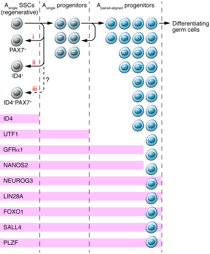
The paper by Aloisio et al. represents a thorough body of work in the field of mammalian male germline stem cell biology. Using state-of-art cutting-edge technologies, they systematically studied a rare subset of PAX7+ Asingle spermatogonia in mouse testes (20). Expression analyses, lineage tracing, germ cell transplantation, germ cell ablation, and a combination of other insults, along with comparative phylogenetic studies, all indicate that PAX7 is a bona fide SSC marker. PAX7 is only the second such exclusive marker for “true” SSCs, the first being the recently reported ID4 (ref. 16 and Figure 1). Several questions remain and need to be addressed in the future.
Figure 1. A summary of known markers in various undifferentiated spermatogonial cells is shown.

Based on the report by Chan and colleagues, ID4 is the only known Asingle spermatogonial marker (16). This ID4+ cell population has the ability to self-renew and to give rise to progenitors from which other transient progenitors and eventually differentiating germ cells are produced. Studies by Aloisio et al. have now identified PAX7+ as another true SSC marker. Three possibilities exist: (i) PAX7+ cells are one subset; (ii) ID4+ cells are another subset of nonoverlapping SSCs; or (iii) PAX7+ID4+ cells are an overlapping subset of Asingle spermatogonia. Known markers of spermatogonia include ID4, undifferentiated embryonic cell transcription factor 1 (UTF1), GDNF family receptor αv1 (GFRα1), nanos homolog 2 (NANOS2), neurogenin 3 (NEUROG3), lin-28 homolog A (LIN28A), FOXO1, spalt-like transcription factor 4 (SALL4), and promyelocytic leukemia zinc finger (PLZF). Figure modified with permission from Genes & Development (16).
First, it would be interesting to determine whether PAX7 and ID4 are expressed in overlapping or nonoverlapping subsets of Asingle spermatogonia. An Id4-Gfp transgenic mouse line is available (16); therefore, evaluation of these two markers should be feasible. Second, development of a Pax7-Gfp mouse line is highly desirable, because SSCs enriched from testes of these mice could prove useful for testing several biological properties of PAX7+ Asingle spermatogonia both in vitro and in vivo. For example, the gene expression signatures and other surface markers that are expressed on these PAX7+ Asingle cells could be tested. Creation of a Pax7-Gfp mouse strain should be technically feasible, because Pax7 promoter sequences that are able to drive expression of a Cre recombinase reporter in mouse testes are already known. Third, it is intriguing to note that robustly dividing PAX7+ spermatogonia are resistant to various toxic, chemotherapy, and radiotherapy insults. Whether these cells have unique antiapoptotic mechanisms or whether there are heterogeneous populations among them is worth exploring. Fourth, although mice lacking PAX7 in the germline are fertile and exhibit normal spermatogenesis, it remains to be tested whether new phenotypes would emerge in these conditional KO mice as a natural consequence of aging or when subjected to stress. Finally, it should be feasible in the future to address the fundamental issue of how and when gonocytes acquire the “stemness.” It is possible that this unique property could also be inherent in primordial germ cells in the XY gonad. It is hoped that with some of the key mouse strains already available, those anticipated in future, and the powerful in vivo genetic linage-tracing methods, we may be able to better understand the forever fascinating male germline stem cells.
Acknowledgments
I thank Kyle Orwig (Magee-Womens Research Institute, University of Pittsburgh) for critically reading this commentary and helpful suggestions. I thank Stan Fernald for helping me with graphic art design. Work done in the author’s laboratory is partially supported by NIH grant 9P20GM104936 from the National Institute of General Medical Sciences.
Footnotes
Conflict of interest: The author has declared that no conflict of interest exists.
Reference information:J Clin Invest. 2014;124(10):4219–4222. doi:10.1172/JCI77926.
See the related article beginning on page 3929.
References
- 1.Yoshida S. Spermatogenic stem cell system in the mouse testis. Cold Spring Harb Symp Quant Biol. 2008;73:25–32. doi: 10.1101/sqb.2008.73.046. [DOI] [PubMed] [Google Scholar]
- 2.Yoshida S. Stem cells in mammalian spermatogenesis. Dev Growth Differ. 2010;52(3):311–317. doi: 10.1111/j.1440-169X.2010.01174.x. [DOI] [PubMed] [Google Scholar]
- 3.Saitou M, Yamaji M. Primordial germ cells in mice. Cold Spring Harb Perspect Biol. 2012;4(11):a008375. doi: 10.1101/cshperspect.a008375. [DOI] [PMC free article] [PubMed] [Google Scholar]
- 4.Cool J, DeFalco T, Capel B. Testis formation in the fetal mouse: dynamic and complex de novo tubulogenesis. Wiley Interdiscip Rev Dev Biol. 2012;1(6):847–859. doi: 10.1002/wdev.62. [DOI] [PubMed] [Google Scholar]
- 5.DeFalco T, Capel B. Gonad morphogenesis in vertebrates: divergent means to a convergent end. Annu Rev Cell Dev Biol. 2009;25:457–482. doi: 10.1146/annurev.cellbio.042308.13350. [DOI] [PMC free article] [PubMed] [Google Scholar]
- 6.Jan SZ, Hamer G, Repping S, de Rooij DG, van Pelt AM, Vormer TL. Molecular control of rodent spermatogenesis. Biochim Biophys Acta. 2012;1822(12):1838–1850. doi: 10.1016/j.bbadis.2012.02.008. [DOI] [PubMed] [Google Scholar]
- 7.Kanatsu-Shinohara M, Shinohara T. Spermatogonial stem cell self-renewal and development. Annu Rev Cell Dev Biol. 2013;29:163–187. doi: 10.1146/annurev-cellbio-101512-122353. [DOI] [PubMed] [Google Scholar]
- 8.Aponte PM, van Bragt MP, de Rooij DG, van Pelt AM. Spermatogonial stem cells: characteristics and experimental possibilities. APMIS. 2005;113(11–12):727–742. doi: 10.1111/j.1600-0463.2005.apm_302.x. [DOI] [PubMed] [Google Scholar]
- 9.De Rooij DG. Regulation of the proliferation of spermatogonial stem cells. J Cell Sci Suppl. 1988;10:181–194. doi: 10.1242/jcs.1988.supplement_10.14. [DOI] [PubMed] [Google Scholar]
- 10.de Rooij DG, Griswold MD. Questions about spermatogonia posed answered since 2000. J Androl. 2012;33(6):1085–1095. doi: 10.2164/jandrol.112.016832. [DOI] [PubMed] [Google Scholar]
- 11.Oatley JM, Brinster RL. Spermatogonial stem cells. Methods Enzymol. 2006;419:259–282. doi: 10.1016/S0076-6879(06)19011-4. [DOI] [PubMed] [Google Scholar]
- 12.Yang QE, Oatley JM. Spermatogonial stem cell functions in physiological and pathological conditions. Curr Top Dev Biol. 2014;107:235–267. doi: 10.1016/B978-0-12-416022-4.00009-3. [DOI] [PubMed] [Google Scholar]
- 13.Oatley JM, Brinster RL. Regulation of spermatogonial stem cell self-renewal in mammals. Annu Rev Cell Dev Biol. 2008;24:263–286. doi: 10.1146/annurev.cellbio.24.110707.175355. [DOI] [PMC free article] [PubMed] [Google Scholar]
- 14.Kanatsu-Shinohara M, Shinohara T. Culture and genetic modification of mouse germline stem cells. Ann N Y Acad Sci. 2007;1120:59–71. doi: 10.1196/annals.1411.001. [DOI] [PubMed] [Google Scholar]
- 15.Yoshida S. Elucidating the identity and behavior of spermatogenic stem cells in the mouse testis. Reproduction. 2012;144(3):293–302. doi: 10.1530/REP-11-0320. [DOI] [PubMed] [Google Scholar]
- 16.Chan F, et al. Functional molecular features of the Id4+ germline stem cell population in mouse testes. Genes Dev. 2014;28(12):1351–1362. doi: 10.1101/gad.240465.114. [DOI] [PMC free article] [PubMed] [Google Scholar]
- 17.Yoshida S, Nabeshima Y, Nakagawa T. Stem cell heterogeneity: actual and potential stem cell compartments in mouse spermatogenesis. Ann N Y Acad Sci. 2007;1120:47–58. doi: 10.1196/annals.1411.003. [DOI] [PubMed] [Google Scholar]
- 18.Oatley JM, Avarbock MR, Telaranta AI, Fearon DT, Brinster RL. Identifying genes important for spermatogonial stem cell self-renewal and survival. Proc Natl Acad Sci U S A. 2006;103(25):9524–9529. doi: 10.1073/pnas.0603332103. [DOI] [PMC free article] [PubMed] [Google Scholar]
- 19.Oatley JM, Oatley MJ, Avarbock MR, Tobias JW, Brinster RL. Colony stimulating factor 1 is an extrinsic stimulator of mouse spermatogonial stem cell self-renewal. Development. 2009;136(7):1191–1199. doi: 10.1242/dev.032243. [DOI] [PMC free article] [PubMed] [Google Scholar]
- 20.Aloisio GM, et al. PAX7 expression defines germline stem cells in the adult testis. J Clin Invest. 2014;124(9):3929–3944. doi: 10.1172/JCI75943. [DOI] [PMC free article] [PubMed] [Google Scholar]
- 21.McCullagh KJ, Perlingeiro RC. Coaxing stem cells for skeletal muscle repair. Adv Drug Deliv Rev. 2014;pii:S0169-409X(14)00148-3. doi: 10.1016/j.addr.2014.07.007. [DOI] [PMC free article] [PubMed] [Google Scholar]
- 22.Motohashi N, Asakura A. Muscle satellite cell heterogeneity and self-renewal. Front Cell Dev Biol. 2014;2(1):pii:00001. doi: 10.3389/fcell.2014.00001. [DOI] [PMC free article] [PubMed] [Google Scholar]


