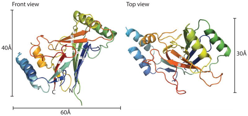
Figure 2. Structure of the wild type C. elegans CTL2 domain.
The structure of the wild type Piezo CTL2 is colored in a rainbow scheme, progressing from the N-terminus (blue) to C-terminus (red). Front view (left): The loop is oriented so that the connections to the putative transmembrane helices are positioned toward the bottom. The β-strands are numbered sequentially from the N-terminus. Top view (right): the front view, rotated 90° around the horizontal axis. See also Figures S2 and S3.

