


Abstract
Langerhans cell histiocytosis (LCH) is a rare disease that can infiltrate various organs. LCH presents with solitary organ involvement or as a multi-system disease. We present a patient who has tonsillary infiltration with LCH. A 74 year-old Caucasian male was admitted for swelling of the neck and difficulty swallowing for 3 months. Physical examination showed submandibular lymph node enlargement of approximately 3 cm and tonsil enlargement. A tonsillectomy and excisional biopsy of the lymph node were done. Histiocyte-like cell infiltration was seen in the tonsil biopsy. CD3, CD20, CD15, CD30, CD5, CD138, Lambda, Kappa, Bcl-2, ALK, CD23, CD10, Bcl-6, keratin, EMA, HMB-45, and Cyl D1 were negative. CD68, S-100, CD1a, and fascin were positive, and the Ki-67 proliferation index was 20 % in immunocytochemical staining. The most commonly infiltrated bones are the skull, femur, lower jaw, pelvis, and vertebrae in LCH. Oral or perioral lesions are present in 30 % of cases. Oral lesions most often involve bone loss, unexpected tooth loss, and gum inflammation. We administered oral prednisolone to our patient due to the presence of lytic lesion of the bone, mild anemia and a higher sedimentation rate, which was from a separate, explained cause. Isolated tonsillar involvement in adult LCH was reported in only 2 cases in the literature. There is no standard recommendation for treatment. Our patient responded well to steroid therapy.
Keywords: Langerhans cell histiocytosis, Tonsillar neoplasms, Prednisolone
Introduction
Langerhans cell histiocytosis (LCH) is a rare disease that can infiltrate various organs. LCH presents with solitary organ involvement or as a multi-system disease. It usually affects the bone, skin, and pituitary gland, and occasionally affects the hematopoietic system, lymph nodes, and lungs [1]. LCH is most often found in the pediatric population and less commonly in adults [1]. In this paper, we present a case report of an adult patient with tonsil infiltration with LCH.
Case Presentation
In December 2013, a 74 year-old Caucasian male was admitted to our hematology clinic for swelling of the neck and difficulty swallowing for 3 months. He had no previous medical history and did not take medications. Physical examination showed submandibular lymph node enlargement of approximately 3 cm and tonsil enlargement. A tonsillectomy and excisional biopsy of the lymph node were done. Histiocyte-like cell infiltration was seen in the tonsil biopsy. CD3, CD20, CD15, CD30, CD5, CD138, Lambda, Kappa, Bcl-2, ALK, CD23, CD10, Bcl-6, keratin, EMA, HMB-45, and Cyl D1 were negative. CD68, S-100, CD1a, and fascin were positive, and the Ki-67 proliferation index was 20 % in immunocytochemical staining (Fig. 1–4). Reactive lymphocytic proliferation was seen in the lymph node biopsy. For the patient’s risk stratification, we conducted cervical-thoracal-abdominal computerized tomography (CT) scanning, whole body positron emission tomography (PET) PET CT, and a bone marrow biopsy. F-18 fluorodeoxyglucose (FDG) uptake positive for lytic lesion (standard uptake value (SUV) max: 9.59) was seen in the subtrochanteric area of the left femur in PET CT. There was no abnormal cell infiltration in the bone marrow biopsy and no lymph node enlargement was found during the thoraco-abdominal CT scanning. His laboratory values were mild normochromic normocytic anemia with mild higher sedimentation rate. We gave him oral prednisolone at a dose of 1 mg/d for 6 week. Steroids were gradually tapered off. Following this treatment schedule, he had no complaints and his laboratory values were normal and FDG uptake were not seen in the whole body PET CT.
Fig. 3.
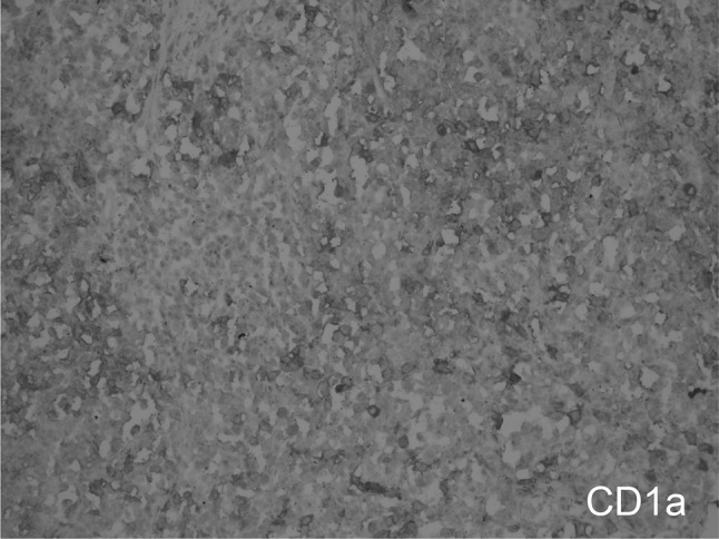
CD1a staining of the sinusoidal LCH cells are demonstrated (immunoperoxidase, CD1a X200)
Fig. 1, 2.

Langerhans cell histiocytosis (LCH) in the tonsil. Low-power view shows sheets of tonsil LCH cells with grooved, folded nuclei; pale eosinophilic cytoplasm; and interspersed eosinophils (Fig. 1; H + E, X50) (Fig. 2; H + E, X200)
Fig. 4.

LCH cells are cytoplasmic positive with fascin (immunoperoxidase, Fascin X200)
Discussion
LCH was renamed by the Histiocyte Society in 1985. Its former names were eosinophilic granuloma, histiocytosis X, Letterer-Siwe disease, and Hand-Schuller-Christian disease [2]. Although the etiology of the disease is not known, it could be caused by an immunological dysregulation subsequent to cytokine and prostaglandin over-production [3].
The disease can be difficult to diagnose. Cases of LCH exhibiting immature morphology and few associated eosinophils should be excluded using appropriate markers (S-100, CD1a, and langerin) [4]. The immuno-histochemical staining feature in our patient’s biopsy material supported the exact diagnosis. LCH is typically a systemic infiltrating disease and can present in 2 forms; non-systemic (localized) disease and systemic (multi-focal) disease. The distribution of affected organs shapes the prognosis of the disease [5]. The frequency of involvement of the bone is 80 %; skin, 25 %; the pituitary gland, 25 %; spleen, 15 %; liver, 15 %; the hematopoietic system, 15 %; lymph nodes, 5–10 %; and cranial involvement excluding the pituitary gland, 2–4 % [1]. The most commonly infiltrated bones are the skull, femur, lower jaw, pelvis, and vertebrae [6]. Oral or perioral lesions are present in 30 % of cases [6]. Oral lesions most often involve bone loss, unexpected tooth loss, and gum inflammation [6]. Isolated infiltration of the tonsil is rare; only 2 case reports were found in the literature review [7, 8].
The first case was reported by Issing et al. The patient was not given medication and remained in remission for 16 months [7]. Hwang et al. reported the case of a 68 year-old man with tonsil enlargement due to the infiltration of LCH concomitantly with acute myeloblastic leukemia. The lymph node enlargement resolved spontaneously with acute leukemia treatment. The authors explained that this unusual case, involving neoplastic histiocytic cells and leukemic cells could have been a member of the same clone or a reaction to the acute leukemia [8]. Treatment recommendations were completed according to the presentation of the disease. In some cases, localized surgical excisional biopsy may be sufficient for treatment, while other cases require the application of multi-agent chemotherapy protocols [9]. Our patient had infiltration of unilateral tonsil, only one lytic lesion of the bone and mild anemia without bone marrow infiltration. We did not find any author advice regarding isolated infiltration of the lymph nodes and tonsils. In these cases, the watch and wait method should be considered because of the potential for spontaneous regression [10]. We administered oral prednisolone to our patient due to the presence of lytic lesion of the bone, mild anemia and a higher sedimentation rate, which was from a separate, explained cause.
Conclusion
Isolated tonsillar involvement in adult LCH was reported in only 2 cases in the literature. There is no standard recommendation for treatment. Our patient responded well to steroid therapy.
References
- 1.Haupt R, Minkov M, Astigarraga I, et al. Langerhans cell histiocytosis (LCH): Guidelines for diagnosis, clinical work-up, and treatment for patients till the age of 18 years. Pediatr Blood Cancer. 2013;60(2):175–184. doi: 10.1002/pbc.24367. [DOI] [PMC free article] [PubMed] [Google Scholar]
- 2.Writing Group of the Histiocyte Society Histiocytosis syndromes in children. Lancet. 1987;1(8526):208–209. [PubMed] [Google Scholar]
- 3.lyeyasu JN, Marcos Vaz AC, Reis F, Altemani J, Queiroz LC, Carvalho KL. Langerhans cell histiocytosis diagnosed in an elderly patient. Radiol Bras. 2012;45(4):241–243. doi: 10.1590/S0100-39842012000400014. [DOI] [Google Scholar]
- 4.Pinkus GS, Lones MA, Matsumura F, Yamashiro S, Said JW, Pinkus JL. Langerhans cell histiocytosis immunohistochemical expression of fascin, a dendritic cell marker. Am J Clin Pathol. 2002;118(3):335–343. doi: 10.1309/N2TW-ENRB-1N1C-DWL0. [DOI] [PubMed] [Google Scholar]
- 5.Shankar U, Prasad M, Chaurasia OP. Gastrointestinal tract involvement in Langerhans cell histiocytosis: case report and literature review. World J Gastroenterol. 2012;18(12):1410–1413. doi: 10.3748/wjg.v18.i12.1410. [DOI] [PMC free article] [PubMed] [Google Scholar]
- 6.Martínez DSM, Villagrán UJ, Ajqui RR, Cervantes CK. Oral manifestations of Langerhans cell histiocytosis (LCH): review of scientific literature and case report. Rev Odontol Mex. 2012;16(2):123–130. [Google Scholar]
- 7.Issing PR, Kaiserling E, Lenarz T. Langerhans cell histiocytosis of the tonsil. Laryngorhinootologie. 1993;72(7):346–349. doi: 10.1055/s-2007-997914. [DOI] [PubMed] [Google Scholar]
- 8.Hwang YY, Tsui P, Leung RY, Kwong YL. Disseminated Langerhans cell histiocytosis associated with acute myeloid leukaemia: complete remission with daunorubicin and cytarabine. Ann Hematol. 2013;92(2):267–268. doi: 10.1007/s00277-012-1555-6. [DOI] [PMC free article] [PubMed] [Google Scholar]
- 9.Girschikofsky M, Arico M, Castillo D. Management of adult patients with Langerhans cell histiocytosis: recommendations from an expert panel on behalf of Euro-Histio-Net. Orphanet J Rare Dis. 2013;14(8):72. doi: 10.1186/1750-1172-8-72. [DOI] [PMC free article] [PubMed] [Google Scholar]
- 10.Lo WC, Chen CC, Tsai CC, Cheng PW. Isolated adult Langerhans cell histiocytosis in cervical lymph nodes: should it be treated? J Laryngol Otol. 2009;123(9):1055–1057. doi: 10.1017/S0022215108004155. [DOI] [PubMed] [Google Scholar]