

Abstract
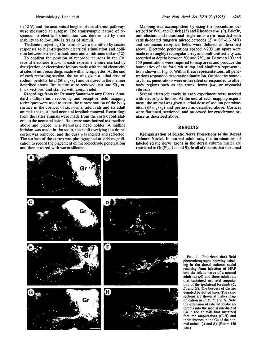
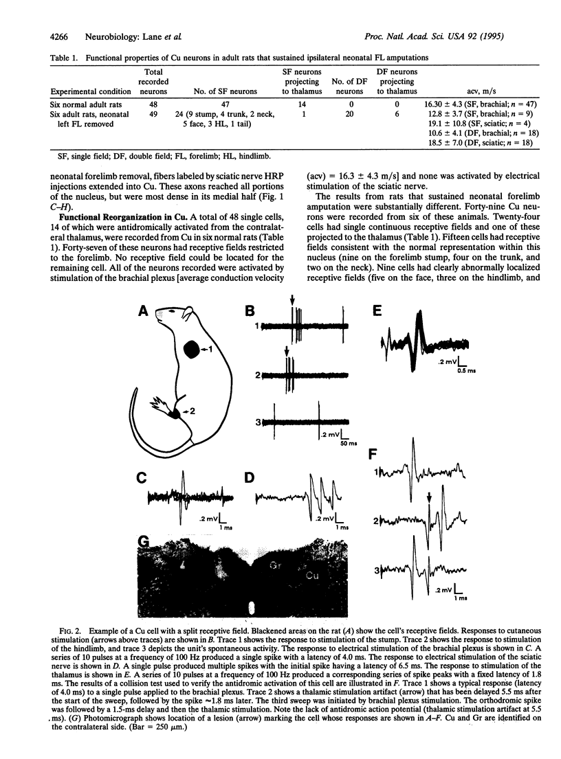
Electrophysiological and neuroanatomical methods were used to determine the extent to which neonatal forelimb removal altered the organization of the cuneate nucleus and representations of the fore- and hindlimbs in the primary somatosensory cortex of adult rats. Neonatal forelimb removal resulted in invasion of the cuneate nucleus by sciatic nerve primary afferents and development of cuneothalamic projection neurons with split receptive fields that included both the hindlimb and forelimb stump. Mapping in the primary somatosensory cortex of the neonatally manipulated adult rats demonstrated abnormalities, but the major change observed in the cuneate nucleus was demonstrable at only a few (5%) cortical recording sites in the remaining stump representation and there were none at all in the hindlimb representation. These results suggest that lesion-induced brainstem reorganization may be functionally suppressed at either the thalamic or cortical level.
Full text
PDF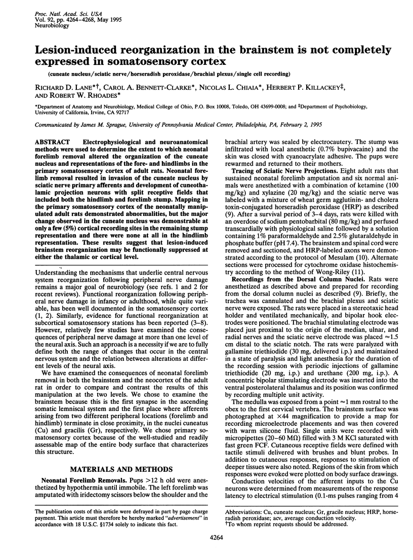
Images in this article

Selected References
These references are in PubMed. This may not be the complete list of references from this article.
- Bates C. A., Killackey H. P. The organization of the neonatal rat's brainstem trigeminal complex and its role in the formation of central trigeminal patterns. J Comp Neurol. 1985 Oct 15;240(3):265–287. doi: 10.1002/cne.902400305. [DOI] [PubMed] [Google Scholar]
- Calford M. B., Tweedale R. Immediate expansion of receptive fields of neurons in area 3b of macaque monkeys after digit denervation. Somatosens Mot Res. 1991;8(3):249–260. doi: 10.3109/08990229109144748. [DOI] [PubMed] [Google Scholar]
- Cusick C. G., Wall J. T., Whiting J. H., Jr, Wiley R. G. Temporal progression of cortical reorganization following nerve injury. Brain Res. 1990 Dec 24;537(1-2):355–358. doi: 10.1016/0006-8993(90)90385-o. [DOI] [PubMed] [Google Scholar]
- Donoghue J. P., Sanes J. N. Organization of adult motor cortex representation patterns following neonatal forelimb nerve injury in rats. J Neurosci. 1988 Sep;8(9):3221–3232. doi: 10.1523/JNEUROSCI.08-09-03221.1988. [DOI] [PMC free article] [PubMed] [Google Scholar]
- Dostrovsky J. O., Millar J., Wall P. D. The immediate shift of afferent drive to dorsal column nucleus cells following deafferentation: a comparison of acute and chronic deafferentation in gracile nucleus and spinal cord. Exp Neurol. 1976 Sep;52(3):480–495. doi: 10.1016/0014-4886(76)90219-3. [DOI] [PubMed] [Google Scholar]
- Dougherty P. M., Willis W. D. Enhanced responses of spinothalamic tract neurons to excitatory amino acids accompany capsaicin-induced sensitization in the monkey. J Neurosci. 1992 Mar;12(3):883–894. doi: 10.1523/JNEUROSCI.12-03-00883.1992. [DOI] [PMC free article] [PubMed] [Google Scholar]
- Fitzgerald M., Woolf C. J., Shortland P. Collateral sprouting of the central terminals of cutaneous primary afferent neurons in the rat spinal cord: pattern, morphology, and influence of targets. J Comp Neurol. 1990 Oct 15;300(3):370–385. doi: 10.1002/cne.903000308. [DOI] [PubMed] [Google Scholar]
- Florence S. L., Garraghty P. E., Carlson M., Kaas J. H. Sprouting of peripheral nerve axons in the spinal cord of monkeys. Brain Res. 1993 Jan 22;601(1-2):343–348. doi: 10.1016/0006-8993(93)91734-a. [DOI] [PubMed] [Google Scholar]
- Garraghty P. E., Hanes D. P., Florence S. L., Kaas J. H. Pattern of peripheral deafferentation predicts reorganizational limits in adult primate somatosensory cortex. Somatosens Mot Res. 1994;11(2):109–117. doi: 10.3109/08990229409028864. [DOI] [PubMed] [Google Scholar]
- Garraghty P. E., Pons T. P., Sur M., Kaas J. H. The arbors of axons terminating in middle cortical layers of somatosensory area 3b in owl monkeys. Somatosens Mot Res. 1989;6(4):401–411. doi: 10.3109/08990228909144683. [DOI] [PubMed] [Google Scholar]
- Jacquin M. F., Rhoades R. W. Effects of neonatal infraorbital lesions upon central trigeminal primary afferent projections in rat and hamster. J Comp Neurol. 1985 May 1;235(1):129–143. doi: 10.1002/cne.902350110. [DOI] [PubMed] [Google Scholar]
- Kaas J. H., Guillery R. W. The transfer of abnormal visual field representations from the dorsal lateral geniculate nucleus to the visual cortex in Siamese cats. Brain Res. 1973 Sep 14;59:61–95. doi: 10.1016/0006-8993(73)90253-9. [DOI] [PubMed] [Google Scholar]
- Kaas J. H. Plasticity of sensory and motor maps in adult mammals. Annu Rev Neurosci. 1991;14:137–167. doi: 10.1146/annurev.ne.14.030191.001033. [DOI] [PubMed] [Google Scholar]
- Kolarik R. C., Rasey S. K., Wall J. T. The consistency, extent, and locations of early-onset changes in cortical nerve dominance aggregates following injury of nerves to primate hands. J Neurosci. 1994 Jul;14(7):4269–4288. doi: 10.1523/JNEUROSCI.14-07-04269.1994. [DOI] [PMC free article] [PubMed] [Google Scholar]
- Landry P., Villemure J., Deschênes M. Geometry and orientation of thalamocortical arborizations in the cat somatosensory cortex as revealed by computer reconstruction. Brain Res. 1982 Apr 8;237(1):222–226. doi: 10.1016/0006-8993(82)90570-4. [DOI] [PubMed] [Google Scholar]
- Mesulam M. M. Tetramethyl benzidine for horseradish peroxidase neurohistochemistry: a non-carcinogenic blue reaction product with superior sensitivity for visualizing neural afferents and efferents. J Histochem Cytochem. 1978 Feb;26(2):106–117. doi: 10.1177/26.2.24068. [DOI] [PubMed] [Google Scholar]
- Millar J., Basbaum A. I., Wall P. D. Restructuring of the somatotopic map and appearance of abnormal neuronal activity in the gracile nucleus after partial deafferentation. Exp Neurol. 1976 Mar;50(3):658–672. doi: 10.1016/0014-4886(76)90035-2. [DOI] [PubMed] [Google Scholar]
- Pubols L. M., Bowen D. C. Lack of central sprouting of primary afferent fibers after ricin deafferentation. J Comp Neurol. 1988 Sep 8;275(2):282–287. doi: 10.1002/cne.902750209. [DOI] [PubMed] [Google Scholar]
- Rasmusson D. D., Turnbull B. G. Immediate effects of digit amputation on SI cortex in the raccoon: unmasking of inhibitory fields. Brain Res. 1983 Dec 12;288(1-2):368–370. doi: 10.1016/0006-8993(83)90120-8. [DOI] [PubMed] [Google Scholar]
- Rhoades R. W., Belford G. R., Killackey H. P. Receptive-field properties of rat ventral posterior medial neurons before and after selective kainic acid lesions of the trigeminal brain stem complex. J Neurophysiol. 1987 May;57(5):1577–1600. doi: 10.1152/jn.1987.57.5.1577. [DOI] [PubMed] [Google Scholar]
- Rhoades R. W., Wall J. T., Chiaia N. L., Bennett-Clarke C. A., Killackey H. P. Anatomical and functional changes in the organization of the cuneate nucleus of adult rats after fetal forelimb amputation. J Neurosci. 1993 Mar;13(3):1106–1119. doi: 10.1523/JNEUROSCI.13-03-01106.1993. [DOI] [PMC free article] [PubMed] [Google Scholar]
- Rodin B. E., Kruger L. Absence of intraspinal sprouting in dorsal root axons caudal to a partial spinal hemisection: a horseradish peroxidase transport study. Somatosens Res. 1984;2(2):171–192. doi: 10.1080/07367244.1984.11800557. [DOI] [PubMed] [Google Scholar]
- Waite P. M. Rearrangement of neuronal responses in the trigeminal system of the rat following peripheral nerve section. J Physiol. 1984 Jul;352:425–445. doi: 10.1113/jphysiol.1984.sp015301. [DOI] [PMC free article] [PubMed] [Google Scholar]
- Waite P. M., Taylor P. K. Removal of whiskers in young rats causes functional changes in cerebral cortex. Nature. 1978 Aug 10;274(5671):600–602. doi: 10.1038/274600a0. [DOI] [PubMed] [Google Scholar]