

Abstract
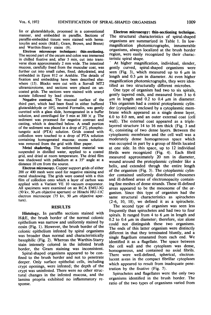
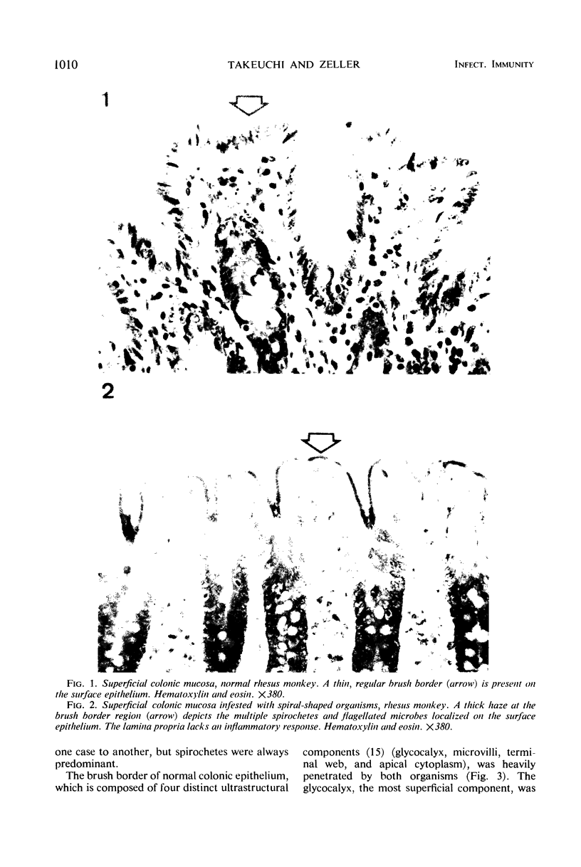
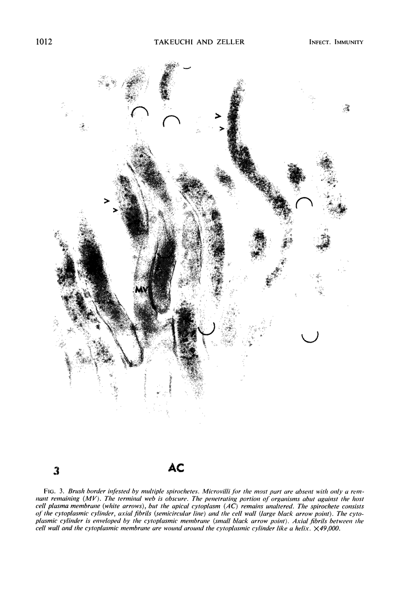
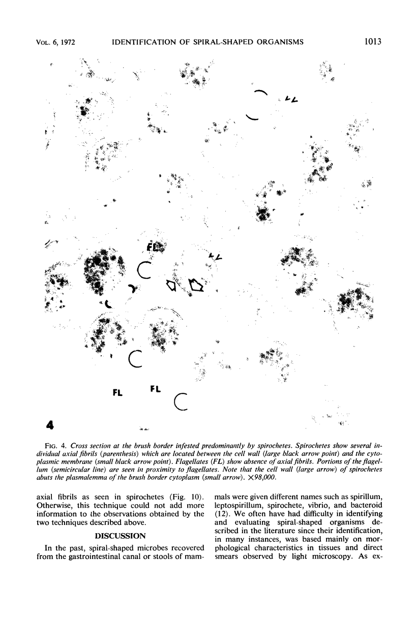
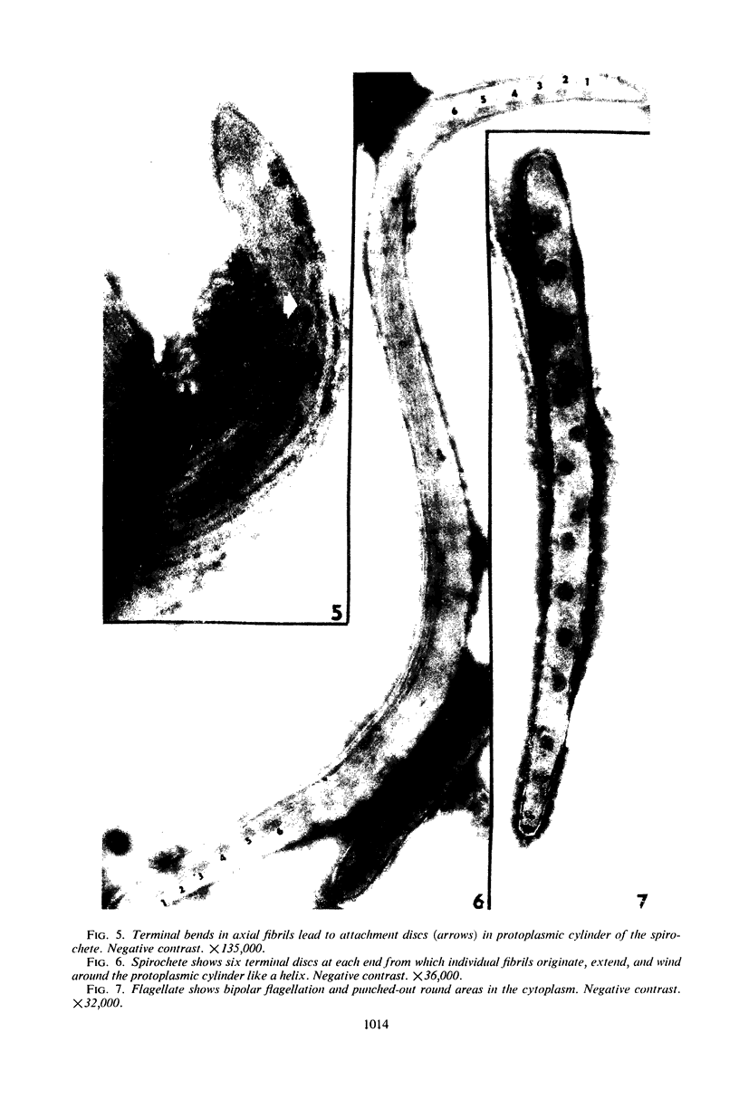
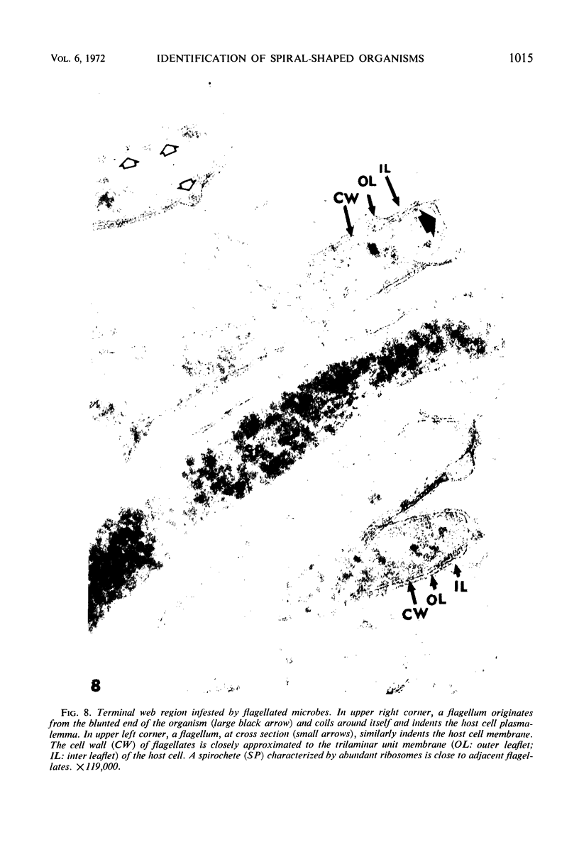
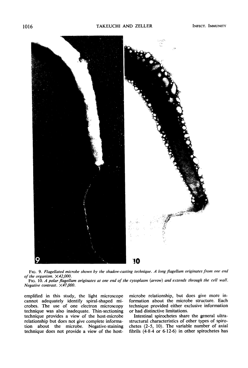
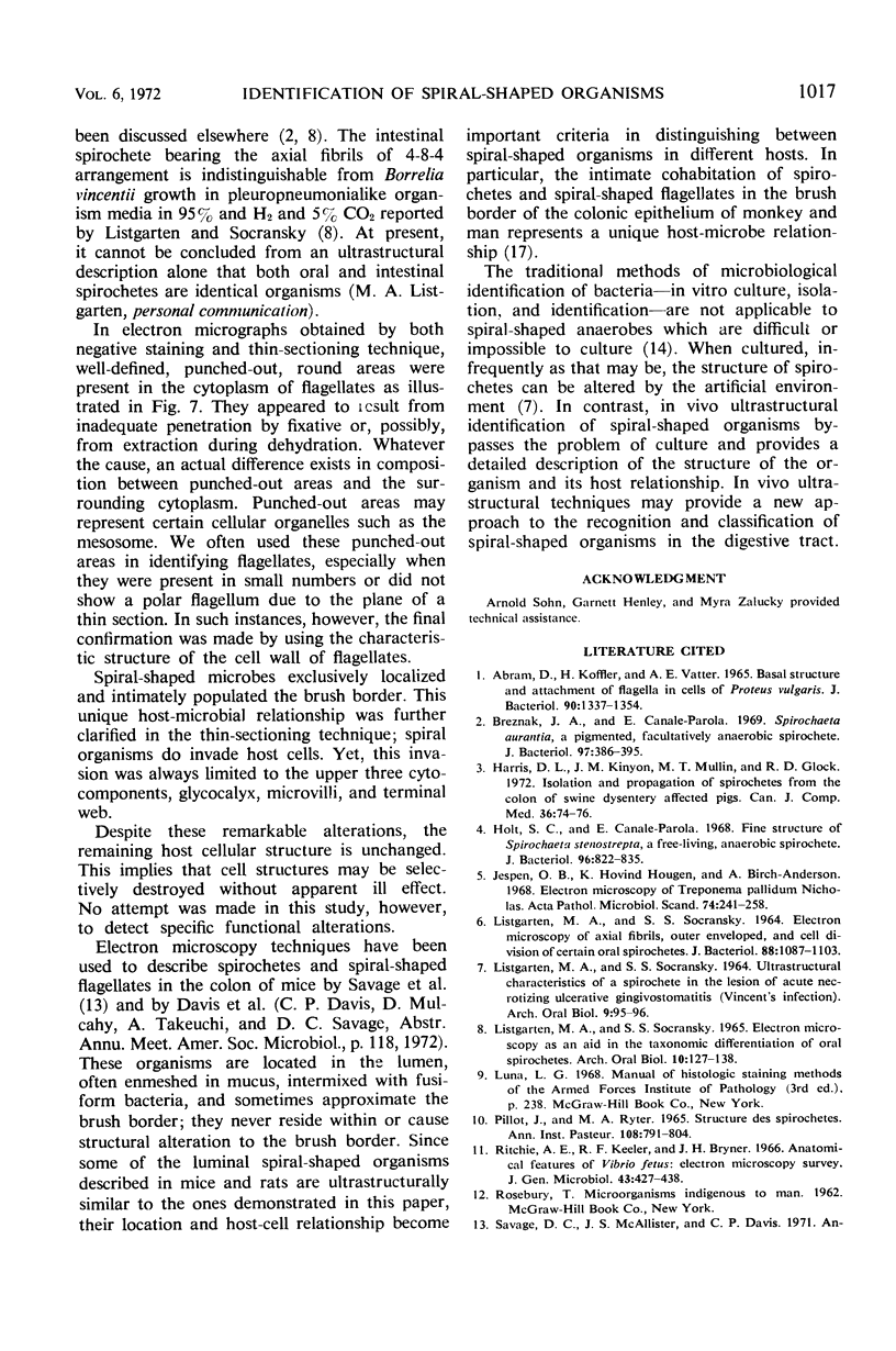
Spiral-shaped organisms exclusively and intimately populate the brush border of the cecal and colonic epithelium of healthy monkeys (Macaca mulatta). These organisms replace the glycocalyx, destroy most microvilli, and attenuate the terminal web of the brush border. Despite these remarkable alterations, the remaining host cellular structure is unchanged. Two structurally distinct microbes, a spirochete and a flagellate, were recognized by electron microscopy. These spirochetes share the general characteristics of other known spirochetes: they are 3 to 6 μm long and 0.2 to 0.4 μm wide, spiral 2 to 6 times, and have axial fibrils of 6-12-6 and 4-8-4 arrangements. Flagellated microbes are 4 to 6 μm long and 0.2 to 0.4 μm wide, spiral 2 to 4 times, and are characterized by a polar flagellum which originates from the terminal button at each end of the cytoplasmic body. This in vivo ultrastructural study of anaerobic spiral-shaped organisms bypasses the difficulties of in vitro culture techniques and provides a detailed description and identification of these organisms in their host environment.
Full text
PDF
Images in this article

Selected References
These references are in PubMed. This may not be the complete list of references from this article.
- Abram D., Koffler H., Vatter A. E. Basal structure and attachment of flagella in cells of Proteus vulgaris. J Bacteriol. 1965 Nov;90(5):1337–1354. doi: 10.1128/jb.90.5.1337-1354.1965. [DOI] [PMC free article] [PubMed] [Google Scholar]
- Breznak J. A., Canale-Parola E. Spirochaeta aurantia, a pigmented, facultatively anaerobic spirochete. J Bacteriol. 1969 Jan;97(1):386–395. doi: 10.1128/jb.97.1.386-395.1969. [DOI] [PMC free article] [PubMed] [Google Scholar]
- Harris D. L., Kinyon J. M., Mullin M. T., Glock R. D. Isolation and propagation of spirochetes from the colon of swine dysentery affected pigs. Can J Comp Med. 1972 Jan;36(1):74–76. [PMC free article] [PubMed] [Google Scholar]
- Holt S. C., Canale-Parola E. Fine structure of Spirochaeta stenostrepta, a free-living, anaerobic spirochete. J Bacteriol. 1968 Sep;96(3):822–835. doi: 10.1128/jb.96.3.822-835.1968. [DOI] [PMC free article] [PubMed] [Google Scholar]
- Jepsen O. B., Hougen K. H., Birch-Andersen A. Electron microscopy of treponema pallidum Nichols. Acta Pathol Microbiol Scand. 1968;74(2):241–258. doi: 10.1111/j.1699-0463.1968.tb03477.x. [DOI] [PubMed] [Google Scholar]
- LISTGARTEN M. A., SOCRANSKY S. S. ELECTRON MICROSCOPY AS AN AID IN THE TAXONOMIC DIFFERENTIATION OF ORAL SPIROCHETES. Arch Oral Biol. 1965 Jan-Feb;10:127–138. doi: 10.1016/0003-9969(65)90064-6. [DOI] [PubMed] [Google Scholar]
- LISTGARTEN M. A., SOCRANSKY S. S. ELECTRON MICROSCOPY OF AXIAL FIBRILS, OUTER ENVELOPE, AND CELL DIVISION OF CERTAIN ORAL SPIROCHETES. J Bacteriol. 1964 Oct;88:1087–1103. doi: 10.1128/jb.88.4.1087-1103.1964. [DOI] [PMC free article] [PubMed] [Google Scholar]
- LISTGARTEN M. A., SOCRANSKY S. S. ULTRASTRUCTURAL CHARACTERISTICS OF A SPIROCHETE IN THE LESION OF ACUTE NECROTIZING ULCERATIVE GINGIVOSTOMATITIS (VINCENT'S INFECTION). Arch Oral Biol. 1964 Jan-Feb;9:95–96. doi: 10.1016/0003-9969(64)90050-0. [DOI] [PubMed] [Google Scholar]
- Pillot J., Ryter A. Structure des spirochètes. 1. Etude des generes Treponema, Borrelia et Leptospira au microscope electronique. Ann Inst Pasteur (Paris) 1965 Jun;108(6):791–804. [PubMed] [Google Scholar]
- Ritchie A. E., Keeler R. F., Bryner J. H. Anatomical features of Vibrio fetus: Electron microscopic survey. J Gen Microbiol. 1966 Jun;43(3):427–438. doi: 10.1099/00221287-43-3-427. [DOI] [PubMed] [Google Scholar]
- Savage D. C., McAllister J. S., Davis C. P. Anaerobic bacteria on the mucosal epithelium of the murine large bowel. Infect Immun. 1971 Oct;4(4):492–502. doi: 10.1128/iai.4.4.492-502.1971. [DOI] [PMC free article] [PubMed] [Google Scholar]
- Syed S. A., Abrams G. D., Freter R. Efficiency of various intestinal bacteria in assuming normal functions of enteric flora after association with germ-free mice. Infect Immun. 1970 Oct;2(4):376–386. doi: 10.1128/iai.2.4.376-386.1970. [DOI] [PMC free article] [PubMed] [Google Scholar]
- Takeuchi A. Electron microscope studies of experimental Salmonella infection. I. Penetration into the intestinal epithelium by Salmonella typhimurium. Am J Pathol. 1967 Jan;50(1):109–136. [PMC free article] [PubMed] [Google Scholar]
- Takeuchi A., Formal S. B., Sprinz H. Exerimental acute colitis in the Rhesus monkey following peroral infection with Shigella flexneri. An electron microscope study. Am J Pathol. 1968 Mar;52(3):503–529. [PMC free article] [PubMed] [Google Scholar]
- Taylor D. J., Blakemore W. F. Spirochaetal invasion of the colonic epithelium in swine dysentery. Res Vet Sci. 1971 Mar;12(2):177–179. [PubMed] [Google Scholar]