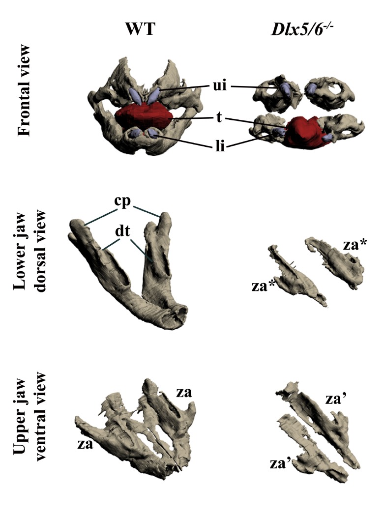
Figure 1. Three-dimensional reconstruction of the dentary and maxillary bones of 18.5dpc wild type and Dlx5/6 -/- mouse embryos.
Upper row: Frontal view of WT and Dlx5/6 -/- oral apparatus. Skeletal elements are grey, the tongue is red and incisors are violet. Middle row: Dorsal view of the dentary bone of WT and Dlx5/6 -/- 18.5dpc mice. Lower row: Ventral view of the maxillary components of WT and Dlx5/6 -/- 18.5dpc mice. Note that the inactivation of Dlx5/6 results in the transformation of both lower and upper jaw skeletal elements into new structures which appear more similar to each other than to their WT counterpart. cp, coronoid processes; dt, dentary bone; li, lower incisor; t, tongue; ui, upper incisor; za, zygomatic arch; za*, zygomatic arch-like structure deriving from lower jaw transformation; za’, zygomatic arch-like structure deriving from upper jaw transformation.

