Abstract
We report that the cytomegalovirus-encoded cell death suppressor vMIA binds Bax and prevents Bax-mediated mitochondrial membrane permeabilization by sequestering Bax at mitochondria in the form of a vMIA–Bax complex. vMIA mutants with a defective mitochondria-targeting domain retain their Bax-binding function but not their ability to suppress mitochondrial membrane permeabilization or cell death. vMIA does not seem to either specifically associate with Bak or suppress Bak-mediated mitochondrial membrane permeabilization. Recent evidence suggests that the contribution of Bax and Bak in the mitochondrial apoptotic signaling pathway depends on the distinct phenotypes of cells, and it appears from our data that vMIA is capable of suppressing apoptosis in cells in which this pathway is dominated by Bax, but not in cells where Bak also plays a role.
The viral mitochondrial inhibitor of apoptosis vMIA is encoded by human cytomegalovirus (CMV) and closely related primate CMVs (1, 2), and appears to be unique: no overt homologues of this gene have been found to date in any other viral or cellular genome, including those of other CMVs and other herpesviruses. The antiapoptotic function of vMIA seems to play a critical role in replication of human and primate CMVs (reviewed in refs. 1–3). vMIA is predominantly localized at mitochondria, and blocks the Fas-mediated apoptotic pathway downstream of caspase-8 activation and conversion of Bid into tBid, but upstream of mitochondrial permeabilization (1), i.e., at or close to the step suppressed by Bcl-2 (4, 5). The amino acid sequence of vMIA lacks any homology with Bcl-2, suggesting that the mechanisms of action of these two proteins differ. The first functional evidence favoring this notion was gained in experiments with mitochondtria isolated from HeLa cells stably transfected with either Bcl-2, Bcl-XL, or vMIA (3). These mitochondria were resistant to GST–tBid-mediated cytochrome c release, compared to control mitochondria, but to a different degree. Although Bcl-2- or Bcl-xL-containing mitochondria became permeable when exposed to an excess of tBid, vMIA-containing mitochondria remained intact even in the presence of a high concentration of tBid. This behavior of Bcl-2- and of Bcl-xL-containing mitochondria is consistent with the notion that Bcl-2 blocks apoptosis, at least in part, through its association with tBid or with another BH3-only death-activating factor (6–8), and, apparently, not through direct association with Bax or Bak: (i) Bcl-2 does not associate with Bax in the absence of Triton X-100 (9, 10); (ii) Bcl-2 and Bcl-xL mutants that lack affinity to Bax retain their cell-death-suppressing activity (11–14); and (iii) Bcl-2 or Bcl-xL localize at mitochondria of cells transfected with these genes, but this does not affect the cytoplasmic distribution of GFP–Bax in these cells (15).
The insensitivity of vMIA-containing mitochondria to GST–tBid suggested that vMIA suppresses mitochondrial permeabilization not through sequestration of tBid, but, perhaps, through an interaction with another protein. Previously, we reported that vMIA contains two amino acid segments that are necessary and sufficient for its antiapoptotic function, Tyr-5–Leu-34, which targets vMIA to mitochondria, and Asn-115–Arg-147 with an as-yet-unidentified function (16). Our experiments reported here reveal the function of this domain and the mechanism of action of vMIA.
Experimental Procedures
Cells. The development and culturing of HeLa/vMIA cells previously denoted as HeLa/pUL37 × 1 #3, BJAB/vMIA cells, HeLa/pcDNA3 cells, and BJAB/pcDNA3 cells have been described (1, 17). HCT116Bax+/– cells (also denoted below as HCT116) and HCT116Bax–/– cells (18) were a generous gift from Bert Vogelstein (Johns Hopkins University, Baltimore). Simian virus 40-transformed murine embryonic fibroblasts (MEFs), with either intact Bax and Bak (MEFwt) or knocked out Bax (MEFBax–/–Bak+/+), Bak (MEFBax+/+Bak–/–), or both (MEFBax–/–Bak–/–) were a generous gift from Stanley Korsmeyer (Harvard University, Boston).
Proteins and Plasmids. Full-length oligomeric recombinant Bax and monomeric recombinant Bax were described elsewhere (19, 20). Recombinant tBid was purchased from R & D Systems. GST–tBid/pGEX-4T-1 expression plasmid was a generous gift of Eric A. Hendrickson (Brown University, Providence, RI). N-biotinylated synthetic peptides vMIA114-150 and vMIA114-150E128P (>95% purity grade) were made at BioSynthesis (Lewisville, TX). Mammalian expression plasmids (based on pcDNA3) encoding vMIA and its deletion mutants were described elsewhere (1, 16), except for pcDNA3vMIAE128P that was constructed by side-directed mutagenesis of pcDNA3vMIAmyc. Anti-adenine nucleotide translocator (ANT) antiserum (21) was generously provided by Patricia Schmid (University of Minnesota, Austin).
Immunoprecipitations, Immunocytochemistry, Subcellular Fractionation, in Vitro Assay for the Efflux of Cytochrome c, Smac/Diablo, and HtrA2/Omi, and in Vitro Binding Assays. These experiments were performed as described (1, 22, 23). Detailed protocols are in Supporting Text, which is published as supporting information on the PNAS web site. For immunohistochemical detection of non-tagged vMIA and endogenous Bax, MRC-5 fibroblasts infected with human CMV were stained with rabbit anti-vMIA antiserum (1) and murine monoclonal anti-Bax (BD Biosciences Pharmingen, clone 3).
Results
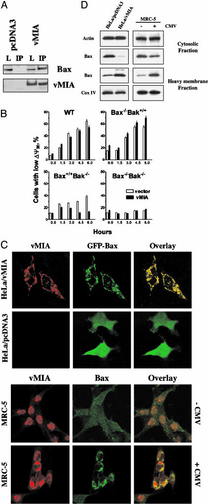
Bax but Not Bak Coimmunoprecipitates with vMIA from Cell Lysates. In an effort to reveal the mechanism of the antiapoptotic function of vMIA, we immunoprecipitated vMIA from lysates of HeLa cells stably transfected with vMIA, and examined these immunoprecipitates for the presence of various apoptosis-related proteins by immunoblotting. These experiments revealed that Bax coprecipitates with vMIA (Fig. 1A). We obtained similar results with lysates of a number of other cells, BJAB cells stably transfected with vMIA, and HeLa, HCT116, and Cos-7 cells transiently transfected with vMIA (data not shown). We also tested whether vMIA binds Bak, a close structural and functional homologue of Bax (24, 25). These experiments were done in a fashion similar to the experiments shown in Fig. 1A, and we could not detect any coimmunoprecipitation of Bak from lysates of vMIA-expressing cells in excess of the nonspecific binding of Bak to beads incubated with lysates of control cells.
Fig. 1.
(A) Bax is associated with vMIA in cells expressing vMIA. Lysates (L) from HeLa cells lysed in a CHAPS-containing buffer (LBC, see Experimental Procedures) constitutively expressing myc-tagged vMIA, or from control cells (pcDNA3 transfected) were immunoprecipitated (IP) with anti-myc beads, bound proteins were separated by SDS/PAGE and examined by Western blotting with either anti-Bax or anti-myc. Equal amounts of HeLa/pcDNA3 and HeLa/vMIA lysates (by total protein) were loaded. The “inputs” represent 10% of the starting materials for IP. (B) vMIA protects Bax-expressing MEFs that lack Bak, but fails to protect Bak-expressing MEFs from staurosporine-induced apoptosis. The loss of the mitochondrial membrane potential ΔΨM was used as an index for apoptosis (see text). The data presented as the means ± SD (three independent experiments). (C) GFP–Bax colocalizes with vMIA in transiently transfected cells. Cells stably transfected with vector only (HeLa/pcDNA3) or myc-tagged vMIA (HeLa/vMIA) were transfected with EGFP–Bax. Eighteen hours after transfection, cells were stained with anti-myc recognizing the myc-tag of vMIA (red). Colocalization of green and red fluorescence is indicated by a yellow blend. Endogenous Bax colocalizes with vMIA in human fibroblasts infected with human CMV (TownevarRIT, 48 h after infection). Cells were stained with rabbit anti-vMIA antiserum (green) and mouse monoclonal anti-Bax (red). Colocalization of green and red fluorescence is indicated by a yellow blend. (D) vMIA induces relocation of endogenous Bax to mitochondria. Cytosolic and heavy membrane fractions of HeLa/pcDNA3 and HeLa/vMIA cells and of MRC-5 human fibroblasts infected or not with human CMV (TownevarRIT, 72 h after infection) were analyzed by Western blotting for the presence of Bax. As control for loading, actin was used in the cytosolic fraction and Cox IV in the heavy membrane fraction. Visual observation of the infected populations of fibroblasts indicated that a fraction of cells remained noninfected, consistent with some remaining cytoplasmic Bax.
vMIA Suppresses Bax- but Not Bak-Mediated Mitochondrial Damage in Murine Fibroblasts. To test whether vMIA could suppress Bax- or Bak-mediated apoptosis, we examined four clones of simian virus 40-transformed MEFs, those that express both Bax and Bak (MEFwt), Bax only (MEFBax+/+Bak–/–), Bak only (MEFBax–/–Bak+/+), or none (MEFBax–/–Bak–/–). It had been established that MEFs expressing Bax and Bak, and MEFs expressing at least one of the proteins, are sensitive to apoptosis, whereas those deficient in both Bax and Bak are resistant (26), indicating that apoptosis in MEFs can be mediated by either Bax or Bak. We cotransfected MEFs with pDsRed-1-mito (for the identification of transfectants) with either vMIA or empty vector, exposed the cells to 10 μM staurosporine 24 h later, and stained with DIOC6(3), a mitochondrial potential probe (27) (40 nM) for flow cytometric analysis. The percentage of DIOC6(3)-low cells that expressed DsRed-1-mito was then used as an index for the extent of cells in the population undergoing apoptosis. We found that vMIA suppressed mitochondrial damage (compared to the empty vector) in neither MEFswt nor MEFsBax–/–Bak+/+, but did protect MEFsBax+/+Bak–/– (Fig. 1B). These data indicate that vMIA suppresses Bax-mediated, but not Bak-mediated, mitochondrial signaling pathway in MEFs. MEFsBax–/–Bak–/– were refractory to apoptosis in accordance with the published results (26).
vMIA Induces Relocation of Bax to Mitochondria. Having established that vMIA coimmunoprecipitates Bax and suppresses Bax-mediated apoptosis, we investigated whether Bax colocalizes with vMIA in intact cells. We examined intracellular distribution of GFP–Bax in transiently transfected cells. It had been established that GFP–Bax is functionally similar to endogenous Bax in its intracellular distribution in healthy and apoptotic cells (15). HeLa/vMIA cells and the control HeLa/pcDNA3 cells were transiently transfected with GFP–Bax, cultured for 24 h, fixed, stained with anti-myc to visualize vMIA, and examined under a confocal microscope (Fig. 1C). Bax (green fluorescence) was, as reported (15), diffusely distributed throughout the cytoplasm of cells that did not express vMIA (red fluorescence), but colocalized (as indicated by yellow in the overlay) with vMIA, which associates with mitochondria, as confirmed by its colocalization with mitochondrial markers (1). Similar results were obtained with HeLa, HCT116, and Bax-deficient HCT116Bax–/– cells transiently cotransfected with vMIA and GFP–Bax (not shown) and with human fibroblasts infected with human CMV (Fig. 1C). Examination of subcellular fractions from HeLa/vMIA cells and from human fibroblasts infected with human CMV (Fig. 1D) for the presence of endogenous Bax confirmed that, in vMIA-expressing transfected or CMV-infected cells, Bax was mainly associated with the mitochondrial fraction, whereas in control cells, Bax was mostly cytoplasmic. vMIA transiently transfected into Bax-deficient HCT116Bax–/– cells was predominantly localized at mitochondria (immunofluorescence observations, data not shown), demonstrating that mitochondrial targeting of vMIA does not require the presence of Bax.
Bax-Binding Domain of vMIA Is Required but Is Not Sufficient for its Cell-Death-Suppressing Activity. To identify the Bax-binding domain of vMIA, we transiently transfected Cos-7 cells and HeLa cells with various deletion mutants of vMIA (all myc-tagged), and examined the affinity of these mutants to Bax in a coimmunoprecipitation assay similar to that shown in Fig. 1A. A summary of these experiments is shown in Table 1. Bax coimmunoprecipitated with vMIAΔ35–112/Δ148–163, consisting essentially of only the two segments required for its antiapoptotic activity, and vMIAΔ2–23 and vMIAΔ23–34, both with an inactivating deletion in the mitochondria-targeting domain (16), but failed to coimmunoprecipitate with vMIAΔ31–163, vMIAΔ115–130, vMIAΔ131–147, and the point mutant vMIAE128P, each containing a functional mitochondrial targeting domain and either completely lacking or containing a mutation in vMIA's Asn-115-Arg-147 segment, which abolished its cell-death suppressing function (Table 1). These observations demonstrated that the Asn-115–Arg-147 segment of vMIA (or most of it) is required for the ability of vMIA to bind Bax. Furthermore, vMIAΔ2–23 and vMIAΔ23–34 retained Bax-binding function, indicating that (i) the mitochondria-targeting domain is not required for this interaction, and, therefore, the Asn-115–Arg-147 segment of vMIA is solely responsible for Bax binding, and (ii) association of Bax with mitochondria-targeting signal-deficient vMIA mutants does not suppress its proapoptotic activity.
Table 1. Coprecipitation of Bax with vMIA and its deletion mutants.
| Bax-binding function
|
Intracellular localization
|
Cell death suppression
|
|||
|---|---|---|---|---|---|
| vMIA construct | Cos-7 | HeLa | HeLa* | Human fibroblasts† | HeLa‡ |
| Full length | Yes | Yes | Mito, ER | Mito, ER | Yes |
| Δ35-112/Δ148-163 | Yes | Yes | Mito, ER | ND | Yes |
| Δ2-23 | Yes | Yes | Cyto | Cyto | No |
| Δ23-34 | Yes | Yes | Cyto, ER | Cyto, ER | No |
| Δ31-163 | No | ND | Mito, ER | ND | No |
| Δ115-130 | No | No | Mito, ER | ND | No |
| Δ131-147 | No | ND | Mito, ER | ND | No |
| E128P | No | Trace | ND | ND | No |
Mito, mitochondria; cyto, cytoplasm; ND, not determined.
The data for localization of vMIA constructs in transfected HeLa cells were taken from refs. 1 and 16.
The data for localization of vMIA constructs in human fibroblasts were taken from ref. 38.
Cells were exposed to anti-Fas plus cycloheximide, then examined as in ref. 16.
Previously, we found that vMIA coprecipitates mitochondrial ANT (1). We tested the immunoprecipitates of various vMIA mutants in Cos-7 cells on the presence of ANT by immunoblotting with anti-ANT, and found that the cell-death-suppressing activity of vMIA mutants did not correlate with their ability to pull down ANT from the cell lysates. vMIA, Δ2–23 (mitochondrial targeting deficient), Δ115–130 and Δ131–147 (both Bax binding deficient) pulled down ANT, whereas Δ35–112/Δ148–163 (a functional mini-vMIA protein containing both domains), Δ23–34 (mitochondrial targeting deficient), and Δ131–147 (mitochondrial targeting only) did not. Thus, there appeared to be no definitive segment of vMIA responsible for its association with ANT, suggesting that this interaction is likely of a nonspecific nature.
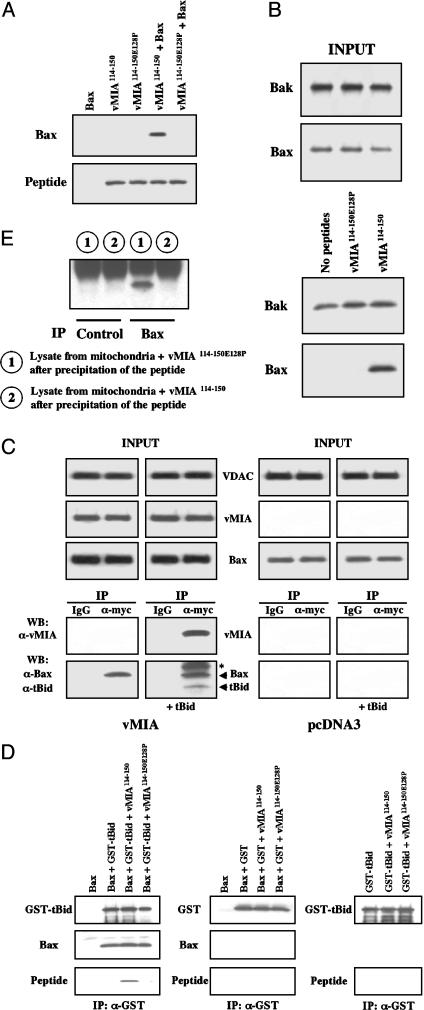
To examine whether vMIA binds Bax directly or through an intermediary, we tested the affinity of a synthetic N-biotinylated peptide with the amino acid sequence of the Val-114–Ala-150 segment of vMIA (denoted below as vMIA114-150) to a bacterially produced recombinant Bax. After incubation with Bax, vMIA114-150 was pulled down with streptavidin–agarose beads, and the polypeptides associated with the beads were then separated by SDS/PAGE and examined by immunoblotting (Bax) or with horseradish peroxidase–streptavidin (vMIA114-150). N-biotinylated peptide with a sequence identical to that of vMIA114-150 except for the substitution of a conserved (2) residue E128P to P (vMIA114-150E128P) was used as a control based on the inability of vMIAE128P to bind Bax or to protect cells from apoptosis (Table 1). Bax bound to vMIA114-150, but not to vMIA114-150E128P (Fig. 2A), confirming that (i) vMIA binds Bax, (ii) the interaction between the two proteins is direct and does not require any intermediary (although it does not rule out that the intracellular vMIA–Bax complex contains other proteins as well), and (iii) Bax-binding domain of vMIA is confined within its Val-114–Ala-150 segment. A shorter N-biotinylated synthetic peptide with a sequence corresponding to the vMIA114-141 segment failed to bind Bax in similar experiments (data not shown), suggesting that at least some of the extra amino acids in vMIA114-150 are required for its Bax-binding function. We also tested whether vMIA114-150 binds either Bax or Bak present in mitochondria isolated from HeLa cells. Purified mitochondria were incubated at 37°C for 30 min with vMIA114-150, vMIA114-150E128P, or buffer alone, then washed, lysed, and pulled down with streptavidin beads. The proteins on the beads were then separated by SDS/PAGE and analyzed by Western blotting for the presence of Bax or Bak. As shown in Fig. 2B, Bax selectively associated with vMIA114-150, whereas we could not detect any vMIA-selective association of Bak to the beads in excess of nonspecific binding.
Fig. 2.
(A) A synthetic peptide vMIA114-150 has specific affinity for Bax. Biotinylated vMIA114-150 (1 μM) and control biotinylated vMIA114-150E128P (1μM), containing a point mutation that inactivates cell-death-suppressing activity of vMIA were incubated with either recombinant Bax (100 nM) or buffer alone, pulled down with streptavidin-agarose, separated by SDS/PAGE, and examined by Western blotting with horseradish peroxidase–streptavidin and anti-Bax. (B) vMIA114-150 binds endogenous Bax, but not endogenous Bak. Isolated mitochondria (500 μg) were incubated with biotinylated vMIA114-150 (10 μg), control biotinylated vMIA114-150E128P (10 μg), or buffer alone, pulled down with streptavidin-agarose, separated by SDS/PAGE, and analyzed by Western blotting. As a control, “input” represents 2% of the starting material. (C) vMIA does not prevent the binding of tBid to Bax. Isolated mitochondria from HeLa/vMIA (vMIA mitochondria) (500 μg) or from HeLa/pcDNA3 (500 μg) were incubated with tBid (100 nM) or buffer alone, then vMIA was immunoprecipitated (IP) with anti-myc by using an irrelevant antibody as a control, the samples were separated by SDS/PAGE, and Bax, tBid, and vMIA were then detected by Western blotting (WB). Asterisk indicates a band that may be either of a nonspecific nature, or a covalently modified form of Bax or tBid. As a control, “input” represents 2% of the starting material. (D) Recombinant Bax (100 nM) was incubated with GST–tBid (100 nM) in the presence or absence of a 10-fold molar excess of vMIA114-150 or vMIA114-150 E128P (each at 1 μM), then GST–tBid was immunoprecipitated (IP) with an anti-GST, and bound proteins were separated by SDS/PAGE and detected by Western blotting (Left). In control samples, Bax (100 nM) was incubated with GST (100 nM) in the presence or absence of the biotinylated peptides (1 μM), and GST–tBid was incubated with the biotinylated peptides (1 μM) to see whether they bind GST–tBid (Right). (E) Mitochondria isolated from HeLa/pcDNA3 were incubated either with vMIA114-150 (1 μM) or vMIA114-150E128P (1 μM) as a control. Peptides were pulled down as in B, then Bax was immunoprecipitated (IP) from the remaining supernatant of the mitochondrial lysate, and the immunoprecipitate was examined for the presence of Bax. An irrelevant antibody was used as a control.
vMIA Does Not Prevent Binding of tBid to Bax. After exposure to tBid, Bax and Bak undergo a conformational change, oligomerize, and then trigger the efflux of mitochondrial death-activating factors into the cytoplasm (28, 29). vMIA blocks mitochondrial membrane permeabilization and suppresses apoptosis in HeLa and HCT116 cells (Figs. 5 and 6A, which are published as supporting information on the PNAS web site). We set out to examine whether vMIA interferes with these phenomena in a cell-free environment. It had been shown that mitochondria isolated from HeLa cells contain loosely associated monomeric (nonactivated) Bax (30). First, we tested whether Bax associated with vMIA-containing mitochondria could bind tBid. We incubated purified mitochondria from HeLa/vMIA cells with recombinant tBid, then extensively washed the mitochondria, lysed them in a Triton X-100-containing buffer, immunoprecipitated vMIA with anti-myc, and tested the immunoprecipitates for the presence of Bax and tBid by Western blotting. We found (Fig. 2C) that, under these cell-free conditions, tBid coimmunoprecipitated with vMIA and Bax, presumably via its binding to Bax. To establish in a better-defined system if vMIA competes with tBid for Bax binding, we examined whether GST–tBid bound Bax in the presence of an excess of vMIA114-150, using GST and vMIA114-150E128P as controls. Upon mixing and incubating the reagents, GST–tBid (or GST) was immunoprecipitated with anti-GST, then the proteins associated with the immunoprecipitates were separated by SDS/PAGE, and examined for the presence of Bax, the vMIA peptides, and GST–tBid or GST (Fig. 2D). Bax associated with GST–tBid, but not with GST. vMIA114-150 associated with the GST–tBid/Bax complex, but not with GST–tBid in the absence of Bax. vMIA114-150E128P did not associate with the GST–tBid/Bax complex. Importantly, even a 10-fold molar excess of vMIA114-150 failed to prevent GST–tBid/Bax association, even though all Bax molecules were associated with vMIA (Fig. 2E). Thus, there is no competition between GST–tBid and vMIA114-150 for Bax, and GST–tBid does not directly interact with vMIA114-150.
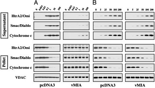
An Excess of Activated Bax, but Not of tBid, Induces Permeabilization of Mitochondria Isolated from vMIA-Expressing Cells. If vMIA protects mitochondria by directly interacting with and sequestering all intracellular Bax, then it could be expected that the protection by vMIA would not be overcome by an excess tBid, but could be overcome by an excess of activated Bax. To test this hypothesis, we incubated mitochondria isolated from either HeLa/vMIA cells or control HeLa/pcDNA3 cells with increasing concentrations of either tBid or oligomeric activated Bax (which had been described in ref. 19). We used the efflux of the three mitochondrial death-activating factors, HtrA2/Omi, Smac/Diablo, and cytochrome c, from mitochondria (pellet) into the buffer (supernatant) as an index of mitochondrial membrane permeabilization. Although control mitochondria were sensitive to the permeabilizing effect of as little as 0.1 nM tBid, vMIA-containing mitochondria remained essentially intact after being exposed to up to 100 nM tBid, the highest concentration tested, with only a minor leakage (Fig. 3A). In contrast, vMIA-containing mitochondria were nearly as sensitive as the control mitochondria in the presence of oligomeric Bax (Fig. 3B). Monomeric Bax activated with tBid (28) was similar to oligomeric activated Bax in that it also overcame the protective effect of vMIA (Fig. 7A, which is published as supporting information on the PNAS web site). We also examined whether vMIA114-150 could protect mitochondria from the permeabilizing effect of tBid. Mitochondria that were isolated from HeLa cells were exposed to vMIA114-150, then to tBid, and examined as above. vMIA114-150 was unable to prevent mitochondrial membrane permeabilization (Fig. 7B), even though all Bax associated with mitochondria was bound to this peptide (Fig. 2E).
Fig. 3.
(A and B) vMIA prevents mitochondrial membrane permeabilization induced by recombinant tBid (concentrations are shown on top in nM) (A), but not by recombinant oligomeric Bax (concentrations are shown on top in nM) (B), see text for explanations. Equal loading of the mitochondrial pellet was verified by using VDAC.
Mitochondria Isolated from Bax-Deficient HCT116 Cells Are Resistant to tBid. Several human and a primate cell lines, HeLa, MRC5 human normal fibroblasts, HCT116, and Cos-7 express both Bax and Bak (easily detectable by Western blot), and yet vMIA protects these cells from apoptosis (Figs. 5 and 6A, data not shown, and ref. 1), suggesting that, in these cells, Bax is dominant over Bak in mediating the mitochondrial apoptotic pathway. This notion is supported by the published observations that Bax-deficient HCT116Bax–/– cells that express Bak are resistant to apoptosis induced by a variety of apoptotic stimuli (31–36). This model predicts that mitochondria isolated from Bax-deficient HCT116Bax–/– cells would be resistant to the permeabilizing activity of tBid. This was indeed confirmed in our experiments with mitochondria isolated from HCT116 and HCT116Bax–/– cells (Fig. 6B): although mitochondria isolated from HCT116 cells were permeabilized after an exposure to 1 nM tBid, those isolated from HCT116Bax–/– cells were intact after an exposure to as high as 100 nM tBid, the highest concentration tested.
Discussion
Here we present evidence that vMIA suppresses the mitochondrial apoptotic signaling pathway by directly binding Bax and sequestering it at mitochondria. In agreement with this mechanism, and within a model (28, 37) that tBid induces mitochondrial membrane permeabilization by activating Bax, mitochondria isolated from vMIA-expressing cells are refractory to the permeabilizing effect of even a vast excess of tBid, but not activated Bax.
vMIA appears to have a dual mitochondria/endoplasmic reticulum (ER) localization signal, and a significant fraction of the intracellular pool of vMIA is localized at the ER (16, 38). The deletion mutants vMIAΔ2–23 and vMIAΔ23–34 are not colocalized with mitochondria, and are found in the cytoplasm and in the cytoplasm and at the ER, respectively (1, 38). These mutants bind Bax but fail to suppress apoptosis. Similarly, a deletion mutant lacking the mitochondria-targeting domain altogether, vMIA114-150, binds Bax but fails to prevent tBid-mediated permeabilization of mitochondria in a cell-free assay. These data suggest that mitochondrial targeting of vMIA-associated Bax is required for its sequestration, although the reason for this requirement is, at present, unclear. Our finding that vMIA associates with mitochondria (most likely with the mitochondrial outer membrane, ref. 1) irrespective of the presence or absence of Bax is consistent with the model in which Bax–vMIA complex is attached to mitochondria, at least in part, via the mitochondria-targeting domain of vMIA. It remains to be elucidated whether the mitochondrial localization signal of Bax is involved as well.
Vieira et al. reported (39) that vMIA did not protect intact cells or isolated mitochondria from the damage induced by peptides containing either a Bax–BH3 or Bcl-2–BH3 sequence. This property of the peptides is phenomenologically similar to the effect of recombinant activated Bax on cell-free mitochondria, but different from that of recombinant tBid. At present, the molecular mechanism of Bax-mediated permeabilization of mitochondrial membranes is not known, and it is not clear whether Bax and the peptides act in a similar manner (6).
Recent data suggest that cells can be divided into at least two distinct phenotypes in regard to the relative contribution of Bax and Bak into the mediation of the mitochondrial apoptotic signaling pathway, those where this pathway is dominated mainly by Bax (BaxD phenotype), and those in which either Bax or Bax can mediate this pathway (Bax/Bak-codominance, Bax/BakcoD). Human colorectal carcinoma HCT116 cells that express both Bax and Bak, represent an example of BaxD cells. In response to a wide variety of apoptotic stimuli, HCT116Bax+/– cells undergo massive apoptosis, whereas most HCT116Bax–/– cells remain viable (31–36). Oligomerization of Bak at mitochondria of HCT116 cells requires the presence of Bax (40), and mitochondria isolated from HCT116Bax–/– cells, unlike those isolated from HCT116 cells, are resistant to the permealizing effect of tBid. MEFs, human glioblastoma cell lines, and transformed baby mouse kidney cells represent a Bax/BakcoD phenotype (26, 41–44).
The apparent inability of vMIA to suppress apoptosis in cells with active Bak suggests that vMIA may be used as a probe to reveal the BaxD phenotype. Ectopic vMIA is functional as a cell death suppressor in several cell types, including HeLa (1), HCT116 (this study), and Cos-7 and MRC5 normal human fibroblasts (L.M.B. and V.S.G., unpublished observations). Interestingly, human and primate CMV encode vMIA (2), suggesting that cell types that are essential for supporting CMV infections in humans and primates may be of the BaxD phenotype. On the other hand, none of the characterized CMVs infecting other species encode vMIA, and MEFs are permissive for murine CMV (which does not have any vMIA homologs) and may be of Bax/BakcoD phenotype.
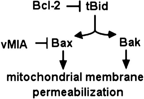
Previously, we reported (17) that the cell-death-suppressing activity of vMIA, like that of Bcl-2, is limited to type II cells (45) and that vMIA was not functional in type I BJAB cells. Our current data suggest that the death-suppressing activity of vMIA is limited even further to only the BaxD subset of type II cells. In this property, vMIA differs from Bcl-2 and Bcl-xL, which suppress both Bax- and Bak-mediated apoptosis (8), which fits the current models for their mechanisms of action (Fig. 4). However, we should take into consideration that the division of cells into type I and type II has been defined only for death-receptor-mediated apoptosis, and it remains to be established whether apoptosis induced by other types of stimuli, such as viral infection, various cytotoxic drugs, or γ-irradiation, in type I cells proceeds through activation of caspase-8 and then through the direct caspase-8-mediated activation of Bid, or through a caspase-8-independent up-regulation of a BH3-only death factor. It may well be that vMIA (and Bcl-2) suppresses apoptosis triggered by such stimuli in type I cells as well. Similarly, the involvement of Bax and Bak in the mitochondrial apoptotic pathway triggered in response to various stimuli may also differ depending on the nature of the stimulus (42).
Fig. 4.
A schematic representation of suppression of the tBid-mediated apoptotic pathway by vMIA.
Taken together, our data support the model in which vMIA and Bcl-2/Bcl-xL block mitochondrial membrane permeabilization by targeting the opposite components of the tBid/Bax pair: vMIA inactivates Bax, whereas Bcl-2/Bcl-xL inactivates tBid. The mechanism of cell death suppression by vMIA is also distinct from that by E1B19K and humanin, two antiapoptotic proteins that directly target Bax. E1B19K inhibits Bax- and Bak-mediated apoptosis, has affinity for Bak and for tBid-activated Bax, and blocks relocation of Bax from the cytoplasm to mitochondria (46, 47). Humanin appears to suppress apoptosis by binding Bax and preventing its activation and relocation to mitochondria (48).
CMV is a major cause of morbidity and mortality in immuno-compromised individuals, such as organ transplant recipients and patients with AIDS, and a major cause of congenital disease during pregnancy. Our finding that vMIA binds Bax establishes a rationale for the development of screening assays to search for anti-CMV drugs that target and inactivate vMIA.
Supplementary Material
Acknowledgments
GST–tBid/pGEX-4T-1 was a generous gift of Eric A. Hendrickson (Brown University). Recombinant oligomeric Bax was a generous gift from Bruno Antonsson (Serono Pharmaceutical Research Institute, Geneva). HCT116 cells and MEFs were a generous gift of Bert Vogelstein (Johns Hopkins University) and Stanley J. Korsmeyer (Harvard University), respectively. Anti-ANT rabbit antiserum was a generous gift from Patricia Schmid (University of Minnesota).
This paper was submitted directly (Track II) to the PNAS office.
Abbreviations: CMV, cytomegalovirus; MEF, murine embryonic fibroblast; ANT, adenine nucleotide translocator; ER, endoplasmic reticulum.
References
- 1.Goldmacher, V. S., Bartle, L. M., Skaletskaya, A., Dionne, C. A., Kedersha, N. L., Vater, C. A., Han, J. W., Lutz, R. J., Watanabe, S., Cahir McFarland, E. D., et al. (1999) Proc. Natl. Acad. Sci. USA 96, 12536–12541. [DOI] [PMC free article] [PubMed] [Google Scholar]
- 2.McCormick, A. L., Skaletskaya, A., Barry, P. A., Mocarski, E. S. & Goldmacher, V. S. (2003) Virology 316, 221–233. [DOI] [PubMed] [Google Scholar]
- 3.Goldmacher, V. S. (2002) Biochimie 84, 177–185. [DOI] [PubMed] [Google Scholar]
- 4.Yang, J., Liu, X., Bhalla, K., Kim, C. N., Ibrado, A. M., Cai, J., Peng, T. I., Jones, D. P. & Wang, X. (1997) Science 275, 1129–1132. [DOI] [PubMed] [Google Scholar]
- 5.Kluck, R. M., Bossy-Wetzel, E., Green, D. R. & Newmeyer, D. D. (1997) Science 275, 1132–1136. [DOI] [PubMed] [Google Scholar]
- 6.Letai, A., Bassik, M. C., Walensky, L. D., Sorcinelli, M. D., Weiler, S. & Korsmeyer, S. J. (2002) Cancer Cell 2, 183–192. [DOI] [PubMed] [Google Scholar]
- 7.Chittenden, T. (2002) Cancer Cell 2, 165–166. [DOI] [PubMed] [Google Scholar]
- 8.Cheng, E. H., Wei, M. C., Weiler, S., Flavell, R. A., Mak, T. W., Lindsten, T. & Korsmeyer, S. J. (2001) Mol. Cell 8, 705–711. [DOI] [PubMed] [Google Scholar]
- 9.Hsu, Y. T. & Youle, R. J. (1997) J. Biol. Chem. 272, 13829–13834. [DOI] [PubMed] [Google Scholar]
- 10.Mikhailov, V., Mikhailova, M., Pulkrabek, D. J., Dong, Z., Venkatachalam, M. A. & Saikumar, P. (2001) J. Biol. Chem. 276, 18361–18374. [DOI] [PubMed] [Google Scholar]
- 11.Simonian, P. L., Grillot, D. A., Merino, R. & Nunez, G. (1996) J. Biol. Chem. 271, 22764–22772. [DOI] [PubMed] [Google Scholar]
- 12.Simonian, P. L., Grillot, D. A. & Nunez, G. (1997) Oncogene 15, 1871–1875. [DOI] [PubMed] [Google Scholar]
- 13.Cheng, E. H., Levine, B., Boise, L. H., Thompson, C. B. & Hardwick, J. M. (1996) Nature 379, 554–556. [DOI] [PubMed] [Google Scholar]
- 14.Zha, H. & Reed, J. C. (1997) J. Biol. Chem. 272, 31482–31488. [DOI] [PubMed] [Google Scholar]
- 15.Wolter, K. G., Hsu, Y. T., Smith, C. L., Nechushtan, A., Xi, X. G. & Youle, R. J. (1997) J. Cell Biol. 139, 1281–1292. [DOI] [PMC free article] [PubMed] [Google Scholar]
- 16.Hayajneh, W. A., Colberg-Poley, A. M., Skaletskaya, A., Bartle, L. M., Lesperance, M. M., Contopoulos-Ioannidis, D. G., Kedersha, N. L. & Goldmacher, V. S. (2001) Virology 279, 233–240. [DOI] [PubMed] [Google Scholar]
- 17.Skaletskaya, A., Bartle, L. M., Chittenden, T., McCormick, A. L., Mocarski, E. S. & Goldmacher, V. S. (2001) Proc. Natl. Acad. Sci. USA 98, 7829–7834. [DOI] [PMC free article] [PubMed] [Google Scholar]
- 18.Zhang, L., Yu, J., Park, B. H., Kinzler, K. W. & Vogelstein, B. (2000) Science 290, 989–992. [DOI] [PubMed] [Google Scholar]
- 19.Antonsson, B., Montessuit, S., Lauper, S., Eskes, R. & Martinou, J. C. (2000) Biochem. J. 345, 271–278. [PMC free article] [PubMed] [Google Scholar]
- 20.Suzuki, M., Youle, R. J. & Tjandra, N. (2000) Cell 103, 645–654. [DOI] [PubMed] [Google Scholar]
- 21.Giron-Calle, J., Zwizinski, C. W. & Schmid, H. H. (1994) Arch. Biochem. Biophys. 315, 1–7. [DOI] [PubMed] [Google Scholar]
- 22.Arnoult, D., Parone, P., Martinou, J. C., Antonsson, B., Estaquier, J. & Ameisen, J. C. (2002) J. Cell Biol. 159, 923–929. [DOI] [PMC free article] [PubMed] [Google Scholar]
- 23.Arnoult, D., Gaume, B., Karbowski, M., Sharpe, J. C., Cecconi, F. & Youle, R. J. (2003) EMBO J. 22, 4385–4399. [DOI] [PMC free article] [PubMed] [Google Scholar]
- 24.Chittenden, T., Harrington, E. A., O'Connor, R., Flemington, C., Lutz, R. J., Evan, G. I. & Guild, B. C. (1995) Nature 374, 733–736. [DOI] [PubMed] [Google Scholar]
- 25.Farrow, S. N., White, J. H., Martinou, I., Raven, T., Pun, K. T., Grinham, C. J., Martinou, J. C. & Brown, R. (1995) Nature 374, 731–733. [DOI] [PubMed] [Google Scholar]
- 26.Wei, M. C., Zong, W. X., Cheng, E. H., Lindsten, T., Panoutsakopoulou, V., Ross, A. J., Roth, K. A., MacGregor, G. R., Thompson, C. B. & Korsmeyer, S. J. (2001) Science 292, 727–730. [DOI] [PMC free article] [PubMed] [Google Scholar]
- 27.Zamzami, N., Marchetti, P., Castedo, M., Decaudin, D., Macho, A., Hirsch, T., Susin, S. A., Petit, P. X., Mignotte, B. & Kroemer, G. (1995) J. Exp. Med. 182, 367–377. [DOI] [PMC free article] [PubMed] [Google Scholar]
- 28.Desagher, S., Osen-Sand, A., Nichols, A., Eskes, R., Montessuit, S., Lauper, S., Maundrell, K., Antonsson, B. & Martinou, J. C. (1999) J. Cell Biol. 144, 891–901. [DOI] [PMC free article] [PubMed] [Google Scholar]
- 29.Wei, M. C., Lindsten, T., Mootha, V. K., Weiler, S., Gross, A., Ashiya, M., Thompson, C. B. & Korsmeyer, S. J. (2000) Genes Dev. 14, 2060–2071. [PMC free article] [PubMed] [Google Scholar]
- 30.Antonsson, B., Montessuit, S., Sanchez, B. & Martinou, J. C. (2001) J. Biol. Chem. 276, 11615–11623. [DOI] [PubMed] [Google Scholar]
- 31.LeBlanc, H., Lawrence, D., Varfolomeev, E., Totpal, K., Morlan, J., Schow, P., Fong, S., Schwall, R., Sinicropi, D. & Ashkenazi, A. (2002) Nat. Med. 8, 274–281. [DOI] [PubMed] [Google Scholar]
- 32.Gillissen, B., Essmann, F., Graupner, V., Starck, L., Radetzki, S., Dorken, B., Schulze-Osthoff, K. & Daniel, P. T. (2003) EMBO J. 22, 3580–3590. [DOI] [PMC free article] [PubMed] [Google Scholar]
- 33.Yamaguchi, H., Bhalla, K. & Wang, H. G. (2003) Cancer Res. 63, 1483–1489. [PubMed] [Google Scholar]
- 34.Theodorakis, P., Lomonosova, E. & Chinnadurai, G. (2002) Cancer Res. 62, 3373–3376. [PubMed] [Google Scholar]
- 35.He, Q., Montalbano, J., Corcoran, C., Jin, W., Huang, Y. & Sheikh, M. S. (2003) Oncogene 22, 2674–2679. [DOI] [PubMed] [Google Scholar]
- 36.Mahyar-Roemer, M., Kohler, H. & Roemer, K. (2002) BMC Cancer 2, 27–35. [DOI] [PMC free article] [PubMed] [Google Scholar]
- 37.Eskes, R., Desagher, S., Antonsson, B. & Martinou, J. C. (2000) Mol. Cell. Biol. 20, 929–935. [DOI] [PMC free article] [PubMed] [Google Scholar]
- 38.Mavinakere, M. S. & Colberg-Poley, A. M. (2004) J. Gen. Virol. 85, 323–329. [DOI] [PubMed] [Google Scholar]
- 39.Vieira, H. L., Boya, P., Cohen, I., El Hamel, C., Haouzi, D., Druillenec, S., Belzacq, A. S., Brenner, C., Roques, B. & Kroemer, G. (2002) Oncogene 21, 1963–1977. [DOI] [PubMed] [Google Scholar]
- 40.Mikhailov, V., Mikhailova, M., Degenhardt, K., Venkatachalam, M. A., White, E. & Saikumar, P. (2003) J. Biol. Chem. 278, 5367–5376. [DOI] [PubMed] [Google Scholar]
- 41.Juin, P., Hunt, A., Littlewood, T., Griffiths, B., Swigart, L. B., Korsmeyer, S. & Evan, G. (2002) Mol. Cell. Biol. 22, 6158–6169. [DOI] [PMC free article] [PubMed] [Google Scholar]
- 42.Cartron, P. F., Juin, P., Oliver, L., Martin, S., Meflah, K. & Vallette, F. M. (2003) Mol. Cell. Biol. 23, 4701–4712. [DOI] [PMC free article] [PubMed] [Google Scholar]
- 43.Cuconati, A., Degenhardt, K., Sundararajan, R., Anschel, A. & White, E. (2002) J. Virol. 76, 4547–4558. [DOI] [PMC free article] [PubMed] [Google Scholar]
- 44.Degenhardt, K., Sundararajan, R., Lindsten, T., Thompson, C. & White, E. (2002) J. Biol. Chem. 277, 14127–14134. [DOI] [PubMed] [Google Scholar]
- 45.Scaffidi, C., Fulda, S., Srinivasan, A., Friesen, C., Li, F., Tomaselli, K. J., Debatin, K. M., Krammer, P. H. & Peter, M. E. (1998) EMBO J. 17, 1675–1687. [DOI] [PMC free article] [PubMed] [Google Scholar]
- 46.Cuconati, A. & White, E. (2002) Genes Dev. 16, 2465–2478. [DOI] [PubMed] [Google Scholar]
- 47.Sundararajan, R., Cuconati, A., Nelson, D. & White, E. (2001) J. Biol. Chem. 276, 45120–45127. [DOI] [PubMed] [Google Scholar]
- 48.Guo, B., Zhai, D., Cabezas, E., Welsh, K., Nouraini, S., Satterthwait, A. C. & Reed, J. C. (2003) Nature 423, 456–461. [DOI] [PubMed] [Google Scholar]
Associated Data
This section collects any data citations, data availability statements, or supplementary materials included in this article.


