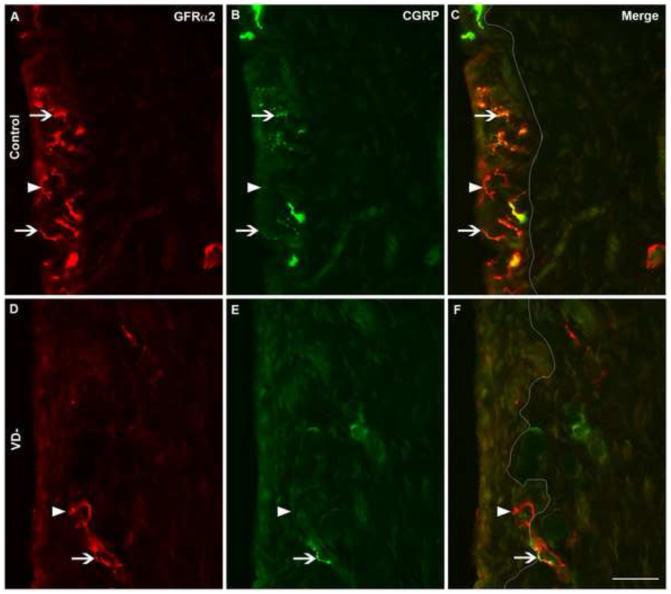
Fig. 4.
Colocalization of CGRP and GFRα2 immunoreactivity in synovial nerves. Representative images are shown of immunofluorescently-labeled nerves in knee synovium (A-F) from control (A-C) and vitamin D deficient (VD-, D-F) rats. Synovial sections were immunofluorescently-labeled for both GDNF family receptor alpha 2 (GFRα2, A&D) and calcitonin gene-related peptide (CGRP, B&E) and shown in merged images (C&F). A white line has been superimposed on images to separate the intima (left) from the subintima (right) (C&F). Arrowheads point toward selected nerves with GFRα2-only labeling, while arrows point toward selected nerves with CGRP and GFRα2 co-labeling. Scale=50μm

