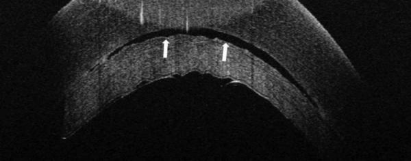
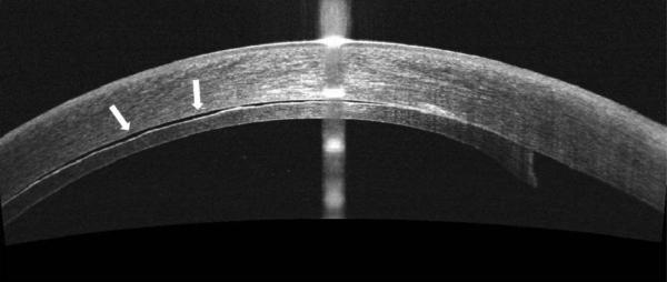
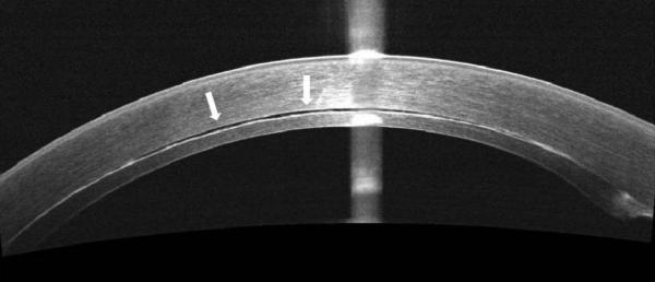
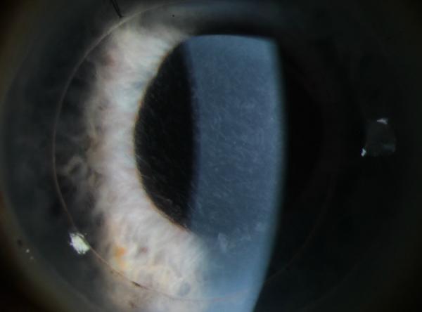
Figure 2.


In some cases, TIF is seen on iOCT, and persists beyond POD 1, also resulting in TIO. This persistent TIF is thought to be secondary to retained viscoelastic.
A. iOCT with interface space. This space is seen in multiple cross-sectional images.
B. POD 1 OCT. Note the areas of hyper-reflectivity along the stromal surface of the donor lenticule.
C. POM 1 OCT showing a stable amount of persistent interface fluid D. POM 1 Photo showing wave-like TIO.

